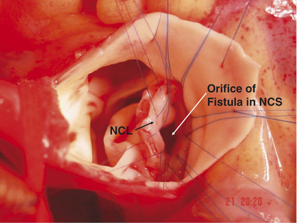
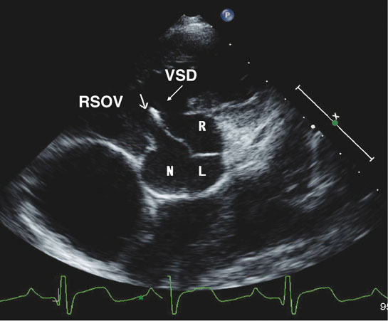
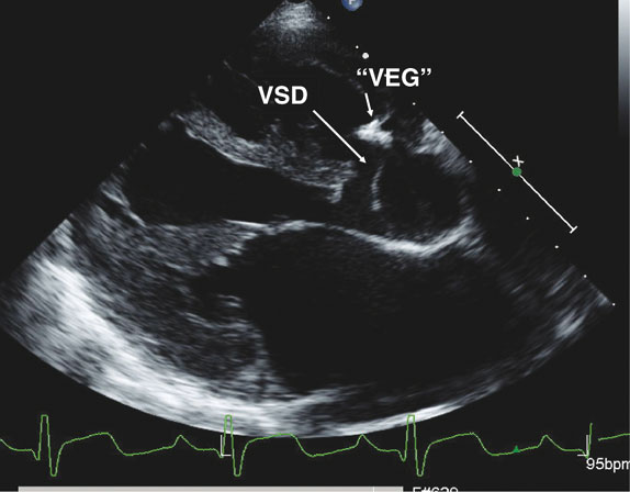
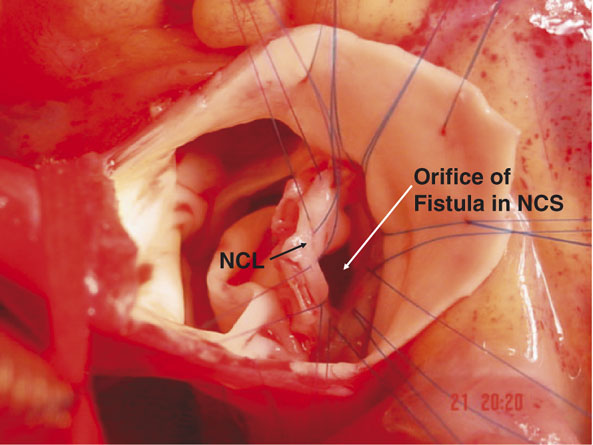
We present the images of a 12 year old boy known to have a ventricular septal defect, who was found to have a loud continuous murmur and a thrill along the left sternal border. Transthoracic echocardiography in the parasternal short axis view (Fig. 1) showed a large perimembranous ventricular septal defect (VSD) with flow from left to right. In addition, the noncoronary aortic sinus of Valsalva (N) had ruptured into the inlet of the right ventricle adjacent to tricuspid valve (arrow). Flow between the aorta and right ventricle was continuous. On this figure, we have also labelled the left (L) and right (R) coronary leaflets of the aortic valve, and the ruptured sinus of Valsalva (RSOV). Thickening of the distal portion of the “windsock” in the right ventricle, seen also in the parasternal long axis view (Fig. 2 – “VEG”), suggested the possibility of a healed vegetation from previous infective endocarditis. At operation (Fig. 3), the non-coronary sinus of Valsalva was dilated. The defect in the sinus, at the hinge point of the valvar leaflet (NCL), measured 1.5 × 1 centimetres. The ventricular septal defect was closed through the right atrium using a Gore-Tex patch. The orifice of the ruptured sinus of Valsalva was exposed through a transverse aortotomy, and closed with a pericardial patch using interrupted sutures. The patient made an uneventful recovery. Postoperative echocardiography confirmed complete closure of both the interventricular communication and the ruptured aneurysm of the sinus of Valsalva. Postoperatively, there was no aortic valvar regurgitation, and the patient remained in sinus rhythm.
Figure 1.
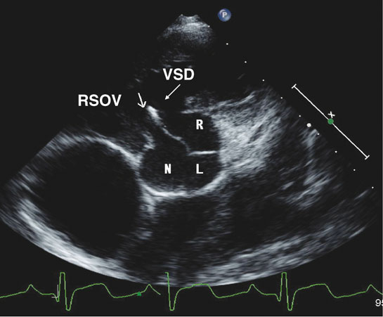
Figure 2.
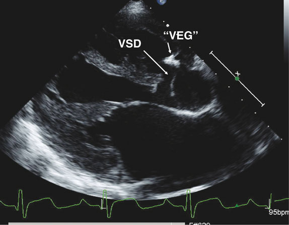
Figure 3.