INTRODUCTION
The fish faunas of the Bellingshausen Sea, Peter I Island, and West Antarctic Peninsula were studied as part of the Spanish Bentart Research Programmes. During the BENTART-03 and BENTART-06 Expeditions many zoarcids were captured with baited traps, mainly from the Bellingshausen Sea where they predominate both in number and biomass in waters deeper than 550 m (Matallanas & Olaso, Reference Matallanas and Olaso2007). With 32 known species in 14 genera, according to recent revisions and descriptions (Eastman, Reference Eastman2005; Anderson, Reference Anderson2006; Moeller & Stewart, Reference Moeller and Stewart2006; Matallanas, Reference Matallanas2008, Reference Matallanas2009a, Reference Matallanasb, Reference Matallanasc), Zoarcidae is one of the most speciose benthic fish families in the Antarctic waters.
Ophthalmolycus, established by Regan (Reference Regan1913), was reviewed by Gosztonyi (Reference Gosztonyi1977), Anderson (Reference Anderson1988, Reference Anderson1990, Reference Anderson1992, Reference Anderson1994), and Anderson & Gosztonyi (Reference Anderson and Gosztonyi1991). According to Anderson & Fedorov (Reference Anderson and Fedorov2004), and Matallanas (Reference Matallanas2009b) it contains seven species: O. amberensis (Tomo et al., Reference Tomo, Marschoff and Torno1977), O. andersoni Matallanas, 2009, O. bothriocephalus (Pappenheim, Reference Pappenheim1912), O. campbellensis Andriashev & Fedorov, Reference Andriashev and Fedorov1986, O. chilensis Anderson, Reference Anderson1992, O. conorhynchus (Garman, Reference Garman1899), and O. macrops (Günther, Reference Günther1880). In the present report, two new species of Ophthalmolycus (Ophthalmolycus eastmani sp. nov., and Ophthalmolycus polylepis sp. nov.) are described, and a key to the species of the genus is provided.
MATERIALS AND METHODS
The type specimens were collected in the Bellingshausen Sea and Gerlache Strait during the Spanish Expeditions BENTART-03 and BENTART-06 (January–February 2003 and January–February 2006) on board of the RV ‘Hespérides’ (Matallanas & Olaso, Reference Matallanas and Olaso2007). All the material has been deposited at the UAB fish collection (Zoología, Universidad Autónoma de Barcelona).
Counts, measurements and general terminology follow Gosztonyi (Reference Gosztonyi1977) and Anderson (Reference Anderson1982, Reference Anderson1988). Measurements were made with ocular micrometer or dial calipers to the nearest 0.1 mm. Pore terminology follows Gosztonyi (Reference Gosztonyi1977) and Anderson (Reference Anderson1982). Meristic and morphometrical data of the seven described species of Ophthalmolycus are in Matallanas (Reference Matallanas2009b). Specimens were X-rayed to record both shape and meristics of axial skeleton and vertical fins. Osteological observations were made from radiographs of all specimens. Suspensorium of Opthalmolycus eastmani sp. nov. was obtained from the dissected paratype UAB:B06BSZ55, stained with alizarin red S. Abbreviations used: SL, standard length; HL, head length; D, dorsal fin; A, anal fin; P, pectoral fin.
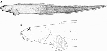
Ophthalmolycus eastmani sp. nov., UAB:B06BSZ54 (holotype), 369 mm standard length male, from Bellingshausen Sea. (A) Left lateral view; (B) left lateral view of head showing pore pattern.

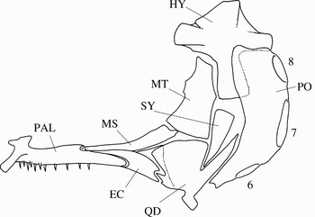
Suspensorium and preopercle of Ophthalmolycus eastmani sp. nov., paratype (UAB:B06BSZ55); external view of the right side: EC, ectopterygoid; HY, hyomandibula; MS, mesopterygoid; MT, metapterygoid; PAL, palatine; QD, quadrate; SY, symplectic; 6–8, preopercular pores.

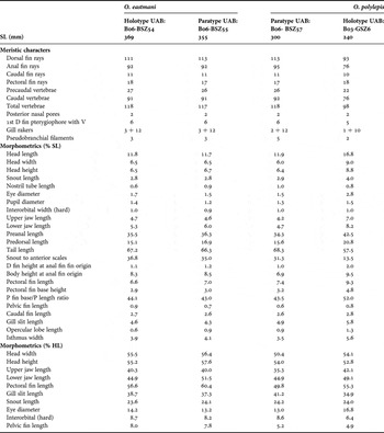
Counts and measurements of Ophthalmolycus eastmani sp. nov. and Ophthalmolycus polylepis sp. nov.

SL, standard length; D, dorsal fin; V, vertebrae; P, pectoral fin; HL, head length.
TYPE MATERIAL
Holotype: male, 369 mm SL (BENTART-06, Bellingshausen Sea, Station MB32N, baited traps; coordinates: 68°48′S 86°38′W; water depth: 1837 m) (UAB:B06BSZ54). Collected by J. Matallanas 28 January 2006.
Paratypes: female, 300 mm SL (UAB:B06BSZ57), and female, 355 mm SL, used for anatomical analysis (UAB:B06BSZ55) captured with the holotype.
COMPARATIVE MATERIAL EXAMINED
Ophthalmolycus andersoni Matallanas, 2009. Holotype male (UAB:B03BSZ25). BENTART-03 Cruise to Antarctic Peninsula and Bellingshausen Sea, Station 22A (Paradise Bay, Antarctic Peninsula; 64°50′37″S 62°58′09″W). Collected 24 February 2003 with Agassiz trawl, 286 m depth. Paratype male (UAB:B03BSZ9), and paratype female (UAB:B03BSZ99) captured with the holotype.
Ophthalmolycus amberensis (Tomo, Marschoff & Torno, Reference Tomo, Marschoff and Torno1977). Specimen (UAB:WSZ29). ANTARKTIS XIII/3 Cruise, Station 4 (Weddell Sea; 71°69′S 12°49′W). Collected 20 February 1997 with Agassiz trawl, 449 m depth.
Ophthalmolycus bothriocephalus (Pappenheim, Reference Pappenheim1912). Specimen (UAB:WSZ30). ANTARKTIS XIII/3 Cruise, Station 92 (Weddell Sea; 71°58′S 12°44′W). Collected 26 February 1997 with Agassiz trawl, 571 m depth.
DIAGNOSIS
A species of Ophthalmolycus as defined by Anderson (Reference Anderson1992, Reference Anderson1994) with the following combination of characters: two posterior nasal pores; epidermal prickles on lips and snout; gill slit extending ventrally to the lower margin of the pectoral fin base; vertebrae asymmetrical 26 − 27 + 91 − 92 = 117 − 118; dorsal fin rays 111–113; dorsal fin origin associated with vertebra 6, with no free pterygiophores; pectoral fin rays 17–18; two postorbital pores; lateral line with ventral and mediolateral branches; scales extending anteriorly to just before anal fin origin; six branchiostegal rays; oral valve nearly reaching the anterior edge of vomer; palatine teeth in two rows anteriorly; 3–5 pseudobranch filaments; two well developed pyloric caeca; pelvic fin and vomerine teeth present.
Counts and proportional measurements are presented in Table 1.
DESCRIPTION (Figures 1–2; Table 1)
Body elongate, ovoid in cross-section. Head ovoid; eye nearly round, not reaching dorsal profile of head. Upper jaw slightly protruding; end of maxilla extending to the posterior margin of pupil; mouth subterminal; lips and snout with small prickles; nasal tube short, unpigmented, not reaching upper lip when pressed forward. Gill slit extending ventrally just to the ventral edge of pectoral fin base; small opercular flap at the upper end of gill slit. Upper end of the pectoral fin base at body midline, its lower end above the ventral profile of body (Figure 1A & B). Flesh and skin firm; scales extending anteriorly to about a snout length anterior to anal fin origin, and covering un-paired fin bases; scales absent on head, nape, pectoral fin, and abdomen.
Teeth on jaws, vomer and palatines conical. Premaxilla with 3 rows of teeth anteriorly and single row posteriorly. Dentary with 3 rows of teeth anteriorly and single row posteriorly. Vomerine teeth in a patch. Palatine teeth in two irregular rows anteriorly and single row posteriorly. Oral valve free of the palate laterally and nearly reaching the anterior edge of vomer. Pyloric caeca well developed and unequal: the anterior, largest, almost equal to eye diameter in length. Gill rakers 2 − 3 + 12, forked with 4–5 cusps. Pseudobranch filaments 3–5, elongated.
Cephalic lateralis pore system with pores small and rounded. Nasal pores three, one located anteromesial to nasal tube, two others posterodorsal to nasal tube, posterior one oval and larger. Postorbital pores two (positions one and four). Suborbital pores 6 + 0 (all on the ventral ramus); preoperculomandibular pores 8 (Figure 1B). Interorbital and occipital pores absent. Lateral line configuration with two branches (Figure 1A); ventral branch beginning just behind the fourth postorbital pore and coursing ventrolaterally to the end of the tail; mediolateral lateral branch, with more scattered neuromasts, originating above the anterior third of the pectoral fin and coursing at mid-body to tail tip.
Neurocranium elongate, depressed. Palatopterygoid series well developed with mesopterygoid and ectopterygoid overlapping more than half of dorsal and anterior surface of quadrate. Anterior surface of quadrate serrated. Ectopterygoid short. Metapterygoid large. Posterior ramus of hyomandibula short (Figure 2). Hyoid bar with six branchiostegal rays. Ceratohyal and epihyal striated, but ceratohyal–epihyal joint smooth. Post-temporal ventral ramus weak.
Vertebrae asymmetrical, 26 − 27 + 91 − 92. Last precaudal vertebra associated with dorsal fin ray 22. Dorsal fin origin associated with vertebra 6, with no free pterygiophores. Terminal dorsal fin ray associated with the second preural vertebrae. Three anal fin pterygiophores, with 3 anal fin rays, inserted anterior to haemal spine of the first caudal vertebra; a free pterygiophore in the holotype. Terminal anal fin ray associated with the second preural vertebra. One epural. Caudal fin rays 11, with two epural, four upper hypural and five lower hypural rays.
Colour in alcohol. Uniformly brownish; pectoral and un-paired fins with darker margins; nasal tube unpigmented; gill cavity light brown; peritoneum black.
REMARKS
Opthalmolycus eastmani agrees with O. campbellensis in meristic counts: vertebrae, D, and A fin rays. However the two species differ in the vertebral centra (asymmetrical in O. eastmani versus symmetrical in O. campbellensis), posterior nasal pores (two versus one), postorbital pore pattern (1 and 4 versus 1, 3, and 4), squamation (scales extending anteriorly to the anal fin origin versus to a vertical through the pectoral fin base), pyloric caeca development (well developed versus small nubs), prickles on lips and snout (present versus absent), pectoral fin ray number (17–18 versus 15), eye diameter (13.2–14.2% HL versus 23.0–27.0), and pelvic fin length (5.2–8.0% HL versus 15.0–20.0). The two species differ also in mouth position and body colour.
Opthalmolycus eastmani differs from the remaining Ophthalmolycus species chiefly by having two posterior nasal pores, higher meristic counts (vertebrae, dorsal and anal fin rays), shorter head and pelvic fins, and in some of the following diagnostic characters: six branchiostegal rays, body lateral line double and prickles on lips and snout.
ETYMOLOGY
The new species is named after Dr Joseph T. Eastman, in honour of his contributions to knowledge of Antarctic fish.
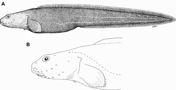
Ophthalmolycus polylepis sp. nov., UAB:B03GSZ6 (holotype), 240 mm standard length female, from Gerlache Strait. (A) Left lateral view; (B) left lateral view of head showing pore pattern.

TYPE MATERIAL
Holotype: female, 240 mm SL (BENTART-03, Gerlache Strait, Station 24N, baited traps; coordinates: 64°19′S 61°58′W; water depth: 1056 m) (UAB:B03GSZ6). Collected by J. Matallanas 26 February 2006.
COMPARATIVE MATERIAL EXAMINED
As for Ophthalmolycus eastmani.
DIAGNOSIS
A species of Ophthalmolycus as defined by Anderson (Reference Anderson1992, Reference Anderson1994) with the following combination of characters: two posterior nasal pores; epidermal prickles on lips; gill slit extending ventrally just to below mid-pectoral fin base; interorbital pore; two postorbital pores; lateral line triple with ventral, mediolateral and dorsolateral branches; scales extending anteriorly to predorsal area, abdomen, pelvic fin base, and pectoral fin base and axil; asymmetrical vertebrae 22 + 76; dorsal fin origin associated with vertebra 5; pectoral fin rays 18; six branchiostegal rays; oral valve overlapping the anterior edge of vomer; two rows of palatine teeth anteriorly; two vestigial pyloric caeca; 2 pseudobranch filaments; pelvic fin and vomerine teeth present.
Counts and proportional measurements are presented in Table 1.
DESCRIPTION (FIGURE 3; TABLE 1)
Body relatively short, ovoid in cross-section. Head small; eyes rounded reaching dorsal profile of head; anterior profile of snout vertical. Upper jaw slightly protruding; end of maxilla extending to the posterior margin of pupil; mouth inferior; lips with small prickles; nasal tube short, pigmented, not reaching upper lip when pressed forward. Gill slit extending ventrally just to below mid-pectoral fin base; triangular opercular flap at the upper end of gill slit. Upper end of the pectoral fin base at body midline, its lower end above the ventral profile of body. Flesh and skin firm; scales extending anteriorly to predorsal area, present on abdomen to pelvic base, pectoral fin base and axil (covering two-thirds of pectoral fin length), and vertical fins; head naked (Figure 3A & B).
All teeth conical. Premaxilla with 3 rows of teeth anteriorly reduced to a single row posteriorly. Dentary with 3–4 rows of teeth anteriorly and a single row posteriorly. Vomerine teeth in a patch. Palatine teeth in two irregular rows anteriorly and in a single row posteriorly. Oral valve overlapping the anterior edge of vomer. Two pyloric caeca barely produced. Gill rakers 1 + 10 forked, with 3 short cusps. Pseudobranch filaments 2, short.
Cephalic lateralis pore system with pores of unequal size. Nasal pores three, one located anteromesial to nasal tube, two others posterodorsal to nasal tube (Figure 3B). Interorbital pore present. Postorbital pores 2 (positions 1 and 4). Suborbital pores 6 + 0 (all on the ventral ramus); preoperculomandibular pores 8. Lateral line configuration with three branches (Figure 3A). Ventral branch beginning just behind the fourth postocular pore and coursing along the anal fin to the end of tail; mediolateral branch originating just before anus, and coursing at mid-body to the tail tip; dorsolateral branch originating behind anal fin origin, with some widely scattered neuromasts on the tail.
Neurocranium elongate, depressed; post-temporal ventral ramus absent. Vertebrae asymmetrical, 22 + 76. Dorsal fin origin associated with vertebra 5, with no free pterygiophores; last precaudal vertebra associated with dorsal fin ray 18. Terminal dorsal fin ray associated with the second preural vertebra. Two anal fin pterygiophores with two anal fin rays anterior to the haemal spine of the first caudal vertebra; a free preanal pterygiophore. Terminal anal fin ray associated with the second preural vertebra. One epural. Caudal fin damaged with two epural, four upper hypural and four (or five ?) lower hypural rays. Hyoid bar with six branchiostegal rays. Ceratohyal–epihyal joint smooth.
Colour in alcohol. Head and body uniform dark brown which contrast with the white colour of the scales; nasal tube pigmented. Lining of mouth pale. Gill cavity and peritoneum black.
REMARKS
Ophthalmolycus polylepis agrees with O. amberensis, O. andersoni, O. bothriocephalus and O. chilensis in having six branchiostegal rays, pelvic fins, and overlapping ranges of vertebrae, dorsal, and anal fin rays. However, O. polylepis differs from all them, and from the other species of the genus including O. eastmani described above, by the following combination of characters: scales present on predorsal area, abdomen, and pectoral fin base and axil; body lateral line with three branches, interorbital pore present, two small pyloric caeca and, excepting O. eastmani, in having two posterior nasal pores.
Some additional characters to distinguish the new species from its congeners are the following. Ophthalmolycus polylepis differs from O. amberensis in pelvic fin length (4.9% HL versus 13, 1–22.2), palatine teeth (2 rows anteriorly versus single row), gill slit (extending ventrally to just below mid-pectoral fin versus to the lower edge of pectoral fin or just above it) and oral valve (overlapping the anterior edge of vomer versus not reaching it), amongst other characters. Ophthalmolyeus polylepis differs from O. andersoni in pelvic fin length (4.9 %HL versus 10.2–12.9), nasal tube (not reaching upper lip when pressed forward versus overhanging it), gill slit (extending ventrally to just below mid-pectoral fin versus well below its lower edge), oral valve (overlapping the anterior edge of vomer versus not reaching it), palatine teeth (2 rows anteriorly versus single row), pyloric caeca (two small bulges versus two well developed), amongst other characters. Ophthalmolycus polylepis differs from O. bothriocephalus mainly in body colour (uniform dark brown versus mottled), pectoral fin rays (18 versus 12–15), dorsal fin origin (associated with vertebra 5 versus vertebrae 2–3), mouth position (inferior versus subterminal), gill slit (extending ventrally to just below mid-pectoral fin versus to its lower edge or just above it), pyloric caeca (two small bulges versus two very long ones), amongst other characters. Ophthalmolycus polylepis differs from O. chilensis in postorbital pore pattern (pores 1 and 4 versus 1, 2, 3, and 4–5), suborbital pores (6 + 0 versus 6 + 1), mouth position (inferior versus subterminal), dorsal fin origin (associated with vertebra 5 versus vertebra 3), nasal tube (not reaching upper lip when pressed forward versus overhanging it), amongst other characters. Ophthalmolycus polylepis differs from O. conorhynchus mainly by having pelvic fins and also in suborbital pore pattern, snout length, gill slit extension and palatine teeth rows. Ophthalmolycus polylepis differs from O. macrops mainly by having 6 branchiostegal rays, pectoral fin rays number, dorsal fin origin, eye diameter, and gill slit extension, amongst other.
Finally, Opthalmolycus polylepis differs from both O. campbellensis and O. eastmani also in meristic counts: precaudal vertebrae (22 versus 26–27 in both O. campbellensis and O. eastmani); caudal vertebrae (76 versus 92–98, and 91–92 respectively), and anal fin rays (76 versus 97–102, and 92–95 respectively).
ETYMOLOGY
The specific name is from the Greek polus, many, and lepis, scales, in reference to the extensive squamation of this species.
DISCUSSION
Questionable characters to include the two new species in Ophthalmolycus are the presence of two posterior nasal pores in both species, the gill slit extension to the lower margin of the pectoral fin base in O. eastmani, and the presence of an interorbital pore in O. polylepis. Both the twin posterior nasal pores and the interorbital pore have not been found in any described species of Ophthalmolycus. However, the main diagnostic characters of Ophthalmolycus given by Anderson (Reference Anderson1994): (suborbital bones 7–8, canal with 6–8 pores; gill slit not reaching ventral end of pectoral base; palatopterygoid series well developed; branchiostegal rays 5–6; scales, pseudobranch, pyloric caeca, pelvic fins, lateral line, vomerine and palatine teeth present; vertebrae 20 − 29 + 71 − 98 = 91 − 125) are present in the two new species. Further, other important characters of Ophthalmolycus given by Anderson (Reference Anderson1994): ‘neurocranium elongate, depressed; posterior ramus of hyomandibula not elongate; ceratohyal–epihyal juncture smooth; posttemporal ventral ramus weak or absent; vertebrae asymmetrical anteriorly (excepting O. campbellensis), and black peritoneum’, are also present in the two new species.
The single posterior nasal pore is apomorphously doubled in twin posterior nasal pores in Nalbantichthys and in a few species of Lycodes (Anderson, Reference Anderson1994), and also in Bentartia (Matallanas Reference Matallanas2009c). This derived state is also found in the two new Ophthalmolycus species described in this paper. A gill slit extending beyond the ventral margin of the pectoral fin base is found in Ophthalmolycus andersoni (Matallanas, Reference Matallanas2009b). The presence of an interorbital pore in O. polylepis can be considered as a reversal, according to Anderson (Reference Anderson1994) who found this condition in three lycodine genera (Lycodapus, Lyconema and Lycenchelys). Thus, none of the aforementioned characters seems to have sufficient weight to prevent the inclusion of the two new species in the genus Ophthalmolycus.
Ophthalmolycus is considered a primitive Lycodinae zoarcid with a relatively low number of vertebrae, in contrast to other genera, most of them having between 109 and 135 vertebrae (Anderson, Reference Anderson1994). However, in relation to this issue, it seems that Ophthalmolycus includes two groups of species: one of them with 20 − 24 + 70 − 81 = 91 − 104 vertebrae (O. amberensis, O. andersoni, O. conorhynchus, O. chilensis, O. macrops and O. polylepis), and the other one with 26 − 27 + 91 − 98 = 117 − 125 vertebrae (O. campbellensis and O. eastmani). Ophthalmolycus bothriocephalus with 20 − 24 + 73 − 88 = 95 − 111 vertebrae belongs to the first group by the number of precaudal vertebrae but it has more caudal vertebrae than the other species of that group.
Finally, the ‘field key’ to the species of Ophthalmolycus given below, follows partially a previous key provided by Anderson (Reference Anderson1992), and it is based on easily observable anatomical characters. Meristic and morphometric data of the seven described species of Ophthalmolycus are in Matallanas (Reference Matallanas2009b); those of O. eastmani and O. polylepis are in this paper.
KEY TO THE SPECIES OF OPHTHALMOLYCUS
1a. Branchiostegal rays 5; eye diameter 2.7–3.3 times in head length…………O. macrops
1b. Branchiostegal rays 6; eye diameter 3.7–7.7 times in head length…………2
2a. Pelvic fins absent; suborbital pores 6 + 1 − 2…………O. conorhynchus
2b. Pelvic fins present; suborbital pore 6 + 0 − 1…………3
3a. Postorbital pores 4–5; suborbital pores 6 + 1…………O. chilensis
3b. Postorbital pores 1–3; suborbital pores 6 + 0(1)*…………4
4a. Posterior nasal pore single…………5
4b. Two posterior nasal pores…………8
5a. Lateral line single, mediolateral; mouth subterminal; pseudobranch absent usually; pectoral fin rays 12–15…………O. bothriocephalus
5b. Lateral line with mediolateral and ventral branches; mouth inferior; pseudobranch present; pectoral fin rays 16–18…………6
6a. Orobranchial cavity pale yellow; postorbital pores 3; P fin rays 15…………O. campbellensis
6b. Orobranchial cavity black; postorbital pores 1–3; P fin rays 16–18…………7
7a. Body dark brown uniform, pectoral fin almost black; pyloric caeca nublike…………O. amberensis (*)
7b. Body light brown, pectoral fin ash; pyloric caeca well developed…………O. andersoni
8a. Scales extending anteriorly to pectoral fin base and axil; lateral line with three branches; pyloric caeca nublike; interorbital pore present…………O. polylepis
8b. Scales extending anteriorly to just before anal fin origin; lateral line with mediolateral and ventral branches; pyloric caeca well developed; interorbital pore absent…………O. eastmani
*Anderson (Reference Anderson1982) reported one specimen of O. amberensis with 6 + 1 suborbital pores.
ACKNOWLEDGEMENTS
I thank the scientific team, the captain, crew and UTM technicians of the RV ‘Hespérides’ for their help during the BENTART-03 and BENTART-06 Cruises, on which the specimens were captured. Special thanks to Ignacio Olaso and José Castro ‘Córdoba’ for helping me onboard with the collection of all type specimens. I am also grateful to Carmen Benito for the X-rays, and Jordi Corbera for the illustrations. I thank my daughter Muriel for the English revision of the manuscript. The BENTART Cruises were supported by the Spanish Antarctic Programme (CICYT), and this study was supported by a personal grant from the Ministerio de Ciencia e Innovación, Spain (CTM2009-07583-E/ANT).