Introduction
Neuroendocrine carcinoma arising from the paranasal sinuses is extremely rare, with fewer than 50 cases previously reported.Reference Grabovac, Prgomet and Begic1, Reference Silva, Butler, MacKay and Geopfert2
Neuroendocrine carcinomas originate from endocrine cells. They are a heterogeneous group of carcinomas found throughout the body, and characterised by the presence of intracellular secretory granules as well as the ability to produce biogenic amines and polypeptide hormones. They arise from endocrine glands such as the adrenal medulla, the pituitary and the parathyroid glands, as well as from endocrine islets within the thyroid and pancreas and from endocrine cells dispersed throughout the respiratory and gastrointestinal tracts. Neuroendocrine carcinomas can be functioning (i.e. hormone-secreting) or non-functioning. They range in behaviour from very slow-growing, well-differentiated tumours, which are the commonest type, to highly aggressive tumours that are poorly differentiated and very malignant. The collective incidence of all classes of neuroendocrine carcinoma is approximately 1 in 10 000 per annum.Reference Warner3
According to recently published guidelines, the optimal treatment of neuroendocrine carcinoma consists of a multidisciplinary approach involving input from surgeons, endocrinologists, oncologists, interventional radiologists and other specialists. Chemotherapy is the most commonly utilised treatment modality in the USA, as neuroendocrine carcinomas tend to present late and are ineligible for surgery.Reference Ramage, Davies, Ardill, Bax, Caplin and Grossman4
As sinonasal neuroendocrine tumours are rare, there is no consensus on their optimum management. To our best knowledge, we describe the first reported case of large cell neuroendocrine tumour of the paranasal sinuses treated with radiotherapy alone.
Case report
An 82-year-old man was referred to the ENT department with a 4-year history of intermittent, right-sided epistaxis and right-sided nasal congestion. He described no facial pain and no ophthalmological symptoms. Interestingly, he had presented to the emergency department 2 years earlier with self-limiting, right-sided epistaxis, but he did not attend his scheduled ENT clinic appointment at that time. The patient was fit apart from well-controlled hypertension.
Examination of the nose revealed a black, necrotic, soft tissue mass in the right nasal cavity, which bled on contact. The left nasal cavity was unremarkable. The oral cavity and palate were normal. Infra-orbital sensation was intact. There was no ophthalmoplegia or exophthalmos. Neck examination failed to demonstrate lymphadenopathy, although there was a small scar on the right side of the neck where the patient had had a lymph node removed 65 years previously. The patient believed this to have been due to tuberculosis, but unfortunately no clinical records were available.
The patient subsequently underwent a computed tomography (CT) scan of the sinuses, neck and chest, as well as magnetic resonance imaging (MRI) of the sinuses.
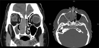
The CT scan revealed a large mass arising from the right ethmoid sinus and eroding both the medial bony wall of the orbit and the cribriform plate. It also extended to the frontal sinus anteriorly, and posteriorly to the sphenoid sinus and nasopharynx (see Figure 1).
Coronal (left image) and axial (right image) computed tomography scans of the paranasal sinuses, showing a large mass arising from the right ethmoid sinus and eroding both the medial bony wall of the orbit and the cribriform plate, and extending to the frontal sinus anteriorly and the sphenoid sinus and nasopharynx posteriorly. A = anterior; R = right; L = left; P = posterior

A possible second primary tumour in the upper lobe of the left lung was identified on the CT scan. However, the reporting radiologist felt that this was representative of old tuberculosis. The patient declined any further investigation of the lung lesion, other than to have interval CT scans. These remained unchanged at 3 and 12 months.
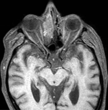
The MRI scan demonstrated no extension to the medial rectus muscle. The tumour was found to be extradural, with only bony erosion of the cribriform plate (See Figure 2).
Axial magnetic resonance imaging scan of the paranasal sinuses, showing the tumour to be extradural, with only bony erosion of the cribriform plate and no extension to the medial rectus muscle.

Examination under anaesthesia confirmed the presence of a large, necrotic mass in the right ethmoid region. The middle turbinate was pushed medially. The tumour was debulked as much as possible and specimens sent for histological analysis.
Histopathological analysis revealed infiltration by a malignant neoplasm composed of sheets, cords and ribbons of malignant cells showing hyperchromatic nuclei, coarse chromatin and inconspicuous nuclei. The cells contained scanty cytoplasm with focal brown pigmentation, and in areas were arranged in small clusters with an attempt at rosette formation. Morphologically, this was suggestive of olfactory neuroblastoma.
However, immunohistochemical analysis revealed strongly positive staining of neuroendocrine markers, with positivity for synaptophysin, chromogranin, cluster of differentiation 56 protein and epithelial membrane antigen, indicating a neuroendocrine carcinoma.
The specimen was sent to a tertiary hospital for a second opinion. This concluded that the tumour was a large cell neuroendocrine carcinoma.
The case was discussed at the multidisciplinary head and neck oncology meeting. It was felt that curative management ought to be possible using a craniofacial approach with adjuvant radiotherapy. However, the patient declined surgery, consenting only to radiotherapy.
He subsequently completed a course of radical radiotherapy, which was tolerated extremely well, on an out-patient basis.
Thirty months after radiotherapy, the patient was asymptomatic with no clinical evidence of any residual tumour in the ethmoid cavity.
Discussion
Neuroendocrine tumours arising in the sinonasal tract are extremely rare. They were initially described in 1982 by Silva et al. Reference Silva, Butler, MacKay and Geopfert2
A literature search using Medline, Embase and the Cochrane library, conducted in October 2009, yielded only 10 cases, reported between January 2004 and October 2009.Reference Grabovac, Prgomet and Begic1, Reference Wang, Hsieh, Chang, Lou, Yang and Ting5–Reference Lee, Cho and Cho13 A further literature search in October 2011 identified only one further case.Reference Vandist, Deridder, Waelput, Parizel, Van de Heyning and Van Laer14 Writing in 2004, Grabovac et al. claimed that only 28 cases had ever been described.Reference Grabovac, Prgomet and Begic1
Of the 11 recent cases identified in our search, intermittent epistaxis was the commonest presenting symptom, affecting eight cases. Nasal congestion was present in three cases, discharge in one case, facial and orbital pain in two cases, and the syndrome of inappropriate antidiuretic hormone secretion in two cases.
Neuroendocrine tumours are rare, malignant neoplasms that contain neurosecretory granules. They can be classified cytologically as well-differentiated, intermediately differentiated and poorly differentiated. Poorly differentiated neuroendocrine tumours are subclassified as small cell, intermediate cell and large cell types. Our patient was diagnosed with a poorly differentiated large cell neuroendocrine tumour, based on light microscopy and immunohistopathology. The clinical relevance of this diagnosis is that olfactory neuroblastomas are characteristically aggressive and invasive, whilst large cell neuroendocrine carcinomas are less so.Reference Warner3, Reference Babin, Rouleau, Vedrine, Toussaint, de Raucort and Malard15 It is impossible to differentiate between these tumours through imaging alone.
No treatment guidelines exist for the management of these rare tumours, and no meta-analysis has ever been conducted for sinonasal neuroendocrine tumours.
Wang et al. published a retrospective case series of 18 patients with neuroendocrine tumours.Reference Wang, Hsieh, Chang, Lou, Yang and Ting5 Fifteen of these patients had small cell neuroendocrine carcinoma. All 18 patients had surgery, three of whom had induction chemotherapy or primary radiotherapy before definitive management. Fifteen patients had surgery first, with or without subsequent adjuvant chemoradiotherapy. Survival rates were between 60 and 70 per cent regardless of treatment modality.
Babin et al. published a similar retrospective case series of 21 patients with small cell neuroendocrine tumours.Reference Babin, Rouleau, Vedrine, Toussaint, de Raucort and Malard15 Eleven underwent surgery, 14 radiotherapy and 12 chemotherapy. Survival rates were similar for all treatment modalities.
• There is no consensus on the management of sinonasal neuroendocrine carcinoma
• The presented case was disease-free 30 months after radiotherapy alone
As stated above, in addition to the 28 previously published cases reported in a 2004 paper, our literature search identified a further 11 neuroendocrine carcinoma cases reported since January 2004.Reference Grabovac, Prgomet and Begic1, Reference Vasan, Medina, Canfield and Gillieis6–Reference Gonzalez-Garcia, Fernandez-Rodriguez, Naval-Gias, Rodriguez-Campo, Nam-Cha and Diaz-Gonzalez12 Six patients underwent surgery, two of whom died. Four patients underwent chemotherapy with or without radiotherapy, one of whom died. This appears to be in keeping with the results of the case series described above. To our best knowledge, in the present paper we describe the only reported case of a large cell neuroendocrine tumour treated with radiotherapy alone.
A 2001 meta-analysis of olfactory neuroblastomas identified the optimal treatment of this related tumour to be surgical intervention with adjuvant radiotherapy.Reference Dulguerov, Allal and Calcaterra16 It remains to be seen if this will prove to be the case for sinonasal neuroendocrine tumours.
Conclusion
No treatment consensus exists for the management of sinonasal neuroendocrine carcinoma. The case presented in this report indicates that curative treatment may be possible utilising radiotherapy alone.