Children with cardiac disease are at an increased risk of in-hospital cardiac arrest; however, several studies have demonstrated better outcomes when compared to non-cardiac patients.Reference Joffe, Lequier and Robertson1–Reference De Mos, Van Litsenberg, McCrindle, Bohn and Parshuram7 In response to arrest refractory to conventional cardiopulmonary resuscitation, many centres utilise extracorporeal cardiopulmonary resuscitation: the initiation of extracorporeal membrane oxygenation during refractory arrest.8 Indeed, the most recent scientific statement from the American Heart Association for cardiopulmonary resuscitation in children with cardiac disease recommends considering extracorporeal cardiopulmonary resuscitation during early resuscitation,Reference Marino, Tabbutt and MacLaren9 and Lasa et al demonstrated increased survival to hospital discharge and better neurological outcomes with extracorporeal cardiopulmonary resuscitation compared to conventional cardiopulmonary resuscitation in children with in-hospital cardiac arrest for >10 minutes.Reference Lasa, Rogers and Localio10
An important factor affecting extracorporeal cardiopulmonary resuscitation outcomes is duration of cardiopulmonary resuscitation prior to extracorporeal membrane oxygenation initiation, as prolonged cardiopulmonary resuscitation has been shown to be a predictor of mortality in children with primary heart disease.Reference Kumar, Zurakowski and Dalton11–Reference Torres-Andres, Fink, Bell, Sharma, Yablonsky and Sanchez-de-Toledo13 Despite this, several case reports describe survival with favourable neurologic outcomes in paediatric cardiac patients who have undergone prolonged periods of cardiopulmonary resuscitation prior to extracorporeal membrane oxygenation.Reference Raymond, Cunnyngham and Thompson5,Reference Kelly, Porter, Meier, Myers and Thomas14 Limited data exist on the upper limit of cardiopulmonary resuscitation duration that will result in favourable survival and neurologic outcomes, and previous authors have called for additional studies to provide clarity on this.Reference Kelly, Porter, Meier, Myers and Thomas14
In summary, data regarding survival and neurological outcomes of paediatric cardiac patients undergoing prolonged extracorporeal cardiopulmonary resuscitation are limited, and risk factors for mortality are unknown. Given this lack of knowledge, the aims of the present study are to characterise the mortality and neurological outcomes of paediatric cardiac patients requiring extracorporeal cardiopulmonary resuscitation greater than 30 minutes and to identify risk factors associated with increased mortality in this population.
Materials and methods
Study setting and design
The study was conducted in a tertiary-level multidisciplinary cardiac ICU at Children’s Health Children’s Medical Center, Dallas (Texas). It was designed as an observational, retrospective cohort study. Approval from the Institutional Review Board was obtained, with waiver of informed consent granted prior to study initiation.
Study population and data collection
All paediatric cardiac patients 1 day to 18 years of age who underwent cardiopulmonary resuscitation for greater than 30 minutes prior to extracorporeal membrane oxygenation cannulation due to refractory in-hospital arrest between July 2000 and July 2013 were included in the study. Exclusion criteria included out-of-hospital arrest, patients who underwent cardiopulmonary resuscitation for less or equal to 30 minutes prior to cannulation, patients without a primary cardiac diagnosis, and children who died during the arrest or had return of spontaneous circulation before establishing extracorporeal membrane oxygenation support. Only data regarding the first extracorporeal cardiopulmonary resuscitation course on patients with multiple runs was included in the study.
Electronic medical records were reviewed to obtain patient characteristics, location and event time (day versus night), cardiac diagnosis category (post-operative versus non-surgical), type of ventricular circulation (univentricular versus biventricular), and history of previous arrest. Investigators collected information regarding arrest characteristics, including presence of pulse and rhythm at onset and during resuscitation, presence of advanced airway and mechanical ventilation at onset of arrest, vital signs during the arrest, and laboratory data during the 24-hour period following arrest. Pharmacological and non-pharmacological interventions utilised during resuscitation were obtained. Cardiopulmonary resuscitation and cannulation duration, interval to extracorporeal membrane oxygenation team activation and response as well as the number of interruptions of chest compressions during cannulation were also noted. Information about extracorporeal membrane oxygenation support, including cannulation site, maximum extracorporeal membrane oxygenation flow, and extracorporeal membrane oxygenation duration were collected. We reviewed post-resuscitation therapies including use of continuous renal replacement therapy to treat acute kidney injury or fluid overload during extracorporeal membrane oxygenation. Neurological assessment, temperature control, and neuroimaging performed during and after extracorporeal membrane oxygenation were recorded. Mortality, causes of death, and neurological outcomes of survivors using Pediatric Cerebral Performance Categories scores at the time of hospital discharge were also documented.
Definitions and extracorporeal cardiopulmonary resuscitation protocol
Extracorporeal cardiopulmonary resuscitation was defined as initiation of extracorporeal membrane oxygenation during ongoing chest compressions for resuscitation due to refractory in-hospital arrest. This is consistent with the definition by the Extracorporeal Life Support Organization: “Extracorporeal Cardiopulmonary Resuscitation, in which extracorporeal life support was used as part of the initial resuscitation from cardiac arrest. Hemodynamically unstable patients placed on extracorporeal life support emergently without cardiac arrest are not considered extracorporeal cardiopulmonary resuscitation”.Reference Thiagarajan, Brediger, Larsen, Fynn-Thompson, Alexander and Brogan15 For the purpose of the study, we defined “prolonged” extracorporeal cardiopulmonary resuscitation as cardiopulmonary resuscitation that lasted for greater than 30 minutes without return of spontaneous circulation prior to extracorporeal membrane oxygenation cannulation. Extracorporeal membrane oxygenation duration was defined as the time from cannulation until decannulation. Daytime was defined as arrest occurring from 7 am to 6 pm. Interval to extracorporeal membrane oxygenation team activation was defined as duration between occurrence of arrest and first call to activate the extracorporeal membrane oxygenation team. Interval to extracorporeal membrane oxygenation team response was defined as duration between first call to activate the extracorporeal membrane oxygenation team and incision for cannulation initiation. Cannulation duration was defined as the time interval from incision to extracorporeal membrane oxygenation initiation.
In our cardiac ICU, the extracorporeal membrane oxygenation team includes cardiac surgeons, cardiac intensivists, and extracorporeal membrane oxygenation specialists. Cardiac surgeons perform all extracorporeal cardiopulmonary resuscitation cannulations in the cardiac ICU. In-house surgical coverage is provided during the day (7 am–6 pm). At night, surgeons provide coverage from home. Activation of the team routinely occurs within 5–10 minutes of no return of spontaneous circulation in a patient with a potentially reversible pathology, without contraindications to mechanical circulatory support. Typical contraindications to extracorporeal membrane oxygenation support at our institution include CHDs not correctable by surgery, severe neurological conditions, and anatomic abnormalities precluding successful cannulation for extracorporeal support. Patients receive an unfractionated heparin bolus of 50–100 units per kilogram after skin incision prior to cannula insertion, followed by a continuous infusion. Once on extracorporeal membrane oxygenation support, inotropic support is weaned to minimal levels or discontinued if haemodynamics are appropriate. Mechanical ventilation is weaned to “rest settings,” typically synchronised intermittent mechanical ventilation, pressure control mode, with a pressure control of 10, rate of 10, and positive end-expiratory pressure of 5–10. Once neurologic function has been adequately assessed, sedation and analgesia are maintained with narcotics and benzodiazepines. Temperature management is per the attending physician’s preference, utilising either therapeutic hypothermia to 32–35 °C for greater than 24 hours or controlled normothermia avoiding fever.
Neuroimaging and neurological follow-up
In our extracorporeal membrane oxygenation patients, head ultrasound is performed soon after cannulation and is repeated routinely in neonates and infants with open fontanel after the arrest. Head CT is performed in children with suspicion for neurologic injury based on neurological assessment or when the head ultrasound results are indeterminate or inconsistent with the patient’s neurologic symptoms. Brain MRI is usually performed after decannulation and before hospital discharge in patients with abnormal findings on head ultrasound or head CT or in patients with clinical neurologic abnormalities. Functional outcomes were assessed using the Pediatric Cerebral Performance Categories score from chart review prior to the arrest and upon hospital discharge. Pediatric Cerebral Performance Categories was developed as a reliable assessment of neurologic disability and has been found to correlate with long-term functional outcomes.Reference Fiser, Long, Roberson, Hefley, Zolten and Brodie-Fowler16,Reference Pollack, Holubkov and Funai17
Statistical analysis
Demographic, clinical and extracorporeal membrane oxygenation characteristics, vital signs, laboratory data, pharmacological and non-pharmacological interventions as well as outcomes for the overall cohort of patients and across mortality status were described using medians and interquartile ranges for continuous variables and frequencies and percentages for categorical variables. Univariate logistic regression was used to ascertain independent associations between each covariate and mortality. Covariates with p < 0.10 were entered into a second model where a backward variable selection was used to ascertain which covariates were highly associated with mortality. Continuous predictors were plotted using a Lowess plot to determine if categorisation was needed to increase the internal validity of the final model. Regarding cardiopulmonary resuscitation duration, there was an inflection point at 50 minutes thus we dichotomised this variable at 50 minutes. The goodness of fit of our model was assessed using the receiver operating characteristic curve, which calculated an area under the curve. The model was determined to be a good model if the area under the curve was 0.70 or greater. In addition, an exploratory analysis was conducted using logistic regression to ascertain the odds of good neurological outcomes relative to all covariates. Odds ratios and 95% confidence intervals were calculated for significant factors. All p-values were two-sided and p < 0.05 was considered statistically significant. Statistical analyses were performed using STATA version 14 (STATACorp; College Station, TX).
Results
Patient and extracorporeal membrane oxygenation characteristics
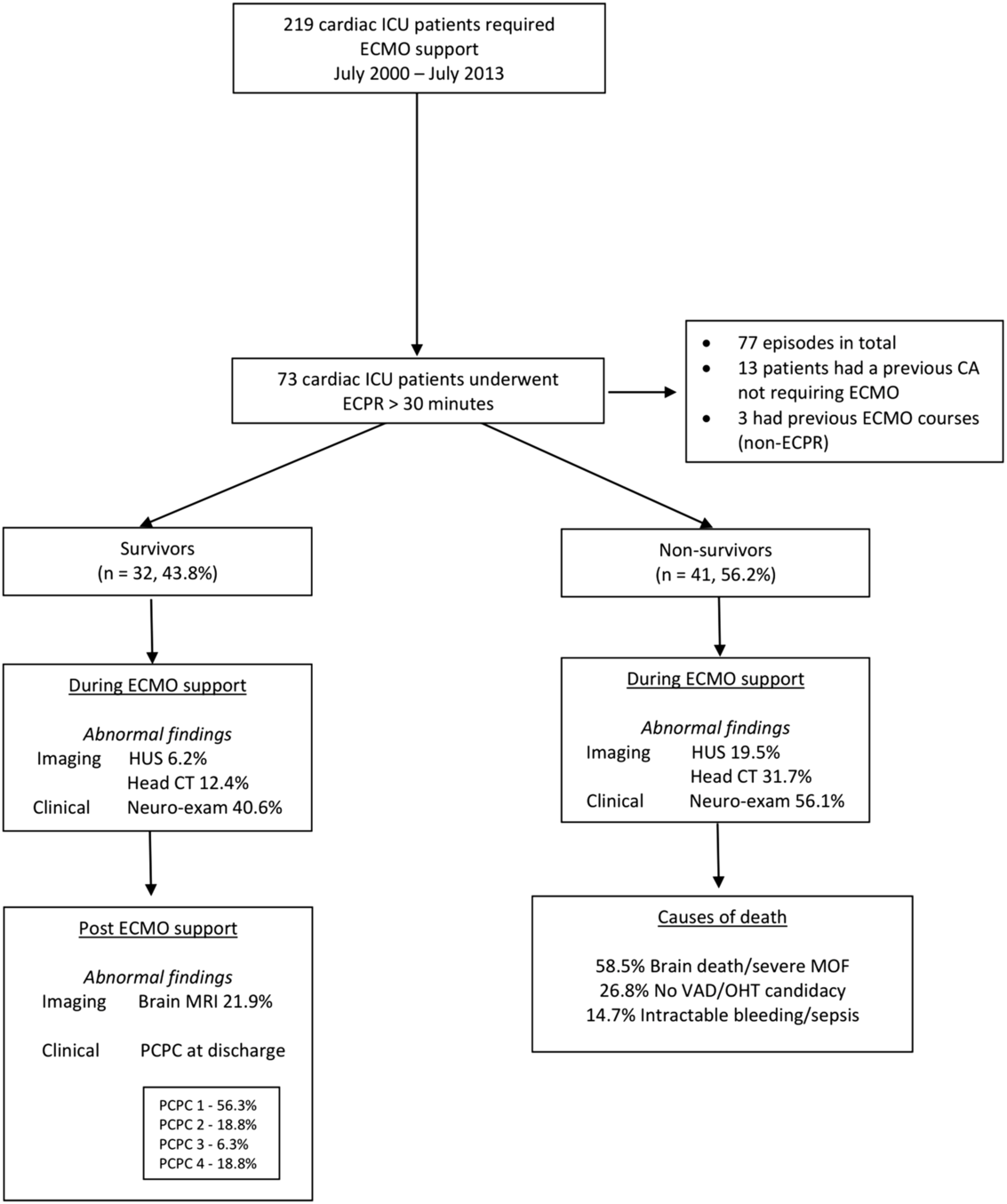
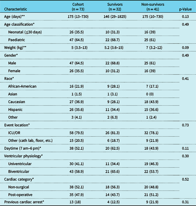
A total of 219 cardiac patients were supported with extracorporeal membrane oxygenation in the cardiac ICU from July 2000 through July 2013. Of them, 73 underwent extracorporeal cardiopulmonary resuscitation for greater than 30 minutes due to refractory in-hospital arrest. Four of these patients had 2 extracorporeal cardiopulmonary resuscitation runs each, resulting in a total of 77 episodes. Previous cardiac arrest not requiring extracorporeal membrane oxygenation occurred in 13 (18%) patients, and 3 (4%) underwent a prior extracorporeal membrane oxygenation run with no cardiopulmonary resuscitation and were successfully decannulated before the extracorporeal cardiopulmonary resuscitation event. Figure 1 shows patient characteristics. Median age was 175 days (interquartile range 13–730) and median weight was 5 kg (interquartile range 3–13). Thirty-eight (52.1%) were non-surgical patients, while 35 (47.9%) were post-operative patients. Non-CHD (dilated cardiomyopathy, myocarditis, and dysrhythmias) accounted for 21.9% of patients. Thirty (41.1%) had single-ventricle physiology. The majority of refractory in-hospital arrest episodes (79.5%) occurred in the operating room or during admission to the cardiac ICU, and 52.1% occurred between 7 am and 6 pm. Sixty-three (86%) patients survived extracorporeal membrane oxygenation decannulation and 32 (43.8%) survived to hospital discharge. Thirty-one of the 73 patients (42%) died after recovering enough organ function to be decannulated. Overall mortality was due to withdrawal of life support due to brain death and/or severe multi-organ dysfunction in 58.5%, non-candidacy for ventricular assist device or heart transplant in 26.8%, and intractable bleeding, sepsis, or mesenteric ischemia in 14.7% of non-survivors. CHD was not associated with mortality (p 0.09). Table 1 represents patient characteristics of the cohort and between groups.
Patient flow chart. CA = cardiac arrest; ECMO = extracorporeal membrane oxygenation; ECPR = extracorporeal cardiopulmonary resuscitation; HUS = head ultrasound; MOF = multi-organ failure; OHT = orthotopic heart transplant; PCPC = paediatric cerebral performance categories; VAD = ventricular assist device.

Pre-arrest characteristics

cath = catheterisation; kg = kilogram; OR = operating room
*n (%), **median (25th, 75th %iles)
p-Values calculated using univariate logistic regression
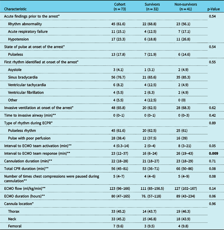
Arrest and extracorporeal cardiopulmonary resuscitation characteristics
Rhythm abnormalities were the most frequent acute finding immediately preceding the event (61.6%). At onset of arrest, sinus bradycardia was the most common rhythm (76.7%), with only 17.8% of patients categorised as pulseless. A total of 34.2% of patients were not invasively ventilated at onset of the arrest, but these patients were quickly placed on mechanical ventilation, with a median interval to advanced airway placement of 0 minutes (interquartile range 0–1). Median interval to extracorporeal membrane oxygenation team activation was 4 minutes with a median interval to extracorporeal membrane oxygenation team response of 23 minutes. Median duration of cardiopulmonary resuscitation was 56 minutes, with median duration of surgical cannulation of 22 minutes. Median maximum extracorporeal membrane oxygenation flow was 123 ml/kg/minute with median extracorporeal membrane oxygenation duration to decannulation of 80 hours. Cannulation site was peripheral in 54.8% and central in 45.2% of patients (Table 2).
Arrest characteristics

CPR = cardiopulmonary resuscitation; ECMO = extracorporeal membrane oxygenation; ECPR = extracorporeal cardiopulmonary resuscitation; min = minutes; ml/kg/min = millilitre per kilogram per minute
*n (%), **median (25th, 75th %iles)
p-Values calculated using univariate logistic regression
p values that are statistically significant (p value < 0.05)
Compared to non-survivors, survivors had a significantly decreased median interval to extracorporeal membrane oxygenation team response (16 versus 26 minutes, p 0.009). Survivors had decreased median interval to extracorporeal membrane oxygenation team activation (2 versus 8 minutes, p 0.05), decreased cardiopulmonary resuscitation duration (53 versus 66 minutes, p 0.08), and decreased extracorporeal membrane oxygenation duration (76 versus 89 hours, p 0.06). Table 2 represents these findings.
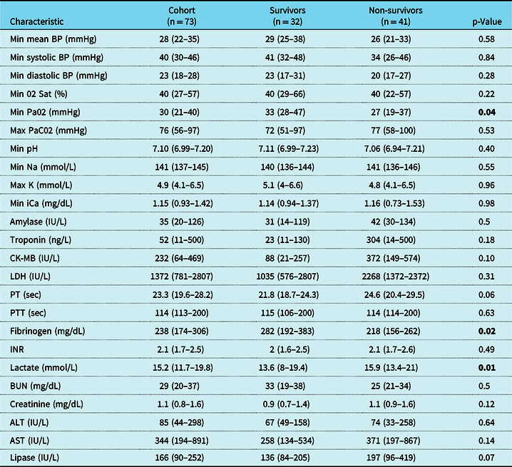
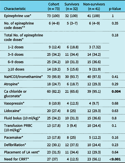
No differences were observed in vital signs during resuscitation or in average laboratory data obtained within the first 24 hours after cannulation between survivors and non-survivors (Table 3), except for survivors having higher minimal Pa02 (33 versus 27 mmHg, p 0.04), lower lactate (13.6 versus 15.9 mmol/L, p 0.01), and higher fibrinogen (282 versus 218 mg/dL, p 0.02). No differences were observed in pharmacological and non-pharmacological interventions during resuscitation, except for increased use of calcium chloride or gluconate (95.1 versus 65.6%, p 0.004) and increased need for continuous renal replacement therapy to treat acute kidney injury and fluid overload post-resuscitation (56.1 versus 12.5%, p < 0.001) in non-survivors (Table 4).
Vital signs during resuscitation and laboratory data within the initial 24 hours after ECMO cannulation

ALT = alanine aminotransferase; AST = aspartate aminotransferase; BP = blood pressure; BUN = blood urea nitrogen; CK-MB = creatine kinase-muscle/brain; iCa = ionised calcium; INR = international normalised ratio; IU/L = international units per litre; K = potassium; LDH = lactate dehydrogenase; max = maximum; mg/dL = milligrams per decilitre; min = minimum; mmHg = millimetres of mercury; mmol/L = millimol per litre; Na = sodium; ng/L = nanograms per litre; 02 = oxygen; PaC02 = partial pressure of carbon dioxide; Pa02 = partial pressure of oxygen; PT = prothrombin time; PTT = partial thromboplastin time; Sat = saturation; sec = seconds
median (25th, 75th %iles). p-Values calculated using Univariate logistic regression
p values that are statistically significant (p value < 0.05)
Pharmacological and non-pharmacological interventions

Epinephrine, NaHCO3/tromethamine, Atropine, Calcium chloride or gluconate, vasopressin, lidocaine administered as code doses per American Heart Association guidelines
Ca = calcium; CRRT = continuous renal replacement therapy; kg = kilograms; LA = left atrium; ml = millilitres; no. = number; PRBC = packet red blood cells
*n (%), **median (25th, 75th percentiles)
p-Values calculated using univariate logistic regression
p values that are statistically significant (p value < 0.05)
Neurological characteristics and imaging
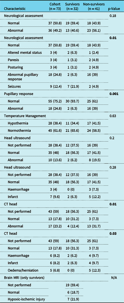
After the code event, non-survivors had a significantly higher incidence of abnormal pupillary response during assessments (39 versus 6.3%, p 0.001) compared to survivors. Therapeutic hypothermia with a median temperature of 33 °C was achieved in 28 patients for a median duration of 53 hours (interquartile range 42–69). No differences were observed in temperature management between survivors and non-survivors. Head ultrasounds were performed in 61.6% of patients while on extracorporeal membrane oxygenation support. Intracranial injuries were observed by head ultrasound in 6.2% of survivors and 19.5% of non-survivors (p 0.2). Head CTs were performed in 41% of patients while on extracorporeal membrane oxygenation. Intracranial injuries were observed by head CT in 12.4% of survivors and 31.7% of non-survivors (p 0.01), with oedema/herniation being the most significant abnormal finding (p 0.03). Brain MRIs were performed in 13 of the 32 survivors (40.6%) after extracorporeal membrane oxygenation decannulation, demonstrating hypoxic-ischemic injury in 7 (21.9%) survivors. Table 5 represents these findings. In patients who survived cardiopulmonary resuscitation greater than 50 minutes, 41% had abnormal neurological assessments which correlated with abnormal neuroimaging in all except for one patient.
Neurological characteristics and imaging after cardiac arrest

N/A = not applicable
n (%). p-Values calculated using univariate logistic regression
p values that are statistically significant (p value < 0.05)
Among survivors, Pediatric Cerebral Performance Categories scores at discharge demonstrated normal neurological function (Pediatric Cerebral Performance Categories 1) in 56.3%, mild neurological disability (Pediatric Cerebral Performance Categories 2) in 18.8%, moderate (Pediatric Cerebral Performance Categories 3) in 6.3%, and severe (Pediatric Cerebral Performance Categories 4) in 18.8%. All patients had normal neurological function (Pediatric Cerebral Performance Categories 1) prior to the arrest, except for two patients with mild neurological disability pre-arrest (Pediatric Cerebral Performance Categories 2) who continued to have the same score at discharge. Brain MRI of all patients with severe neurological disability (Pediatric Cerebral Performance Categories 4) demonstrated extensive severe hypoxic-ischemic injury. Of note, 66.6% of survivors treated with therapeutic hypothermia versus 80% of survivors treated with controlled normothermia had normal neurological function or mild neurological disability (Pediatric Cerebral Performance Categories 1 and 2), but this difference did not achieve statistical significance (p 0.39).
Effect of time on survival and neurological outcomes
Given the prolonged research period, the study time was divided into two eras with similar patient distribution. Era 1 (2000–2006) accounted for 38 patients and era 2 (2007–2013) accounted for 35 patients. No significant differences were observed in era 1 compared to era 2 in survival to hospital discharge (39.4 versus 48.5%, p 0.43) or in the fraction of survivors reaching Pediatric Cerebral Performance Categories 1–2 at discharge (80 versus 64.7%, p 0.33).
Factors associated with hospital mortality and survival with good neurological outcomes
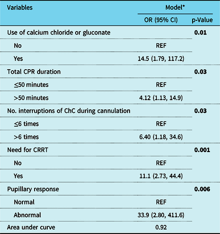
Multivariable logistic regression analysis was performed to evaluate predictors of mortality and survival with good neurological outcomes. After adjusting for confounders, increased use of calcium during resuscitation (odds ratio 14.5, p 0.01), cardiopulmonary resuscitation duration greater than 50 minutes (odds ratio 4.12, p 0.03), increased interruptions of chest compressions (>6 times) during cannulation (odds ratio 6.40, p 0.03), the need for continuous renal replacement therapy (odds ratio 11.1, p 0.001), and abnormal pupillary response (odds ratio 33.9, p 0.006) were independently associated with increased mortality (Table 6). The only predictor of survival with good neurological outcomes was higher minimal Pa02 within 24 hours post-arrest (odds ratio 1.08, p 0.01), while male gender (odds ratio 0.12, p 0.03) and abnormal pupillary response (odds ratio 0.04, p 0.001) were associated with bad neurological outcomes and/or mortality.
Logistic regression to ascertain the associations between selected covariates and mortality

ChC = chest compressions; CI = confidence interval; CPR = cardiopulmonary resuscitation; CRRT = continuous renal replacement therapy; ECPR = extracorporeal cardiopulmonary resuscitation; No. = number; N/A = not applicable; OR = odds ratio; REF = reference
p values that are statistically significant (p value < 0.05)
* Multivariable logistic regression adjusting for all other variables within the model following a backward variable selection
Discussion
This study is the first to date to examine the outcomes of paediatric cardiac patients requiring cardiopulmonary resuscitation for more than 30 minutes until extracorporeal membrane oxygenation initiation. The primary finding of our study is that our cohort of patients had an overall survival to hospital discharge of 43.8%, with 75% of these patients having either normal neurologic function or only mild disability. Our reported survival is comparable to previous paediatric cardiac extracorporeal cardiopulmonary resuscitation studies that reported a survival of 20–46%,Reference Morris, Ittenbach and Godinez2,Reference Kumar, Zurakowski and Dalton11,Reference Sivarajan, Best, Brizard, Shekerdemian, d’Udekem and Butt12,Reference Shah, Shankar and Churchwell18–Reference Guo, Yang and Zhang24 but differs from other studies with more favourable extracorporeal cardiopulmonary resuscitation survival rates of 51–79%.Reference Kane, Thiagarajan and Wypij6,Reference Allan, Thiagarajan, Armsby, del Nido and Laussen25–Reference Shakoor, Pedroso, Jacobs, Okochi, Zenilman, Cheung and Middlesworth30 Many prior studies of extracorporeal cardiopulmonary resuscitation include patients with wide ranges of duration of chest compressions prior to cannulation, some of them including patients receiving less than 10 minutes of cardiopulmonary resuscitation prior to placement on mechanical support.Reference Morris, Ittenbach and Godinez2,Reference Kane, Thiagarajan and Wypij6,Reference Kumar, Zurakowski and Dalton11,Reference Sivarajan, Best, Brizard, Shekerdemian, d’Udekem and Butt12,Reference Shah, Shankar and Churchwell18–Reference Shakoor, Pedroso, Jacobs, Okochi, Zenilman, Cheung and Middlesworth30 Our focus on those patients receiving prolonged cardiopulmonary resuscitation provides information about a more discrete and potentially higher risk population.
We focused on patients with primary cardiac disease because prior studies demonstrated increased survival with extracorporeal cardiopulmonary resuscitation and conventional cardiopulmonary resuscitation compared to non-cardiac patients.Reference Morris, Ittenbach and Godinez2–Reference Tajik and Cardarelli4,Reference Kane, Thiagarajan and Wypij6,Reference Matos, Watson and Nadkarni31,Reference Ortmann, Prodhan and Gossett32 Proposed mechanisms for this cardiac survival benefit are the lower cardiopulmonary reserve and baseline hypoperfusion which may lead to less arrest-related multi-organ dysfunction,Reference Morris, Ittenbach and Godinez2–Reference Tajik and Cardarelli4,Reference Kane, Thiagarajan and Wypij6,Reference Matos, Watson and Nadkarni31 or to the readily available deployment of extracorporeal cardiopulmonary resuscitation in the cardiac ICU.Reference Tajik and Cardarelli4,Reference Conrad and Rycus33 We compared our findings with previous literature that only focus on paediatric cardiac extracorporeal cardiopulmonary resuscitation or paediatric cardiac extracorporeal membrane oxygenation with subgroup analysis of extracorporeal cardiopulmonary resuscitation patients.Reference Morris, Ittenbach and Godinez2,Reference Kane, Thiagarajan and Wypij6,Reference Kumar, Zurakowski and Dalton11,Reference Sivarajan, Best, Brizard, Shekerdemian, d’Udekem and Butt12,Reference Shah, Shankar and Churchwell18–Reference Shakoor, Pedroso, Jacobs, Okochi, Zenilman, Cheung and Middlesworth30 The Supplementary Table provides a review of paediatric extracorporeal cardiopulmonary resuscitation studies in the cardiac ICU literature. Several factors may account for differing results between our study and previous ones. First, several of these studies concentrated on different populations, such as post-cardiotomy or cath lab patients.Reference Kumar, Zurakowski and Dalton11,Reference Shah, Shankar and Churchwell18,Reference Alsoufi, Al-Radi and Gruenwald20,Reference Erek, Aydın and Suzan22,Reference Guo, Yang and Zhang24,Reference Allan, Thiagarajan, Armsby, del Nido and Laussen25,Reference Polimenakos, Rizzo, El-Zein and Ilbawi29 Second, others relied on subgroup analysis of larger reviews of cardiac extracorporeal membrane oxygenation supportReference Morris, Ittenbach and Godinez2,Reference Kumar, Zurakowski and Dalton11,Reference Shah, Shankar and Churchwell18,Reference Alsoufi, Al-Radi and Gruenwald20,Reference Allan, Thiagarajan, Armsby, del Nido and Laussen25,Reference Thourani, Kirshbom and Kanter26 rather than our study, with its specific focus on cardiac extracorporeal cardiopulmonary resuscitation. Third, our cohort focused on prolonged extracorporeal cardiopulmonary resuscitation and as such had a longer median duration of cardiopulmonary resuscitation (56 minutes) compared to the previous cardiac extracorporeal cardiopulmonary resuscitation reports where median duration ranged from 16 to 47.5 minutesReference Morris, Ittenbach and Godinez2,Reference Kane, Thiagarajan and Wypij6,Reference Kumar, Zurakowski and Dalton11,Reference Sivarajan, Best, Brizard, Shekerdemian, d’Udekem and Butt12,Reference Shah, Shankar and Churchwell18–Reference Alsoufi, Al-Radi and Gruenwald20,Reference Allan, Thiagarajan, Armsby, del Nido and Laussen25–Reference Polimenakos, Rizzo, El-Zein and Ilbawi29 with only four studies reporting a prolonged median duration similar to our study.Reference Huang, Wu and Chen21,Reference Guo, Yang and Zhang24,Reference Thourani, Kirshbom and Kanter26,Reference Shakoor, Pedroso, Jacobs, Okochi, Zenilman, Cheung and Middlesworth30 This factor would be expected to portend worse survival to our patients as prolonged cardiopulmonary resuscitation duration has been noted to be a predictor of mortality in paediatric cardiac patients undergoing extracorporeal cardiopulmonary resuscitation.Reference Kumar, Zurakowski and Dalton11,Reference Sivarajan, Best, Brizard, Shekerdemian, d’Udekem and Butt12,Reference Erek, Aydın and Suzan22
In our study, we observed moderate to severe neurological disability (Pediatric Cerebral Performance Categories 3 and 4) in 25% of survivors. We also observed a strong correlation between poor neurological outcomes and evidence of injury on brain MRI (87.5% of survivors with moderate to severe neurological disabilities had severe hypoxic-ischemic injury on brain MRI after decannulation). Data on neurological outcomes of cardiac extracorporeal cardiopulmonary resuscitation survivors are limited and based on different diagnostic modalities.Reference Joffe, Lequier and Robertson1,Reference Morris, Ittenbach and Godinez2,Reference Kane, Thiagarajan and Wypij6,Reference Sivarajan, Best, Brizard, Shekerdemian, d’Udekem and Butt12,Reference Erek, Aydın and Suzan22,Reference Philip, Burgman and Bavare23,Reference Allan, Thiagarajan, Armsby, del Nido and Laussen25,Reference Polimenakos, Rizzo, El-Zein and Ilbawi29 A meta-analysis by Joffe et al described 9 paediatric extracorporeal cardiopulmonary resuscitation studies with a total of 287 survivors and a 22% prevalence of neurological disability.Reference Joffe, Lequier and Robertson1 Four of these studies exclusively evaluated cardiac extracorporeal cardiopulmonary resuscitation patients and reported variable neurological outcomes using different assessment tools: Morris et al and Allan et al demonstrated moderate clinical neurological deficits in 29 and 20% of patients, respectively, while Kane et al and Sivarajan et al described Pediatric Cerebral Performance Categories scores of 3–4 in 21 and 67% of survivors, respectively.Reference Morris, Ittenbach and Godinez2,Reference Kane, Thiagarajan and Wypij6,Reference Sivarajan, Best, Brizard, Shekerdemian, d’Udekem and Butt12,Reference Allan, Thiagarajan, Armsby, del Nido and Laussen25 Philip et al reported Pediatric Overall Performance Category scores of 3–4 (correlating with moderate to severe neurological impairment) in 32% of their cardiac extracorporeal cardiopulmonary resuscitation survivors. Interestingly, 75% of these patients with Pediatric Overall Performance Category 3–4 had cardiopulmonary resuscitation duration to extracorporeal membrane oxygenation initiation >30 minutes.Reference Philip, Burgman and Bavare23 Other studies demonstrated lower incidence of severe neurologic deficits (18Reference Huang, Wu and Chen21 and 10%Reference Wolf, Kanter, Kirshbom, Kogon and Wagoner28) in their cardiac extracorporeal cardiopulmonary resuscitation survivors but did not clearly identify their diagnostic tools.Reference Erek, Aydın and Suzan22,Reference Polimenakos, Rizzo, El-Zein and Ilbawi29
Our study describes several risk factors associated with increased mortality in our cohort. Cardiopulmonary resuscitation lasting greater than 50 minutes was found to be a predictor of mortality. Three of our patients survived with mild neurological deficits (Pediatric Cerebral Performance Categories ≤ 2) after very prolonged cardiopulmonary resuscitation episodes (118, 134, and 139 minutes) before extracorporeal membrane oxygenation cannulation. Data on duration of cardiopulmonary resuscitation before extracorporeal membrane oxygenation initiation as a predictor for survival in paediatric cardiac patients are conflicting. While some investigators identified cardiopulmonary resuscitation duration as a predictor of mortality,Reference Kumar, Zurakowski and Dalton11,Reference Sivarajan, Best, Brizard, Shekerdemian, d’Udekem and Butt12 others did not find this association.Reference Morris, Ittenbach and Godinez2,Reference Kane, Thiagarajan and Wypij6 Two previous paediatric studies demonstrated “adequate” neurological outcomes after cardiopulmonary resuscitation episodes lasting >90 minutes before extracorporeal membrane oxygenation initiation.Reference Raymond, Cunnyngham and Thompson5,Reference Kelly, Porter, Meier, Myers and Thomas14 However, the upper limit of cardiopulmonary resuscitation duration before extracorporeal membrane oxygenation cannulation resulting in acceptable neurological outcomes continues to remain unknown.
Our findings demonstrate increased mortality when chest compressions were paused >6 times during cannula insertion. The American Heart Association 2015 guidelines for “excellent cardiopulmonary resuscitation” include as a quality metric for improved survival the reduction of pauses during chest compressions.Reference Atkins, Berger and Duff34 Adult cardiopulmonary resuscitation studies have demonstrated that minimising interruptions during chest compressions is associated with increased return of spontaneous circulation and survival.Reference Christenson, Andrusiek and Everson-Stewart35,Reference Gundersen, Kvaløy, Kramer-Johansen, Steen and Eftestøl36 We believe this is the first paediatric study demonstrating reduced interruptions of chest compression during extracorporeal membrane oxygenation cannulation being associated with survival.
Additionally, we observed that calcium chloride or gluconate were more frequently used among non-survivors during resuscitation. This finding could be related to patients with poor cardiovascular function requiring higher calcium levels to maintain cardiac output, or to higher calcium levels damaging myocardium and impairing heart function until cannulation. Calcium is essential for myocardial excitation–contraction coupling, and as such for myocardial contractility, especially in neonates due to limited intracellular stores.Reference Kimura, Iwasaki and Oe37,Reference Srinivasan, Morris, Helfaer, Berg and Nadkarni38 However, calcium also has detrimental effects on myocardial function after ischemia-reperfusion injury (such as during bypass and cardiopulmonary resuscitation), likely due to the reduced capacity of the injured myocytes to handle intracellular calcium load.Reference Kimura, Iwasaki and Oe37,Reference Srinivasan, Morris, Helfaer, Berg and Nadkarni38 Our study adds data to the previous literature demonstrating increased mortality with calcium use during paediatric cardiopulmonary resuscitation.Reference De Mos, Van Litsenberg, McCrindle, Bohn and Parshuram7,Reference Sivarajan, Best, Brizard, Shekerdemian, d’Udekem and Butt12
Finally, we demonstrated that both the need for continuous renal replacement therapy and abnormal pupillary response during extracorporeal membrane oxygenation predicted mortality in our cohort. Previous studies have shown that renal and neurological injury, as part of post-arrest multi-organ dysfunction, are important predictors for poor outcomes in children undergoing extracorporeal cardiopulmonary resuscitation.Reference Thiagarajan, Laussen, Rycus, Bartlett and Bratton3,Reference Tajik and Cardarelli4,Reference Kane, Thiagarajan and Wypij6,Reference Chan, Thiagarajan, Frank and Bratton19,Reference Huang, Wu and Chen21,Reference Shakoor, Pedroso, Jacobs, Okochi, Zenilman, Cheung and Middlesworth30 We did not find a relationship between abnormal neuroimaging during extracorporeal membrane oxygenation and mortality in our cohort; however, abnormal pupillary response had a significant strong correlation with poor outcome.
Our study has a few limitations. First, it is a retrospective database review of a single-centre experience with a limited sample, so the results cannot be extrapolated to all extracorporeal cardiopulmonary resuscitation patients. Second, we were not able to adjust for factors associated with mortality, such as severity of illness, comorbidities, and extracorporeal membrane oxygenation-related complications. Third, the absence of data on quality of cardiopulmonary resuscitation (specifically, lack of end tidal carbon dioxide data and time to first dose of epinephrine), as well as absence of standardised extracorporeal cardiopulmonary resuscitation guidelines in our patients, may be an important confounder. Finally, our neurodevelopmental outcome assessments had a limited follow-up to hospital discharge. These limitations leave us unable to draw conclusions regarding the long-term impact of prolonged extracorporeal cardiopulmonary resuscitation in paediatric cardiac patients.
In conclusion, survival after prolonged extracorporeal cardiopulmonary resuscitation in our paediatric cardiac patients was 43.8%, with 75% of these patients having either normal neurologic function or only mild disability. Mortality is associated with increased use of calcium during resuscitation, longer cardiopulmonary resuscitation duration, increased number of interruptions of chest compressions during cannulation, the need for continuous renal replacement therapy, and abnormal pupillary response during extracorporeal membrane oxygenation support. Prospective studies evaluating the factors associated with prolonged extracorporeal cardiopulmonary resuscitation in paediatric cardiac patients may help further define these relationships.
Supplementary Material
To view supplementary material for this article, please visit https://doi.org/10.1017/S1047951120000591
Acknowledgements
We acknowledge the team of extracorporeal membrane oxygenation specialists at Children’s Health Dallas for the great care provided to the patients and for the support to their families.
Financial Support
The authors received no financial support for the research and/or authorship of this manuscript.
Conflict of Interest
The authors declared no conflicts of interest with respect to the authorship and/or publication of this manuscript.