Introduction
The superior sinus venosus atrial septal defect is situated immediately inferior to the junction of the superior caval vein (SCV) and the right atrium. It is usually associated with partial anomalous pulmonary venous drainage of the right upper and/or right middle pulmonary veins. The defect results from a deficiency in folding of the atrial wall that forms the posterior wall of the SCV and the anterior wall of the right upper pulmonary vein. The right upper pulmonary vein is no longer committed to the left atrium and drains into the right atrium at the mouth of the SCV. Reference Naqvi, McCarthy and Ho1,Reference Li, al Zaghal and Anderson2
Surgical correction is the standard of care and, until recently, was the only treatment option. Reference Iyer, Somanrema, Pathak, Manjunath, Pradhan and Krishnan3,Reference Stewart, Bailliard, Kelle, Backer, Young and Mavroudis4 Transcatheter approach using a covered stent deployed in the SCV-right atrium junction was first published by Garg et al. Reference Garg, Tyagi and Radha5 in 2014, having originally been presented during a conference in 2013 by Abdullah et al. Reference Abdullah, Alsalkhi and Khalid6 Since then, this technique has evolved substantially, and more recently, a few larger series of more than 20 patients have been reported with encouraging results, so that covered stent correction is now becoming an acceptable alternative to surgical repair in carefully selected cases. Reference Baruteau, Jones, Butera, Qureshi and Rosenthal7,Reference Hansen, Duong and Jivanji8 Moreover, continued technical modifications along with new stent designs and novel techniques have driven the procedure forward, Reference Rosenthal and Jones9 adding to the promising results from multicentre international registry, making the procedure a very attractive future alternative to surgical repair. Reference Sivakumar, Sagar and Puthiyedath10,Reference Rosenthal, Qureshi and Sivakumar11
In this study, we report our experience of transcatheter correction of superior sinus venosus atrial septal defects and describe the innovative approach that we adopted in selected patients and the evolution of an effective and reproducible transcatheter sinus venosus atrial septal defect closure technique, including our early to medium-term outcomes.
Patients and methods
This is a single-centre retrospective analysis of all patients who underwent transcatheter correction of sinus venosus atrial septal defect in our institution between January 2024 and January 2026. Data was collected from the Adult CHD multidisciplinary meeting records and patients’ medical records. The hospital records, including case notes and other electronic data, were reviewed. Due to the small number of cases, data were presented in a descriptive manner without detailed statistical analysis, as this was felt to be insignificant. The project was registered as a service evaluation project with the audit department under registration number (13963).
Transcatheter correction of sinus venosus atrial septal defect and partial anomalous pulmonary venous drainage by implantation of a covered stent was accepted as an innovative procedure by the University Hospitals of Leicester NHS Trust Risk and Quality Assurance Committee in 2023 after an application was approved by the trust’s New Interventional Procedures Advisory Group. Data was also sent to the Medicines and Healthcare products Regulatory Agency in order to approve custom-made stents. When consents were obtained, all patients were made aware of the novel nature of this procedure. All adult patients with an indication for surgical sinus venosus atrial septal defect and partial anomalous pulmonary venous drainage repair were also considered for transcatheter correction, and suitable cases were offered the treatment. All patients were formally discussed in the Adult CHD multidisciplinary meeting. The indications for treatment included symptoms, right heart volume overload and a pulmonary-to-systemic blood flow ratio > 1.5:1, determined by cardiac MRI.
Patients’ selection and pre-procedural planning
Suitability for transcatheter sinus venosus atrial septal defect closure was assessed by a multimodality stepwise approach, including evaluation with 2-dimensional and 3-dimensional cross-sectional imaging and ex vivo simulation of stent implantation with printed or virtual 3-dimensional models. Inclusion criteria for patient selection were: pulmonary vein to be confined near the SCV-right atrium junction, confluence of pulmonary vein contributing to the sinus venosus atrial septal defect, and preferably the presence of a patent foramen ovale. Exclusion criteria were: significant caudal extension more than 10 mm, the majority of pulmonary veins or large single pulmonary vein connecting higher at the SCV near the innominate vein, significant (>50%) SCV overriding, or a large defect with deficient margins.
Cross-sectional imaging
Cross-sectional imaging played a very important role in pre-procedural planning. Reference Velasco Forte, Byrne and Valverde12,Reference Batteux, Gkogkou and Albenque13 It allowed virtual simulation with predesigned stent prototypes with different stent dimensions and shapes. It also helped in the prediction of potential areas of residual shunts or obstruction to the pulmonary vein after virtual stent placement.
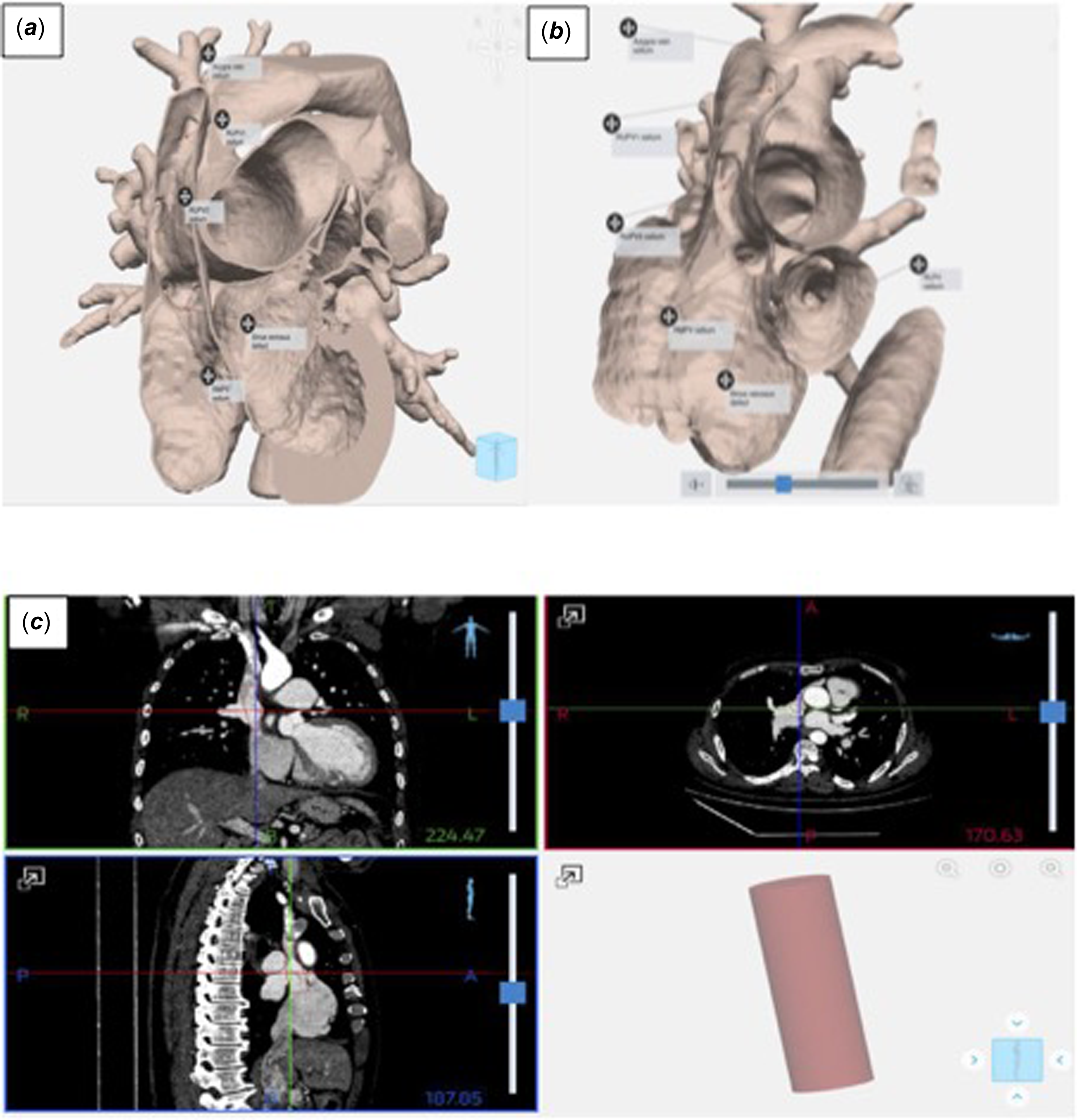
Cardiac MRI or CT images were reviewed to delineate the anatomy of the pulmonary venous return, and their anatomic relationship with the sinus venosus atrial septal defect (Figure 1).
( a ), ( b ): Delineating anatomy with 3D models. ( c ): simulation of stent implantation with printed or virtual 3D model.

Figure 1. Long description
The image consists of three parts: two medical illustrations labeled (a) and (b), and a set of three computed tomography (CT) scans labeled (c). The illustrations depict the superior sinus venosus atrial septal defect, highlighting its position immediately below the junction of the superior caval vein and the right atrium. The defect is associated with partial anomalous pulmonary venous drainage of the right upper and/or right middle pulmonary veins. The right upper pulmonary vein is shown draining into the right atrium at the mouth of the superior caval vein. The CT scans provide cross-sectional views of the chest, showing the anatomical structures involved in the defect. The illustrations and CT scans together provide a comprehensive view of the defect’s location and associated anatomical features.
Catheter procedure technique
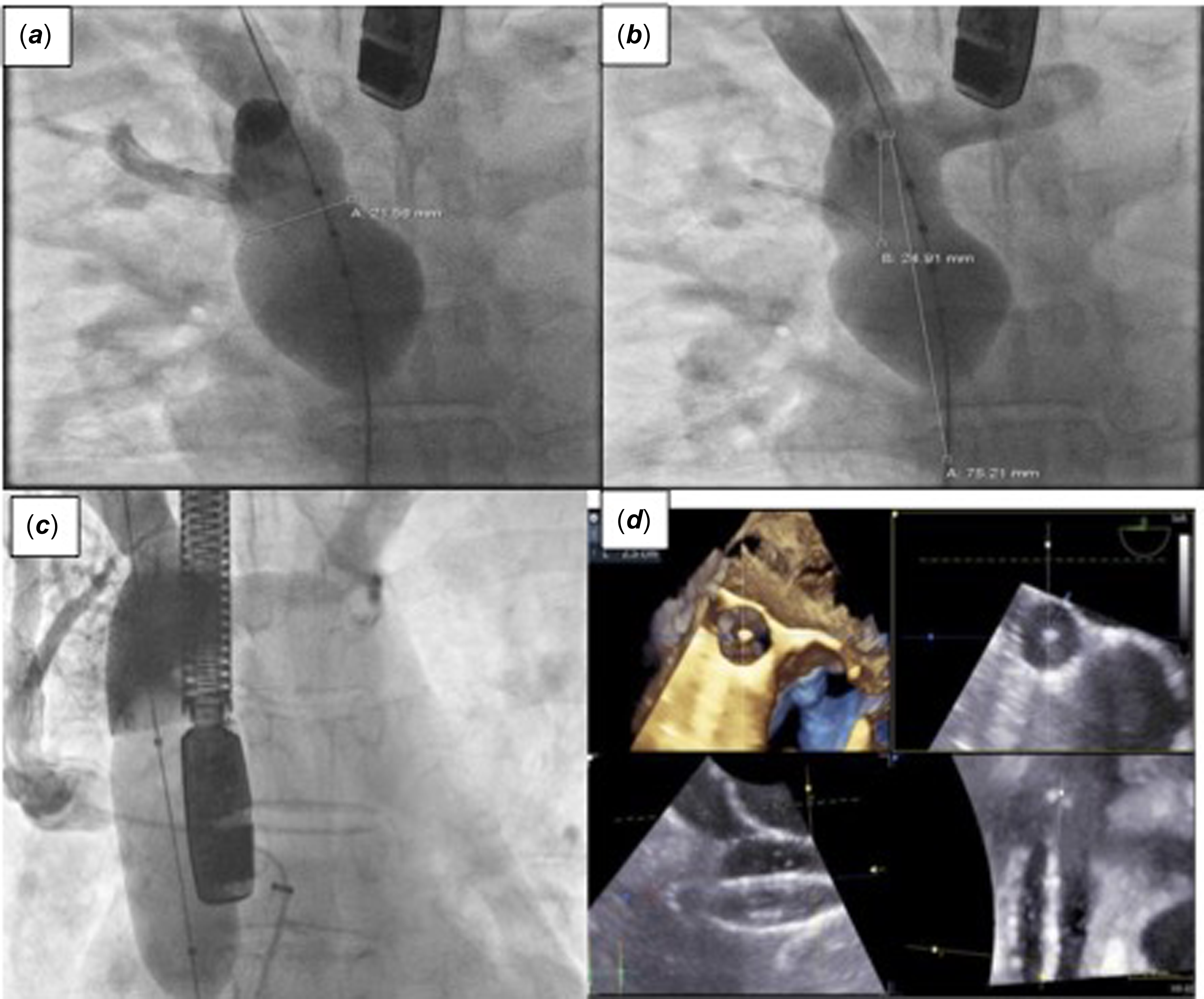
Procedures were performed under general anaesthesia or local anaesthesia with deep sedation, all with transesophageal echocardiographic guidance. Vascular access was obtained, with ultrasound guidance, in the right internal jugular vein, the left and right femoral veins, and the left femoral artery. Heparin was given to maintain an activated clotting time of >250 s. Antibiotics were administered as prophylaxis. A 10-F or 12-F long sheath (Cook Medical, Bloomington, Indiana) was passed from left femoral vein to right atrium and then through the patent foramen ovale to left atrium, and through the long sheath, a 5-F Multipurpose or 5-F Right Coronary Judkins catheter 3.5 (Cordis corporation, Miami Lakes, Florida) was placed in the right upper pulmonary vein to allow serial pulmonary venography as well as simultaneous monitoring of pressure gradient between the right upper pulmonary vein/right middle pulmonary vein and left atrium throughout the procedure. In cases where there was no patent foramen ovale, trans-septal puncture was performed using a Brockenbrough trans-septal needle (St. Jude Medical, St. Paul, Minnesota) in an 8-F Swartz™ Braided Transseptal Guiding Introducers (St. Jude Medical, St. Paul, Minnesota). A venovenous guidewire circuit was established, with a 0.035-inch Amplatzer Extra Stiff wire (Cook Medical, Bloomington, Indiana) from the right femoral vein to the right internal jugular vein, to facilitate balloon test occlusion/sizing and, eventually, stent implantation. Balloon testing was performed in all patients. A 40-mm PTS-X Sizing balloon (NuMED, Hopkinton, New York) or a 34-mm Amplatzer sizing balloon (St. Jude Medical, St. Paul, Minnesota) were used. The balloon was inflated in the anticipated stent landing zone until transesophageal echocardiography confirmed elimination of the interatrial shunt. A SCV angiogram was obtained to document SCV flow occlusion. During balloon inflation, right upper pulmonary vein flow was assessed by transesophageal echocardiography, angiography from the right upper pulmonary vein catheter, and, simultaneously, right upper pulmonary vein and left atrium pressure measurements. Following balloon sizing, repeat balloon interrogation of the stent landing zone with a semi-compliant balloon, either a 23-mm Z-Med balloon (NuMED, Hopkinton, New York) or a 25-mm Cristal Balloon (Balt Extrusion SAS, Montmorency, France), was done to assess for potential pulmonary venous obstruction with stent deployment (Figure 2). Pre-procedural imaging data and measurements obtained during balloon testing were used to choose the stent length and the appropriate balloon diameter for implantation. For all patients, Optimus-CVS polytetrafluoroethylene (PTFE)-Covered Stent XXL of 80 or 100 mm (AndraTec GmbH, Koblenz, Germany) mounted on AltoSa-XL-GEMINI balloon-in-balloon catheters with outer balloon diameters between 24 and 28 mm (AndraTec GmbH, Koblenz, Germany), were used. The stents were introduced through an 18-F or 20-F DrySeal sheath (Gore, Flagstaff, Arizona) from the right femoral vein. The stents were deployed using simultaneous transesophageal echocardiographic imaging and angiography from the right internal jugular vein and right upper pulmonary vein. The “uncovered“ cranial end of the stent was deployed to sit sufficiently high in the SCV to allow secure anchoring without compromising azygous or innominate vein flow, while the caudal end of the stent was placed sufficiently below the inferior rim of the defect. Flaring of the right atrium portion of the stent was done using a compliant balloon, PTS-X Sizing balloon of 40 mm or a Coda balloon of 46 mm (Cook Medical, Bloomington, Indiana) to ensure complete coverage of the interatrial communication in a “bell-shaped” configuration. A high-pressure balloon, Armada of 8 or 10 mm (Abbott Vascular, Santa Clara, California), was inflated in the right upper pulmonary vein during stent placement to form a “mould” and prevent obstruction of the pulmonary venous return behind the stent when this was considered to be at risk of compression during deployment or flaring of the stent.
( a ) and ( b ): Balloon sizing of SCV stent landing zone with compliant balloon and simultaneous RUPV (A) and SCV (B) angiograms. ( c ): semi-compliant balloon inflated in SCV with simultaneous SCV and RUPV angiograms, catheter in RUPV and long sheath in LA to document pressure gradient between RUPV and LA. ( d ): TOE with 3D display during balloon sizing. RUPV; right upper pulmonary vein, SCV; superior caval vein.

Figure 2. Long description
The image contains four separate images related to medical procedures and scans. The first image shows a measurement of a structure within the body, with a label indicating a length of 31.69 millimeters. The second image displays another measurement, with a label indicating a length of 24.91 millimeters and a width of 75.21 millimeters. The third image shows a medical device inserted into the body, likely a stent or catheter. The fourth image consists of two ultrasound scans, one in color and one in grayscale, showing detailed views of internal structures.
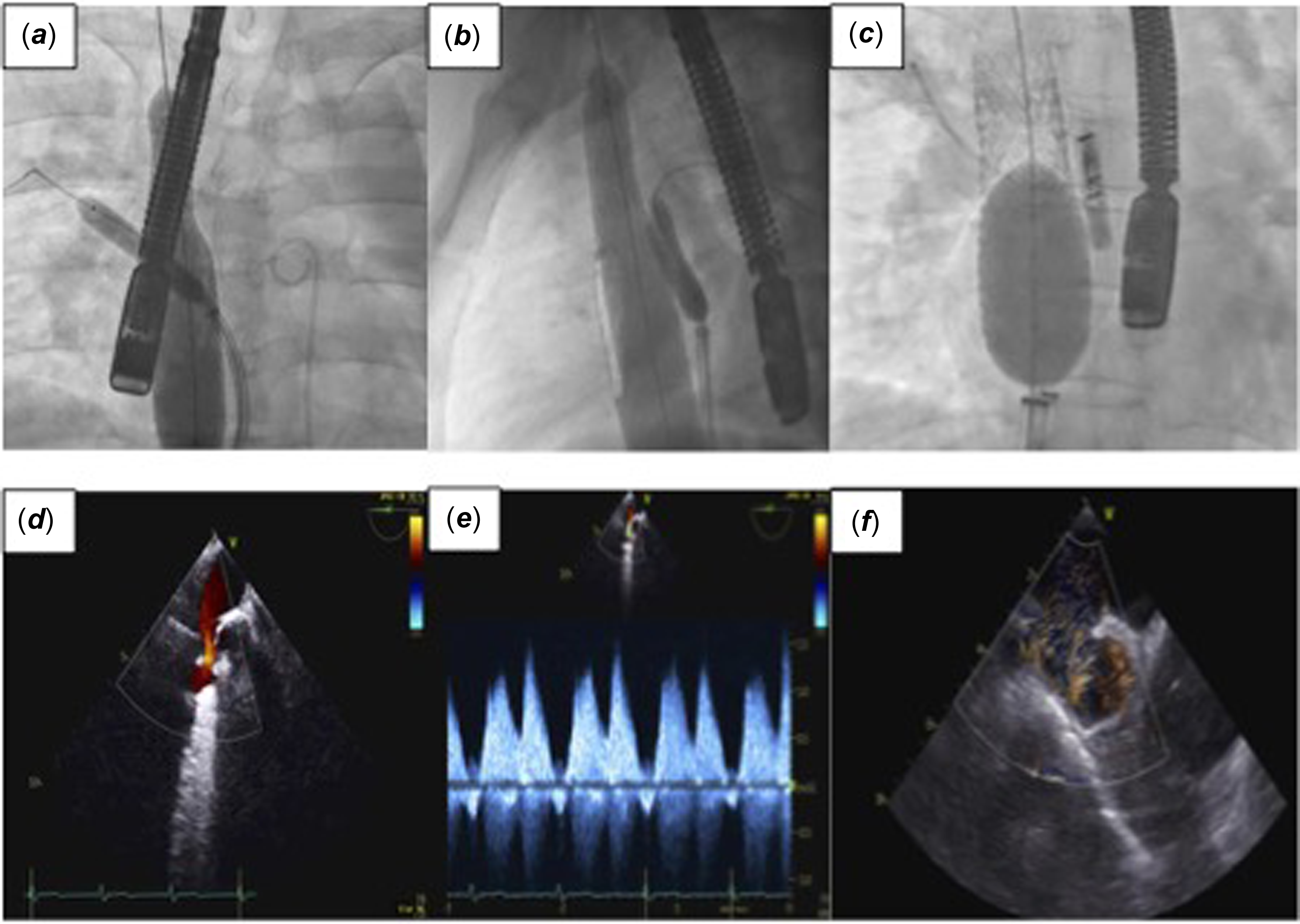
Final angiograms from the SCV and right upper pulmonary vein; pressure measurements in the right upper pulmonary vein, left atrium, and right atrium; and transesophageal echocardiography were used to assess for residual shunting and absence of pulmonary venous compression (Figure 3).
Armada balloon inflated on RUPV during stent expansion; antero-posterior projection ( a ), lateral projection ( b ). Flaring of the caudal end of the stent with a PTSX balloon to oppose stent to RA wall ( c ). TOE documenting no RUPV obstruction on colour doppler ( d ) and pulse-wave doppler ( e ), and blood speckle imaging ( f ).

Figure 3. Long description
The composite image consists of six distinct images arranged in two rows. The top row features three X-ray images labeled (a), (b), and (c), depicting a medical procedure involving the placement of a covered stent in the superior caval vein-right atrium junction. The bottom row contains three ultrasound images labeled (d), (e), and (f), showing different stages or aspects of the same procedure. The X-ray images illustrate the positioning and deployment of the stent, while the ultrasound images provide additional diagnostic information, likely including blood flow and structural details. The images collectively demonstrate the transcatheter approach to correcting a vascular issue, highlighting the evolution and refinement of this technique as an alternative to surgical repair.
Post-procedural management and follow-up
Dual antiplatelet therapy was commenced with clopidogrel 75 mg and aspirin 75 mg for 6 months, followed by aspirin alone for a further 6 months. Patients receiving anticoagulant agents continued receiving them (without additional antiplatelets) for at least 6 months. Routine post-procedural investigations included 12-lead electrocardiogram, chest X-ray and transthoracic echocardiography the day after the procedure. Clinical follow-up with
transthoracic echocardiography and 12-lead electrocardiogram was scheduled at 6 weeks, 6 months and 12 months. A cardiac CT was performed after 3 months to confirm stent position and exclude pulmonary vein obstruction or residual shunts.
Results
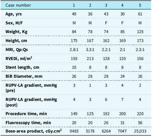
Five patients were treated with transcatheter correction of sinus venosus atrial septal defect and partial anomalous pulmonary venous drainage in our institution between January 2024 and January 2026. The anatomy was considered suitable for a transcatheter approach, based on evaluation of cross-sectional imaging in our adult CHD multidisciplinary meeting. Demographic and procedural data of the five patients who underwent transcatheter correction are summarised in Table 1.
Demographic and procedural data of the five patients who underwent transcatheter correction of SVASD

Table 1. Long description
The table presents demographic and procedural data for five patients treated with transcatheter correction of sinus venosus atrial septal defect and partial anomalous pulmonary venous drainage. The table includes five columns: Case number, Age in years, Sex, Weight in kilograms, Height in centimeters, MRI Qp:Qs ratio, RVEDi in milliliters per square meter, Stent length in centimeters, BiB Diameter in millimeters, RUPV-LA gradient in millimeters of mercury pre and post procedure, Procedure time in minutes, Fluoroscopy time in minutes, and Dose-area product in centigray square centimeters. The table lists data for five patients, with ages ranging from 30 to 61 years, weights from 74 to 125 kilograms, and heights from 162 to 173 centimeters. The MRI Qp:Qs ratios vary between 2.2:1 and 3.3:1, and the RVEDi values range from 128 to 213 milliliters per square meter. Stent lengths are consistently 8 centimeters, while BiB Diameters range from 24 to 28 millimeters. Pre-procedure RUPV-LA gradients range from 1 to 4 millimeters of mercury, and post-procedure gradients range from 3 to 7 millimeters of mercury. Procedure times vary from 125 to 220 minutes, fluoroscopy times from 20 to 56 minutes, and dose-area products from 5178 to 25033 centigray square centimeters.
No patent foramen ovale was found in 2 patients (cases 1 and 4), and smaller right upper pulmonary vein branches draining into the SCV high above the right atrial junction were present in 1 patient (case 5). Pre-procedural planning involved ex vivo simulation using a virtual or printed 3-dimensional model in all patients. Diagnostic cardiac catheterisation with balloon testing was performed in 2 patients (cases 2 and 3), where balloon sizing showed that the anticipated 100-mm long stent would compromise the tricuspid valve and right atrium structure, and an 80-mm stent would be optimal. Those two patients subsequently returned for the definitive procedure.
All patients were adults with an age range of 30–61 years. Three were males. Right upper pulmonary vein access was established with an antegrade approach either through a patent foramen ovale or by TSP using the standard approach. The first patient received a 100-mm Optimus-CVS PTFE-Covered Stent XXL, whereas all remaining 4 patients received an 80-mm stent. The outer diameter of the balloon-in-balloon used ranged from 24 to 28 mm as none of our patients had left SCV to the coronary sinus. The caudal end of the stent was flared to form a “bell-shaped” configuration to oppose the stent to the crest of the defect, thereby occluding the defect and redirecting the right upper pulmonary vein to the left atrium.
Pulmonary vein obstruction was anticipated by balloon sizing in all but one patient (case 2), and an Armada balloon of 8 or 10 mm was inflated in the right upper pulmonary vein for pulmonary vein protection during stent deployment and flaring. No significant gradient between RPV and left atrium was noted post-stent deployment (mean PG of 1–3 mmHg).
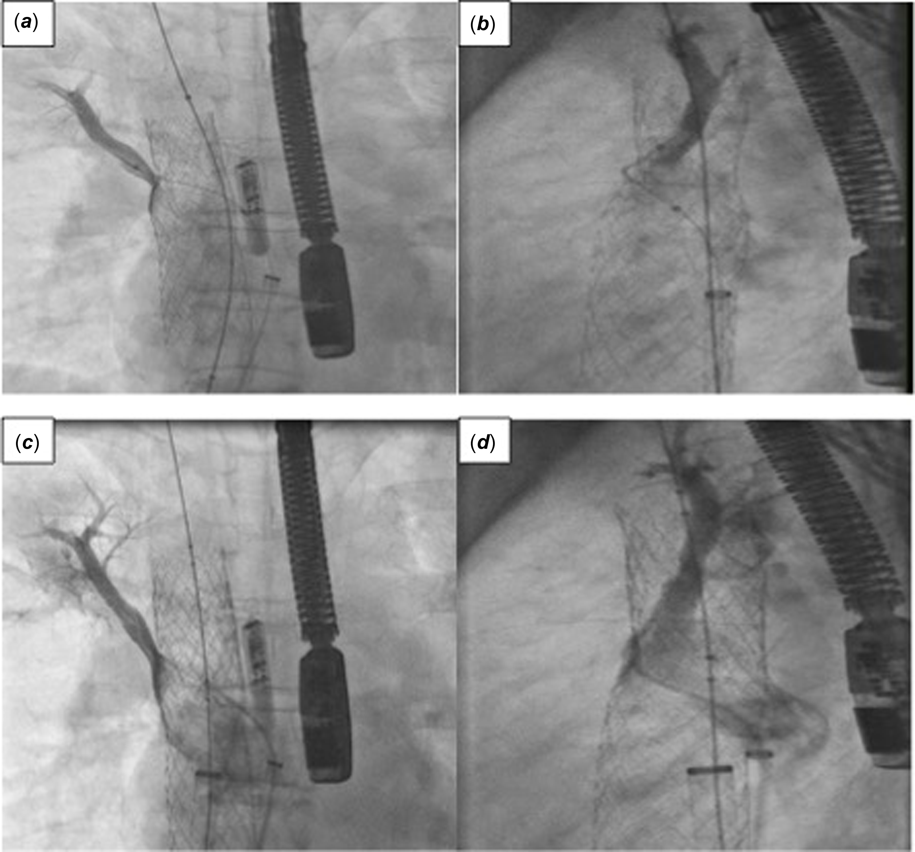
In one patient (case 3), transesophageal echocardiography documented a residual shunt from the cranial end of the stent due to a slightly lower placement of the stent. This was also confirmed with a pulmonary vein angiogram, showing persistent flow from the pulmonary vein to the SCV through the uncovered cranial part of the stent. A second Optimus-CVS PTFE-Covered Stent XXL of 43 mm was then mounted on a 24 × 45 mm balloon-in-balloon, which covered the pulmonary vein completely (Figure 4).
RUPV angiogram showing residual shunt through the cranial “uncovered” end of the stent to SCV; antero-posterior ( a ) and lateral projection ( b ). Same angiogram after placement of second stent showing redirection of flow from RUPV behind the stent to LA on anter-posterior view ( c ) and lateral view ( d ). RUPV; right upper pulmonary vein, SCV; superior caval vein.

Figure 4. Long description
The image displays a series of medical images related to a transcatheter procedure. The images show the deployment of a covered stent at the junction of the superior caval vein and the right atrium. The stent is visible in different stages of the procedure, with the surrounding vascular structures and the positioning of the stent within the body. The images highlight the precise placement and the effectiveness of the stent in the vascular system.
No serious adverse events occurred in any patient. None had any other significant intra- or immediate post-procedural complications. All patients were recovered to the ward and discharged the next day. Pre-discharge chest X-ray, 12-lead electrocardiogram and trans-thoracic echocardiogram were done on all patients. No patient had post-procedural complications such as stent migration, residual shunt, pulmonary vein obstruction, pericardial effusion or sinus node dysfunction.
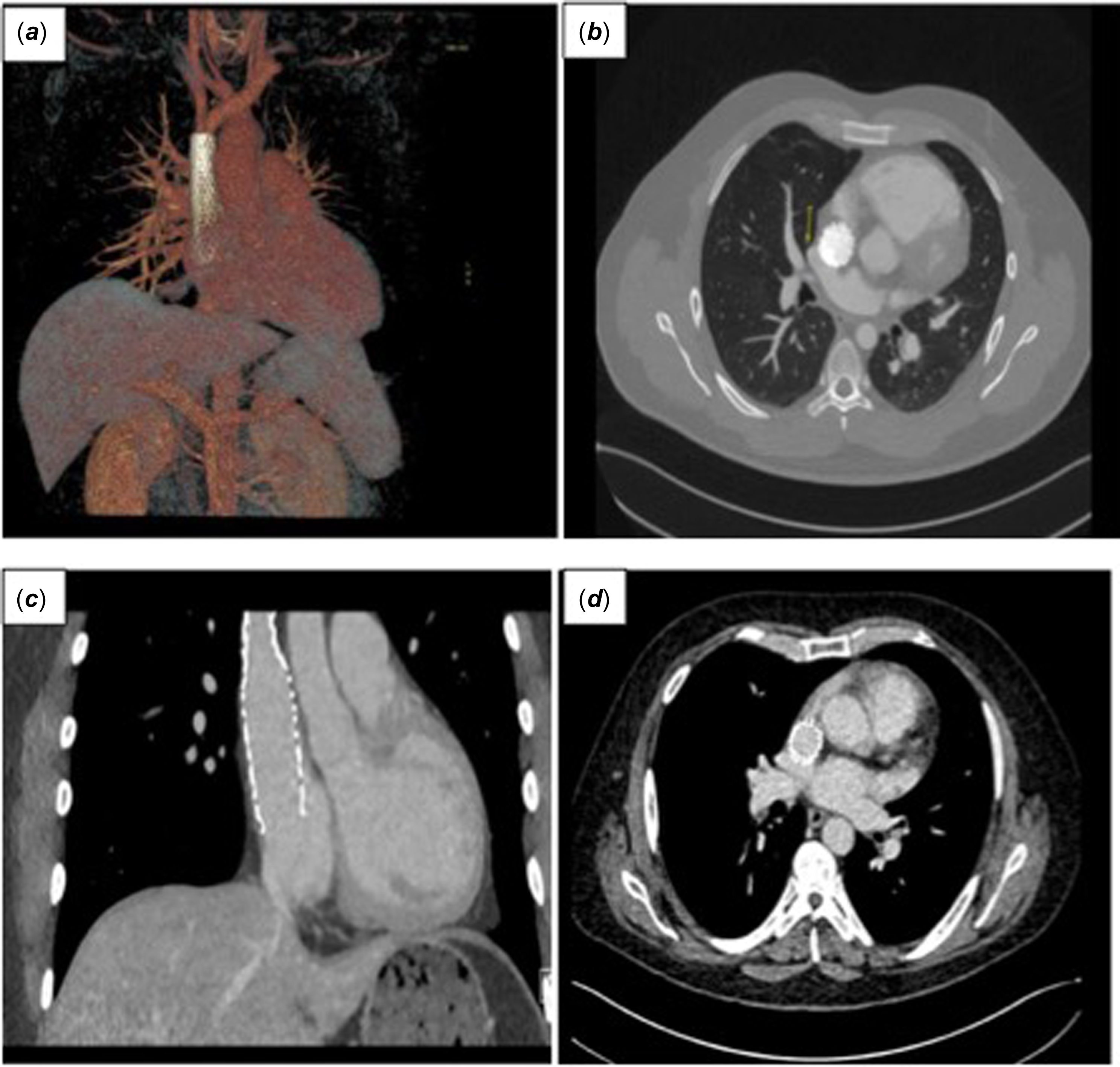
The follow-up period at the time of manuscript submission was 3–24 months. No late complications have been observed. Cardiac CT was performed in all patients at 3 months post-procedure. In all patients, the SCV stent was well-positioned and patent, with unobstructed pulmonary venous drainage and no residual shunts (Figure 5).
Cardiac CT scan done at 3 months showing well positioned stent on 3D volume rendered image ( a ), axial views ( b and d ) and coronal view ( c ).

Figure 5. Long description
The composite image consists of four panels labeled (a), (b), (c), and (d), each depicting different medical imaging techniques. Panel (a) shows a three-dimensional reconstruction of the thoracic vasculature, highlighting the superior caval vein and surrounding structures. Panel (b) presents a cross-sectional computed tomography (CT) scan of the chest, illustrating the placement of a covered stent at the junction of the superior caval vein and the right atrium. Panel (c) displays a sagittal CT scan, providing a side view of the stent and its position relative to the heart and surrounding anatomy. Panel (d) offers an axial CT scan, showing a horizontal cross-section of the chest with the stent visible within the superior caval vein. These images collectively demonstrate the transcatheter approach using a covered stent as an alternative to surgical repair in the treatment of superior caval vein syndrome.
Discussion
Transcatheter correction of sinus venosus atrial septal defect was initially viewed as an experimental approach in highly selected patients with unusual anatomic variants, but unlikely to be applicable to most patients. Reference Garg, Tyagi and Radha5,Reference Abdullah, Alsalkhi and Khalid6,Reference Crystal, Vincent and Gray14 Over the past few years, due to the better understanding of the anatomy of these defects, there has been an exponential rise in the volume of procedures and the number of units offering it. Reference Pascual-Tejerina, Sánchez-Recalde, Cantador, López, Gómez-Ciriza and Gutiérrez-Larraya15–Reference Thejaswi, Sagar and Sivakumar19 The results from the individual centres that performed the procedure are very promising, and this is now backed by data from an international registry. Reference Rosenthal, Qureshi and Sivakumar11 We implemented transcatheter correction of sinus venosus atrial septal defect as an innovative procedure in our centre in carefully selected patients. We went through extensive multidisciplinary discussions and preprocedural planning, along with the administrative and approval process, to finally deliver the care. In this article, we provide our experience with the transcatheter closure of sinus venosus atrial septal defect with our short to medium-term outcomes.
The success of the procedure relies on careful patient selection. This is achieved by multi-modality imaging tools such as transesophageal echocardiography, cardiac CT or MRI. Significant caudal extension of the defect was the main exclusion criterion in our short experience due to increased risk for residual shunt. Reference Sivakumar, Qureshi, Pavithran, Vaidyanathan and Rajendran20 Advanced 3-dimensional printing and heart virtual reality Reference Stephenson, Rosenthal and Jones21 helps assess the patients by showing stent positioning in correlation to the defect and to higher draining of significant pulmonary veins in SCV. In addition, balloon sizing/ test occlusion under transesophageal echocardiography, angiography and haemodynamic assessment, according to our experience, remains the cornerstone for the suitability or not for stent implantation.
Variable stent types have been reported to be used by different institutions since this procedure was first adopted. In earlier series, several telescoping stents were used. Reference Garg, Tyagi and Radha5,Reference Abdullah, Alsalkhi and Khalid6,Reference Batteux, Gkogkou and Albenque13,Reference Riahi, Velasco Forte and Byrne22 As the learning curve and preoperative assessment tools developed, numerous stents came into use. These are mostly balloon-expandable covered stents; however, self-expandable stents were also reported in some centres. To date, the most commonly used stent is the 10-Zig covered CP stents (NuMED, Hopkinton, New York) in 62% of cases. Reference Rosenthal, Qureshi and Sivakumar11 The stent we used in our series was the Optimus-CVS PTFE-Covered Stent XXL (AndraTec GmbH, Koblenz, Germany). This extra-large stent is especially designed for large lumen vessels and offers high radial strength combined with a safe PTFE covering. It has a unique hybrid cell design with high-performance PTFE coating combined with the material cobalt-chromium, providing extra strength and stability. Integrity of the PTFE coating is documented in anecdotal reports up to a diameter of 30 mm.
Pulmonary venous obstruction is reported to be around 1.3–2.1%. Reference Hansen, Duong and Jivanji8,Reference Rosenthal, Qureshi and Sivakumar11 Again, patient selection with pre-operative multimodality imaging helped predict potential pulmonary venous stenosis. We adopted balloon interrogation of the sinus venosus atrial septal defect with both compliant and semi-compliant balloons with contemporary monitoring of the pressure gradient between the right upper pulmonary vein and the left atrium. Whenever right upper pulmonary vein obstruction was anticipated, a high-pressure balloon was inflated in the right upper pulmonary vein throughout stent implantation and dilatation. This helped form a “mould” behind the stent, allowing redirection of the right upper pulmonary vein to the left atrium.
Transcatheter correction of sinus venosus atrial septal defect is a less invasive procedure that avoids surgical sternotomy and reduces recovery time; however, complications still happen. These include stent embolisation (2.6%), other major complications such as cardiac tamponade, sinus node dysfunction needing pacemaker implantation and surgery for pulmonary vein obstruction were reported in 1.3%. In addition, residual leak needing repeat intervention occurred in 1.8%. Reference Rosenthal, Qureshi and Sivakumar11 We did not observe any complications in our case series. The follow-up is still ongoing, and thus far. In all patients, cardiac CT scan at 3 months after procedure did not document any residual shunt or pulmonary vein stenosis. We appreciate that this is a small series with only 5 cases, and the previously mentioned data is from a large volume multicentre international registry.
Finally, it is worth mentioning that there is still a learning curve to gain for transcatheter correction of sinus venosus atrial septal defect, and that surgical repair of sinus venosus atrial septal defect is still a very viable option with good results. A recent systematic review and meta-analysis of the outcomes showed an in-hospital mortality rate of 0.24% and a 30-day mortality of 0.5%, with a 2–6.5% risk of major adverse events. Reference El-Andari, Moolla and John23 Transcatheter correction of sinus venosus atrial septal defect should not be the complete replacement of the surgical option, as the procedure is technically demanding. The SCV into which the cranial part of the stent is lodged is not stenosed and is distensible, unlike in other lesions, such as coarctation of the aorta, which can grip the stent. The caudal part of the stent needs to be inflated in the SCV to occlude the defect; shortening of the lower half of the stent is different from the upper part, and predicting the final position of the caudal end of the stent is not easy. The funnel-shaped stent is not always stable in the SCV. Adding this to the risks of pulmonary vein obstruction and the presence of significant veins draining higher up in the SCV makes surgery a more preferable option, especially in younger patients. Reference Rosenthal and Jones9
Limitations
This is a small centre retrospective study with a small number of patients. The retrospective nature of the analysis put it at risk of selection bias. The small number of cases is not enough to perform an analysis that can reach statistical significance to compare our results with the results from large-volume or multicentre studies. Furthermore, a learning curve is still there, and more modifications to patients’ selection and procedural techniques are yet to come.
Conclusions
Transcatheter closure of sinus venosus atrial septal defect is a feasible and safe procedure in selected patients with good results both from individual and multicentre international cohorts. The learning curve is still to be completed, and surgical options are not to be completely abolished. Future high-quality randomised studies and meta-analyses are required to help select the right treatment options.