


Introduction
Candida auris is a concerning fungal pathogen that causes invasive infections such as bloodstream infections (BSIs) and a significant challenge in healthcare settings due to its propensity to cause outbreaks and persist on environmental surfaces. Reference Ahmad and Alfouzan1–Reference Dire, Ahmad, Duze and Patel5 Invasive fungal infections are associated with significant mortality, ranging from 30-60%, with rates for C. auris BSIs reportedly up to 80%. Reference Bradley6–Reference Vu, Jimenez, Anjan and Abbo8 Additionally, invasive infections may develop in 5-20% of patients previously colonized with C. auris. Reference Schelenz, Hagen and Rhodes9,Reference Rossow, Ostrowsky and Adams10 Risk factors for invasive infection include severe underlying comorbidities, invasive medical devices, immunocompromised status, prior antimicrobial exposure, intensive care unit (ICU) exposure, and recent surgery. Reference Wasylyshyn and Stoneman11 One of the leading risk factors for C. auris acquisition is exposure to skilled nursing facilities (SNFs), long-term acute care hospitals (LTACHs), or acute inpatient rehabilitation facilities (AIRs). 2 As a result, the Centers for Disease Control and Prevention (CDC) recommend that facilities consider active screening of patients with previous healthcare encounters at the aforementioned high-risk facilities. 2
C. auris rates and antimicrobial resistance are increasing in the United States (US) with a reported 200% increase in surveillance samples and 95% increase in clinical isolates from 2020 to 2021. Reference Noble, Jurcic Smith, Jones, Galvin and Timbrook12,Reference Lyman, Forsberg and Sexton13 Similarly, three times the number of echinocandin-resistant isolates were reported in 2021 compared to that in each of the prior two years. Reference Lyman, Forsberg and Sexton13,Reference Jacobs, Jacobs and Dennis14 Furthermore, certain regions may experience greater prevalence. Notably, Illinois carries a heavy burden of C. auris, with data from the CDC indicating it as having the second highest number of clinical cases from 2016 to 2023 and fifth highest number of clinical cases in 2023 in the US. 15
We report an outbreak of C. auris in 2023 in our cardiothoracic transplant intensive care unit (CTICU). This study describes the outbreak investigation, implementation of mitigation strategies, and the potential long-term implications for infection prevention practices amidst an evolving landscape.
Methods
Care setting
Northwestern Memorial Hospital (NMH) is a 943-bed academic medical center in Chicago, Illinois. The CTICU houses cardiothoracic surgical patients including heart and lung transplant recipients and patients requiring advanced mechanical circulatory support devices. The CTICU also shares a patient population with its stepdown unit. All inpatient units in our facility have single, private rooms. The Northwestern University Institutional Review Board (IRB) reviewed this study and deemed it exempt from IRB review.
Clinical and epidemiologic definitions
C. auris colonization was defined as isolation of C. auris during admission or weekly screening from a bilateral nares, composite axilla/groin, and/or endotracheal aspirate specimen. C. auris infection was defined as isolation of C. auris from a clinical specimen coupled with documented administration of targeted antifungal therapy.
A hospital-onset (HO) case of C. auris was defined as isolation of C. auris on day four of hospital admission or later in accordance to criteria set forth by the CDC National Healthcare Safety Network (NHSN). 16 Nosocomial transmission was defined as a HO case of C. auris that had geo-temporal overlap with another C. auris positive patient.
An outbreak was defined as the finding of three or more patients with C. auris isolated from clinical or screening samples whose epidemiology indicated shared time, space, and/or clinical care providers within a month. Outbreak resolution was defined as four consecutive weeks without new identification of a positive HO C. auris case in the CTICU.
Laboratory identification and antifungal susceptibility testing of C. auris
Specimens collected from admission and weekly surveillance were processed and polymerase chain reaction (PCR) was performed using previously described methods and reagents. Reference Malczynski, Dowllow and Rezaeian17
Clinical and environmental cultures were collected using eSwabs® (Copan Diagnostics, Murrieta, CA). Specimens were set to inhibitory mold agar (IMA) Media (Remel, San Diego, CA) plates and held for five days at 300C. C. auris identification was performed using the Vitek® MS Instrument (bioMérieux, Balmes-les-Grottes, France) with the software V3 2.0.
One clinical isolate per patient was tested for antifungal susceptibility testing (AST). Minimum inhibitory concentration (MIC) values (µg/mL) were determined using Sensititre™ YeastOne™ YO3IVD AST Plates (Thermo Scientific, Waltham, MA) for fluconazole, itraconazole, voriconazole, caspofungin, micafungin, and flucytosine. The CDC tentative AST breakpoints for fluconazole and micafungin were utilized for resistance interpretation. 15
C. auris surveillance and isolation protocols
Our institution created a questionnaire in the electronic medical record (EMR) to identify the population to be screened upon admission in 2019. The questionnaire assesses exposure to an SNF, LTACH, or AIR within six months prior to admission and queries whether the patient is on mechanical ventilation or has a tracheostomy. Patients remain on standard precautions while the admission test is in process.
Once a C. auris case is identified from a surveillance or clinical sample, the patient is placed on “contact plus” isolation indefinitely. At our institution, contact plus isolation directs all individuals, including visitors, to don a gown and gloves in the room. The room is cleaned daily with a bleach-containing solution. Upon patient transfer or discharge, the discharge clean consists of a bleach-containing solution, a curtain change, and ultraviolet-C disinfection (Surfacide®, Waukesha, WI).
If a HO-case of C. auris is identified, IP directs a point prevalence survey (PPS) on the affected unit(s). Patients previously positive for C. auris are not further screened. PPS continues weekly until no additional positive cases are identified.
Whole genome sequencing and single nucleotide polymorphism (WGS-SNP) analyses
WGS-SNP analyses of C. auris isolates were performed using previously published methodologies. Reference Mitchell, Kling and Bolon18
Outbreak investigation
Following the detection of a C. auris outbreak, IP convened a multidisciplinary team (MT) that included the following members: CTICU clinical leadership, hospital executive leadership, environmental services (EVS), laboratory, infectious diseases, antimicrobial and diagnostic stewardship, and ancillary teams. The MT met weekly from October 2023 until the cessation of the outbreak to discuss outbreak data and multimodal mitigation strategies.
IP performed a systematic lookback to identify additional patients who may have acquired C. auris while in CTICU to be included in the outbreak. The EMR was used to extract patient characteristics and clinical data points as well as to retrospectively identify cases for constructing an epidemic curve. Patients were included as part of the systematic lookback if they had a newly identified C. auris result and prior exposure to CTICU between July 2023 and February 2024. These additional patients were not initially thought to be part of the outbreak as they were: (1) housed in different inpatient units, (2) discharged to an outside healthcare facility then readmitted to our facility, or (3) seen at an outpatient clinic at the time of their C. auris lab results.
Results
Outbreak detection
Prior to the outbreak, C auris was seen very infrequently among the CTICU population. From 2020 through 2022, there was only a single HO case of C. auris identified in the CTICU and none from its stepdown floor. C. auris BSIs occurred in two patients one month apart in July and August 2023 in the CTICU. A HO-case of C. auris was seen in a third patient in September 2023 who had geo-temporal overlap with the patient from August. Two rounds of PPS one week apart yielded four additional positive cases of C. auris colonization, triggering an outbreak investigation. A total of 20 cases of C. auris were identified from weekly screening in the CTICU during the outbreak and 14 additional cases identified from the retrospective review, raising the total to 34 cases (Figure 1). The 34 isolates were saved for WGS-SNP analysis. Of the 34 isolates, five could not be recovered in culture for WGS-SNP analysis despite being positive on PCR. Of the 29 remaining isolates, WGS-SNP analysis revealed two distinct outbreak strain types (Figure 2). AST of 29 patients revealed an elevated MIC to fluconazole among the two outbreak strains whereas that was not the case for isolates unrelated to the outbreak (Supplemental Table 1). Additionally, a clade analysis revealed all isolates belonged to Clade IV (Supplemental Figure 1).
C. auris outbreak epidemic curve showing cases identified from weekly screening during the outbreak and from the retrospective analysis. *For cases identified by retrospective analysis, some patients were admitted to the CTICU weeks prior to outbreak resolution. The index patient in Cluster I underwent left ventricular assist device implantation and had prior OSH exposure (no SNF/LTACH/AIR) with 23 antibiotic-days. The index patient in Cluster II underwent coronary artery bypass graft and similarly had prior OSH exposure (no SNF/LTACH/AIR) with 21 antibiotic-days.

Dendogram constructed using WGS-SNP analysis detailing patient and environmental isolates as they relate to the outbreak. *Indicates index cases in the outbreak clusters. Prior exposure to SNF/LTACH/AIR is represented by a green box, and those without SNF/LTACH/AIR exposure are represented by a red box.

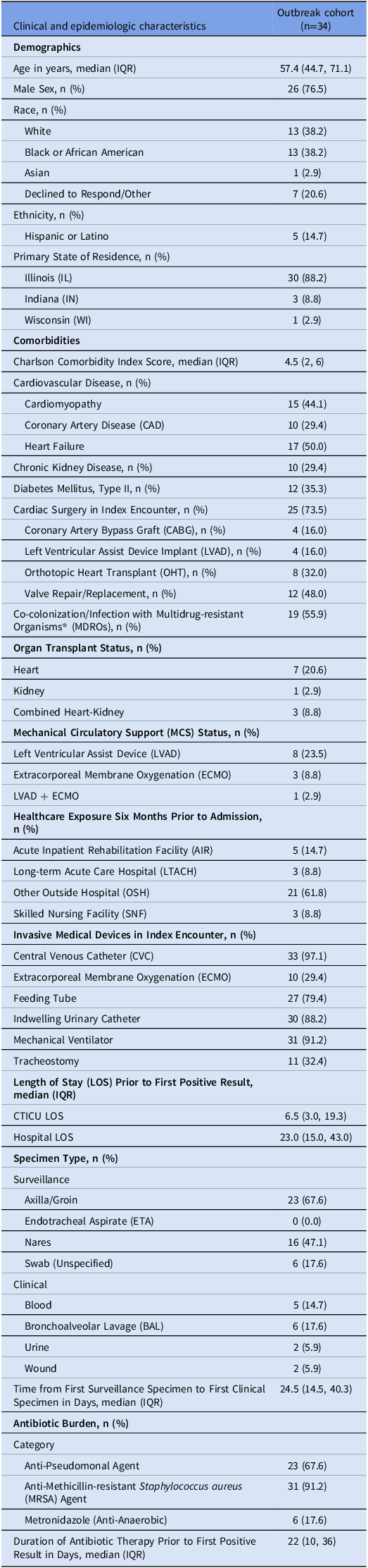
Clinical and epidemiologic characteristics of the outbreak cohort

*MDRO defined as one or more of the following: Methicillin-resistant Staphylococcus aureus (MRSA), Vancomycin-resistant Enterococcus (VRE), Extended-spectrum beta-lactamase-producing Enterobacterales (ESBL), Carbapenem-resistant Enterobacterales (CRE), and Carbapenem-resistant Acinetobacter baumannii (CRAB).
Patient characteristics
Of the cohort, 11/34 (32.3%) had prior exposure to high-risk healthcare facilities within six months prior to admission, as follows: AIRs (n = 5, 14.7%), SNFs (n = 3, 8.8%), and LTACHs (n = 3, 8.8%). A significant proportion of patients had prior exposure to other outside hospitals (n = 21, 61.8%). A majority of the cohort underwent cardiac surgery in their index inpatient encounter (n = 25, 73.5%) in which the two most common procedures were valve repair/replacement (n = 12, 48.0%) and orthotopic heart transplantation (OHT) (n = 8, 32.0%) (Table 1). Many of the identified patients were considered high-risk populations for invasive fungal infections, including those who had undergone solid organ transplant (SOT) (n = 11, 32.3%) or received mechanical circulatory support (MCS) (n = 12, 35.3%). Over half of the cohort had co-colonization/infection with multidrug-resistant organisms (MDROs) (n=19, 55.9%). The median length of stay (LOS) was 23.0 days and 6.5 days for the hospital and CTICU, respectively. Patients had a substantial antimicrobial burden prior to their C. auris results with a median duration of 22.0 antibiotic-days, with anti-pseudomonal (n = 23, 67.6%) and anti-methicillin-resistant Staphylococcus aureus (MRSA) agents (n = 31, 91.2%) being most frequently utilized.
Patient outcomes
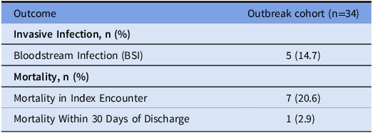
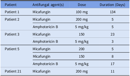
Five (14.7%) patients had C. auris BSIs, three of whom expired likely due to infection after review by infectious diseases and the primary care teams (Table 2A). All five patients were treated for BSI with micafungin monotherapy or a combination of micafungin and amphotericin B (Table 2B). Seven (20.6%) total patients expired during their index inpatient encounter.
A: Patient outcomes in the outbreak cohort

B: Antifungal therapy regimen for the five patients with C. auris bloodstream infections in the outbreak cohort

Outbreak mitigation
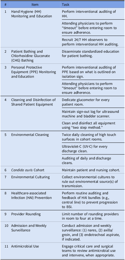
Upon detecting the outbreak, IP formed a MT and implemented several outbreak measures (Table 3) (Figure 1). The CTICU formed a C. auris cohort by sequestering C. auris-positive patients in one section of the unit and enforcing a 1:1 nurse-to-patient ratio. In addition to ongoing weekly PPS, the MT convened universal admission C. auris testing for all new admits and transfers to the CTICU. Patients negative on admission received weekly screening for the duration of their CTICU stay. As weekly testing continued to identify additional positive results, the MT implemented universal contact isolation precautions and centralized cleaning and disinfection of shared equipment in the CTICU.
Infection Prevention outbreak mitigation measures

The investigation identified two frequently utilized pieces of shared equipment on the unit that lacked documentation of disinfection between use: the ultrasound machine and bladder scanner. To ensure both were thoroughly cleaned and disinfected after use, the MT developed a sign-out log maintained by the unit secretary. Clinicians signed each instrument out before use and were required to attest to its cleaning and disinfection after use. The EVS team switched their standard cleaning agent (Alpha-HP®, Solenic LLC, Wilmington, DE) to one with a kill claim against C. auris (Oxivir™, Solenis LLC, Wilmington, DE) institution-wide rather than limit it to rooms with C. auris positive patients along with moving from once to twice daily cleans. This was accompanied by strict oversight and auditing of their daily and discharge cleans.
To rule out environmental sources of transmission, IP conducted two rounds of environmental culturing: one on 11/28/2023 and another on 12/13/2023. In total, IP cultured 349 surfaces, of which one room doorknob was positive for C. auris (Supplemental Table 2). However, this environmental isolate was unrelated to all patient isolates identified during the outbreak (Figure 2). Lastly, IP implemented a strategy to deploy healthcare personnel on the CTICU 24/7 as interventional auditors of hand hygiene (HH) and personal protective equipment (PPE) practices which was maintained until outbreak resolution. This strategy was comprised of recruiting individuals to sign up for one-to-four-hour shifts in the CTICU to audit HH/PPE practices and perform real-time coaching during all shifts throughout the week. The median number of weekly HH audits during October–November was 115.5 audits/week with 96.3% recorded compliance. This number increased during the 24/7 auditing period to a median of 797.5 audits/week with 99.7% recorded compliance. The outbreak was ultimately resolved per our defined criteria.
Discussion
Our experience detailing a C. auris outbreak in the CTICU highlights the implications of an evolving landscape of C. auris epidemiology, patient care challenges, and infection prevention practices.
As stated, patients presenting from facilities such as SNFs, LTACHs, and AIRs have an increased risk for C. auris acquisition. Reference Pacilli, Kerins and Clegg19–Reference Sexton, Bentz and Welsh21 As such, prior exposure in these facilities is the rationale for admission screening in many medical centers. We were not able to determine the sources of C. auris acquisition among the two index patients for the two outbreak clusters; neither had exposure to a SNF, LTACH, or AIR. Furthermore, several patients unrelated to the outbreak similarly lacked prior exposure to those high-risk facilities. Many patients unrelated to the outbreak had prior exposure to an OSH. The alternative risk factors we considered include selection pressure from antimicrobial use and nosocomial transmission facilitated by healthcare personnel or shared equipment across the institution; a third possibility is that these patients were colonized with C. auris upon admission due to OSH exposures but were not tested due to not meeting admission screening criteria. Taken overall, our findings may suggest that the epidemiology of C. auris is changing, pointing towards a rising prevalence in other acute care settings.
These findings may warrant an expansion of institutional surveillance programs. One consideration includes expanding the high-risk healthcare exposure definition to include OSHs. A second consideration may be to pursue admission and weekly C. auris screening in “high-risk” inpatient areas to rapidly identify cases, initiate timely isolation precautions, and prevent nosocomial transmission. Following the outbreak, we continued admission and weekly screening in the CTICU. Further study is needed to optimize early, universal identification of C. auris and inform future surveillance recommendations.
It is important to acknowledge several risk factors present in this cohort that likely contribute to C. auris acquisition and candidemia. A significant proportion of patients underwent cardiac transplantation or left ventricular assist device (LVAD) implantation, both of which are associated with increased antimicrobial exposure and poor clinical outcomes due to C. auris candidemia. Reference Vu, Jimenez, Anjan and Abbo8,Reference Kerai, Doshi, Laleker and Majumdar22 Of the cohort, 73.5% of patients underwent cardiac surgery during the index encounter prior to acquiring C. auris. While candidemia risk factors specific to cardiac surgery are not extensively described in the literature, associated risk factors such as prolonged ICU stay, broad-spectrum antibiotic use, and the presence of central venous catheters (CVCs) may predispose these critically ill patients to a higher risk of developing candidemia after surgery. Reference Pasero, De Rosa and Rana23,Reference Giacobbe, Salsano and Del Puente24 This cohort demonstrated further risk factors for developing invasive infection including frequent use of invasive medical devices.
There was additionally a heavy antibiotic burden, notably anti-pseudomonal and anti-MRSA agents. Exposure to these agents may lead to increased disruption of host microbiomes and reduce protective microdiversity, making patients more susceptible to fungal colonization, invasive infections, and poor clinical outcomes. In addition to an increased risk of invasive fungal infections, antibiotics have also been associated with increased risk of death after invasive candidiasis. Reference Drummond, Desai and Ricotta25 The relationship between antimicrobial exposure and subsequent C. auris colonization/infection should be explored along with enhanced understanding of microbiome changes in these patients. The high rate of antimicrobial use among these patients suggests a need for enhanced antimicrobial stewardship practices to avoid unnecessary exposure and prolonged durations to prevent microbiome disruption and risk for fungal infections.
Our team-based, multimodal approach allowed for enhanced transparency, understanding, and priority communication throughout the outbreak. Among the number of IP interventions implemented to curb nosocomial spread, a select few may have been particularly effective. The process of centralizing the cleaning and disinfection of the ultrasound machine and bladder scanner with a sign-out log increased accountability among healthcare personnel. Prior to centralization, the cleaning and disinfection practices were noted to be inconsistent, which may have facilitated transmission via shared equipment as this pathway is well described in the literature. Reference Eyre, Sheppard and Madder26–Reference Ahmad and Asadzadeh28 The enhanced environmental cleaning protocol of twice daily cleaning of high touch surfaces along with strict auditing and oversight was essential in efforts to reduce persistent environmental contamination, as some literature indicates that C. auris may re-contaminate an environmental surface within four hours after disinfection. Reference Sansom, Gussin and Schoeny4
Similarly, our noted increase in hand hygiene compliance and unit-wide contact precautions strategy may have hastened the resolution of the outbreak. Data collected from the World Health Organization (WHO) shows an average hand hygiene compliance as low as 20% without specific interventions and approximately 64% in ICUs in high-income countries. 29 As such, there is a gap in understanding factors influencing hand hygiene behavior. Traditional approaches to performing interventional hand hygiene and PPE auditing may be limited in capturing a lower volume of audits during off-shift hours. This limitation was addressed via our strategy to deploy external interventional hand hygiene and PPE auditors 24/7 in the CTICU to increase audit volume and scrutiny. Real-time coaching and feedback along with an accountability model for noncompliance may have driven a culture shift on the unit to curtail further transmission.
Healthcare personnel hands, shared patient equipment, and contaminated environmental surfaces all may have contributed to propagate spread due to documented practice gaps. A multidisciplinary approach in employing aggressive IP strategies was instrumental in resolving the outbreak and outlined the framework for a sustainable IP plan in the CTICU.
This study has several limitations. It was a single-center experience with a limited sample size. We acknowledge the need for further study to evaluate a possibly changing C. auris epidemiology, including the evolution of a “high risk” C. auris population or setting. Regarding the WGS analysis, five of the 34 isolates were not typed as they could not be recovered in culture, limiting our full understanding of the outbreak strains. Lastly, the environmental culturing was performed several weeks into the outbreak when the C. auris patient burden on the unit was lower. Since a majority of cases were identified prior to these collection dates, our findings do not necessarily indicate the absence of an environmental source as there were several practice improvements made prior to environmental culturing.
In conclusion, our outbreak experience suggests a possible shift in the epidemiology of C. auris and may warrant an expansion of C. auris surveillance protocols to encompass additional risk factors for both acquiring C. auris and developing invasive infection. A robust multidisciplinary approach to enhance IP practices, including improving compliance with cleaning and disinfection of shared equipment, environmental cleaning, and hand hygiene, may have contributed to outbreak resolution and formulated a sustainability plan in a complex critical care setting.
Supplementary material
For supplementary materials referred to in this article, please visit https://doi.org/10.1017/ice.2025.10217
Acknowledgments
We would like to extend our sincere gratitude to the multidisciplinary team stakeholders in the cardiac surgery, anesthesia critical care, and CTICU nursing teams for their hard work and vigilance during the outbreak investigation and mitigation efforts.
Author contributions
Shardul N. Rathod: Conceptualization, Investigation, Writing – Original Draft, Writing – Review & Editing, Visualization. Grace Barajas: Investigation, Writing – Review & Editing, Supervision. Brooks I. Mitchell: Formal analysis, Investigation, Writing – Review & Editing, Visualization. Michael Malczynski: Investigation, Writing – Review & Editing. W. Justin Moore: Investigation, Writing - Review & Editing. Erin Weslander: Investigation, Writing - Review & Editing. Christine Bulger: Investigation, Writing - Review & Editing. Andrea Stone: Investigation, Writing - Review & Editing. Abbas Al-Qamari: Investigation, Writing - Review & Editing. Christie M. Bertram: Investigation, Writing - Review & Editing. Sarah H. Sutton: Investigation, Writing - Review & Editing. Valentina Stosor: Investigation, Writing - Review & Editing. Chao Qi: Formal analysis, Investigation, Writing – Review & Editing, Visualization. Teresa R. Zembower: Investigation, Writing - Review & Editing, Supervision. Maureen K. Bolon: Conceptualization, Investigation, Writing – Original Draft, Writing – Review & Editing, Visualization, Supervision.
Financial support
None reported.
Competing interests
Shardul N. Rathod reports no conflicts of interest relevant to this article.
Grace Barajas reports no conflicts of interest relevant to this article.
Brooks I. Mitchell reports no conflicts of interest relevant to this article.
Michael Malczynski reports no conflicts of interest relevant to this article.
W. Justin Moore reports no conflicts of interest relevant to this article.
Erin Weslander reports no conflicts of interest relevant to this article.
Christine Bulger reports no conflicts of interest relevant to this article.
Andrea Stone reports no conflicts of interest relevant to this article.
Abbas Al-Qamari reports no conflicts of interest relevant to this article.
Christie M. Bertram reports no conflicts of interest relevant to this article.
Sarah H. Sutton reports no conflicts of interest relevant to this article.
Valentina Stosor reports no conflicts of interest relevant to this article.
Chao Qi reports no conflicts of interest relevant to this article.
Teresa R. Zembower reports no conflicts of interest relevant to this article.
Maureen K. Bolon reports no conflicts of interest relevant to this article.