Introduction
Brittle stars experience remarkable morphological changes through their early development, after they have completed their metamorphosis, and during the transition to adulthood (Clark, Reference Clark1914; Stöhr, Reference Stöhr2005). Within the class Ophiuroidea, the genus Ophioderma (Müller and Troschel, Reference Müller and Troschel1840) is noteworthy for the pronounced changes observed between its juvenile and adult forms. As they reach their maximum size as adults, members of Ophioderma exhibit modifications in several morphological characters, including the shape and extent of the granules on the body, the shape of the dorsal and ventral arm plates, the fragmentation of the dental plate and dorsal arm plates, the number of arm spines, colouration, among others (Clark, Reference Clark1939; Granja-Fernández et al., Reference Granja-Fernández, Herrero-Pérezrul, López-Pérez, Hernández, Rodríguez-Zaragoza, Jones and Pineda-López2014, Reference Granja-Fernández, Pineda-Enríquez, Solís-Marín and Laguarda-Figueras2020; Hendler, Reference Hendler2018; Hendler et al., Reference Hendler, Miller, Pawson and Kier1995; Humara-Gil et al., Reference Humara-Gil, Granja-Fernández, Bautista-Guerrero and Rodríguez-Troncoso2022a; Koehler, Reference Koehler1914; Landschoff and Griffiths, Reference Landschoff and Griffiths2015; Monteiro et al., Reference Monteiro, Reis and Pardo1992; Prata et al., Reference Prata, de Castro Manso and Christoffersen2018; Stöhr et al., Reference Stöhr, Boissin and Chenuil2009; Tommasi, Reference Tommasi1970; Varela-Sánchez et al., Reference Varela-Sánchez, Templado and Machordom2020; Weber et al., Reference Weber, Stöhr and Chenuil2014; Ziesenhenne, Reference Ziesenhenne1955). These changes have been documented in nine of the 35 valid species of the genus (Stöhr et al., Reference Stöhr, O’Hara and Thuy2024), but their description is usually partial and brief, merely mentioned as passing observations.
To date, the most comprehensive accounts of growth changes in Ophioderma are available for three species: Ophioderma januarii Lütken, Reference Lütken1856, Ophioderma panamense Lütken, Reference Lütken1859, and Ophioderma rubicundum Lütken, Reference Lütken1856. In his revision of the genus, Ziesenhenne (Reference Ziesenhenne1955) additionally studied the growth changes of O. panamense, examining 284 specimens from the Gulf of California with disc diameters (DD) ranging from 2.3 to 21 mm. Subsequently, Monteiro et al. (Reference Monteiro, Reis and Pardo1992) investigated the external morphological variation with size and the bathymetric distribution of O. januarii, including 211 individuals within a size range of 10–21.5 mm DD. The most recent study to provide information on the growth changes of an Ophioderma is that of Hendler (Reference Hendler2018), which included the description of the skeletal ontogenesis of O. rubicundum, as well as other species, based on scanning electron micrographs of specimens ranging from 1.3 to 15.1 mm DD. The aforementioned studies are notable for their detailed descriptions and the use of growth series to document the morphological variations of the species at different sizes (Hendler, Reference Hendler2018; Monteiro et al., Reference Monteiro, Reis and Pardo1992; Ziesenhenne, Reference Ziesenhenne1955).
Notwithstanding the limited attention given to growth changes in Ophioderma, as for Ophiuroidea in general (da Silva et al., Reference da Silva, Prata and Christoffersen2023; Humara-Gil et al., Reference Humara-Gil, Granja-Fernández, Montoya-Márquez and López-Pérez2022b), their investigation merits further scrutiny. Historically, growth changes have been a source of misidentification of Ophioderma due to two main points: (1) the taxonomy of the genus is based on adult characters (Stöhr et al., Reference Stöhr, O’Hara and Thuy2012) and (2) juvenile and adult forms of the same species show marked morphological differences (Hendler et al., Reference Hendler, Miller, Pawson and Kier1995; Stöhr et al., Reference Stöhr, Boissin and Chenuil2009). For example, the following have been considered to be distinct entities despite belonging to the same species: Ophiocryptus dubius H.L. Clark, Reference Clark1918 and Ophioderma sp. (Clark, Reference Clark1918; Ziesenhenne, Reference Ziesenhenne1955; KJHG, pers. obs.); Ophiocryptus hexacanthus H.L. Clark, Reference Clark1915 and Ophioderma cinereum (Müller and Troschel, Reference Müller and Troschel1842; Clark, Reference Clark1918; Koehler, Reference Koehler1914; Ziesenhenne, Reference Ziesenhenne1955); Ophiocryptus granulosus Nielsen, Reference Nielsen1932 and Ophioderma panamense Lütken, Reference Lütken1859 (sensu Ziesenhenne, Reference Ziesenhenne1955). Contemporary species descriptions have attempted to prevent the misidentification problem by reporting and illustrating juveniles as part of the variation of the species (Granja-Fernández et al., Reference Granja-Fernández, Pineda-Enríquez, Solís-Marín and Laguarda-Figueras2020; Humara-Gil et al., Reference Humara-Gil, Granja-Fernández, Bautista-Guerrero and Rodríguez-Troncoso2022a, Reference Humara-Gil, Granja-Fernández, Bautista-Guerrero, Solís-Marín and Rodríguez-Troncoso2024). However, a complete characterisation based on growth series remains lacking for almost all species.
Recent visits to scientific collections allowed the identification of specimens of various sizes of three common eastern Pacific Ophioderma: Ophioderma aija Humara-Gil et al., Reference Humara-Gil, Granja-Fernández, Bautista-Guerrero, Solís-Marín and Rodríguez-Troncoso2024, Ophioderma occultum Humara-Gil et al., Reference Humara-Gil, Granja-Fernández, Bautista-Guerrero and Rodríguez-Troncoso2022a, and O. panamense. Thus, this study aimed to comprehensively characterise the external morphological variations with size in these three species based on growth series, incorporating morphology, morphometrics, and DNA barcoding for their analysis. The findings in the present contribution will facilitate the identification of the species at different sizes, prevent confusion among congeners, and provide one of the first accounts of growth changes in Ophioderma.
Materials and methods
The studied material of O. aija, O. occultum, and O. panamense consisted of Mexican Pacific individuals (Supplementary Table S1) deposited in the Colección de Equinodermos, Universidad Autónoma Metropolitana-Iztapalapa, Mexico City, Mexico (CE-UAM), the Colección Nacional de Equinodermos “Dra. Ma. Elena Caso Muñoz”, Instituto de Ciencias del Mar y Limnología, Universidad Nacional Autónoma de México, Mexico City, Mexico (ICML-UNAM), the Museum of Comparative Zoology, Harvard University, Cambridge, USA (MCZ), and the Smithsonian National Museum of Natural History, Washington DC, USA.
Twenty-five specimens of each species (2.5–26.7 mm DD) were selected to establish a growth series, following the criteria outlined by Humara-Gil et al. (Reference Humara-Gil, Granja-Fernández, Montoya-Márquez and López-Pérez2022b). Their identity was validated by comparing with the type material of the species to which they presumably corresponded, deposited in the Colección Regional de Invertebrados Marinos, Unidad Académica Mazatlán, Universidad Nacional Autónoma de México, Mazatlán, Sinaloa, Mexico (ICML-EMU) (O. occultum), ICML-UNAM (O. aija, O. occultum), and the Natural History Museum of Denmark, University of Copenhagen, Copenhagen, Denmark (NHMD) (O. panamense).
All specimens in the growth series were examined morphologically using a stereo microscope and photographed with an adapted Canon EOS Rebel T8i camera. The following data were obtained from each individual: (1) quantitative: disc diameter (DD), oral shield width and length, jaw length, number of lateral oral papillae (LOPas), total arm length (AL), total number of free arm segments, number of pieces forming the dorsal arm plates (DAPs) (three segments from the proximal and median arm sections), first ventral arm plate (1VAP) width and length, VAP width and length, adradial tentacle scale length (median arm section), number of arm spines (three segments from the proximal arm section and one from the median arm section), number of arm segments with pores between the proximalmost VAPs; (2) qualitative: granulation presence or absence on the radial shields, type of ornamentation on the distal genital slit (naked scales and/or granule-bearing scales), granulation presence or absence on the oral shields, presence or absence of divided DAPs, type of ornamentation on the arm base (scales and/or granules), granulation presence or absence on the arm, and general colour pattern (Supplementary Table S2). Morphometric measurements followed Humara-Gil et al. (Reference Humara-Gil, Granja-Fernández, Montoya-Márquez and López-Pérez2022b, Reference Humara-Gil, Granja-Fernández, Bautista-Guerrero, Solís-Marín and Rodríguez-Troncoso2024).
The data were subjected to an exploratory analysis to identify size groups within each species and to describe their morphological characteristics. Quantitative variables were first evaluated with a correlation matrix to avoid redundancy; those with correlation coefficients of 0.9 or higher were excluded from subsequent analyses. The statistical significance of the detected size groups by species was tested with a one-way permutational multivariate analysis of variance (PERMANOVA) (Anderson, Reference Anderson2001) with 9,999 permutations based on a Euclidean distance matrix (function ‘adonis2’, package ‘vegan’) (Oksanen et al., Reference Oksanen, Simpson, Blanchet, Kindt, Legendre, Minchin, O’Hara, Solymos, Stevens, Szoecs, Wagner, Barbour, Bedward, Bolker, Borcard, Carvalho, Chirico, De Caceres, Durand, Evangelista, FitzJohn, Friendly, Furneaux, Hannigan, Hill, Lahti, McGlinn, Oullete, Cunha, Smith, Stier, Ter Braak and Weedon2022). Pairwise comparisons were afterwards assessed, considering the Bonferroni correction (function ‘pairwise.adonis2’, package ‘pairwiseAdonis’) (Martinez-Arbizu, Reference Martinez-Arbizu2020). Analyses were performed in R v.4.3.1 (R Core Team, 2023).
To validate the identity of the studied specimens, particularly those lacking fully developed diagnostic characters (i.e. covered or naked radial shields, division of dorsal arm plates, and colour pattern), six representatives of each species series were used for molecular analysis. DNA extraction, amplification, sequencing, and editing of partial sequences of the cytochrome c oxidase subunit I (COI; >500 bp) and 16S rRNA (16S; <500 bp) genes were performed as described by Humara-Gil et al. (Reference Humara-Gil, Granja-Fernández, Bautista-Guerrero and Rodríguez-Troncoso2022a). All sequences were deposited in GenBank (see Supplementary Table S3 for accession numbers).
The COI and 16S datasets, comprising 20 generated sequences and 15 additional sequences from previous studies (Humara-Gil et al., Reference Humara-Gil, Granja-Fernández, Bautista-Guerrero and Rodríguez-Troncoso2022a, Reference Humara-Gil, Granja-Fernández, Bautista-Guerrero, Solís-Marín and Rodríguez-Troncoso2024), were independently aligned using the MUSCLE algorithm (Edgar, Reference Edgar2004). Two sequences of Ophiopeza fallax Peters, Reference Peters1851 were used as an outgroup (Hoareau et al., Reference Hoareau, Boissin, Paulay and Bruggemann2013; Supplementary Table S2). Neighbour-joining trees for COI and 16S datasets were constructed by applying 1,000 bootstrap replicates and the Kimura two-parameter model (Kimura, Reference Kimura1980). Younger (=smaller) specimens lacking developed diagnostic characters were expected to cluster in the trees with their previously identity-validated conspecifics. In addition, mean intraspecific and interspecific genetic distances were calculated using p-distances. All analyses were performed in MEGA v. 11.0.13 (Tamura et al., Reference Tamura, Stecher and Kumar2021).
Results
Identification and characterisation of size groups
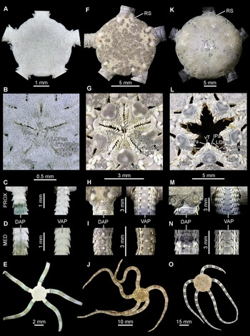
Twenty of the 25 specimens examined per species presented the morphological diagnostic characters that unequivocally identified them a126 s O. aija, O. occultum, or O. panamense, coinciding with the respective type material and original description (Humara-Gil et al., Reference Humara-Gil, Granja-Fernández, Bautista-Guerrero and Rodríguez-Troncoso2022a, Reference Humara-Gil, Granja-Fernández, Bautista-Guerrero, Solís-Marín and Rodríguez-Troncoso2024; Lütken, Reference Lütken1859) or redescription (Granja-Fernández et al., Reference Granja-Fernández, Pineda-Enríquez, Solís-Marín and Laguarda-Figueras2020). The remaining five in each series, the smallest (2.5–6.8 mm DD), lacked developed diagnostic features and presented granulation on otherwise bare structures (Figures 1A–E, 2A–E, 3A–E; Tables 1–3). However, they showed a similar colour pattern to the larger specimens, allowing them to be included in the corresponding series.
Ophioderma aija Humara-Gil et al., Reference Humara-Gil, Granja-Fernández, Bautista-Guerrero, Solís-Marín and Rodríguez-Troncoso2024: (A–E) Group 1 (3.6 mm DD, ICML-UNAM 18469); (F–J) Group 2 (12.0 mm DD, CE-UAM 6); (K–O) Group 3 (26.3 mm DD, ICML-UNAM 11137). (A, F, K) dorsal disc; (B, G, L) jaws; (C, H, M) proximalmost arm section; (D, I, N) median arm section; (E, J, O) dorsal view. Abbreviations: 1VAP, first ventral arm plate; 2°AdShSp, secondary adoral shield spine; AdShSp, adoral shield spine; DAP, dorsal arm plate; IPa, infradental papilla; LOPas, lateral oral papillae; LyOs, Lyman’s ossicle; MED, median arm section; OPRSp, oral plate ridge spine; OSh, oral shield; PROX, proximal arm section; RS, radial shield; TPa, tooth papilla; VAP, ventral arm plate; vT, ventralmost tooth.

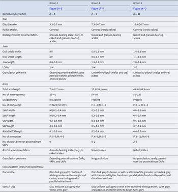
Ophioderma occultum Humara-Gil et al., Reference Humara-Gil, Granja-Fernández, Bautista-Guerrero and Rodríguez-Troncoso2022a: (A–E) Group 1 (3.3 mm DD, CE-UAM 678); (F–J) Group 2 (12.0 mm DD, CE-UAM 320); (K–O) Group 3 (25.5 mm DD, ICML-UNAM 11593). (A, F, K) dorsal disc; (B, G, L) jaws; (C, H, M) proximalmost arm section; (D, I, N) median arm section; (E, J, O) dorsal view. Abbreviations: 1VAP, first ventral arm plate; 2°AdShSp, secondary adoral shield spine; AdShSp, adoral shield spine; DAP, dorsal arm plate; IPa, infradental papilla; LOPas, lateral oral papillae; LyOs, Lyman’s ossicle; MED, median arm section; OSh, oral shield; PROX, proximal arm section; TPa, tooth papilla; VAP, ventral arm plate; vT, ventralmost tooth.

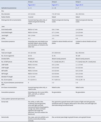
Ophioderma panamense Lütken, Reference Lütken1859: (A–E) Group 1 (3.2 mm DD, CE-UAM 699); (F–J) Group 2 (12.1 mm DD, CE-UAM 284); (K–O) Group 3 (25.6 mm DD, CE-UAM 288). (A, F, K) dorsal disc; (B, G, L) jaws; (C, H, M) proximalmost arm section; (D, I, N) median arm section; (E, J, O) dorsal view. Abbreviations: 1VAP, first ventral arm plate; 2°AdShSp, secondary adoral shield spine; AdShSp, adoral shield spine; DAP, dorsal arm plate; IPa, infradental papilla; LOPas, lateral oral papillae; LyOs, Lyman’s ossicle; MED, median arm section; OPRSp, oral plate ridge spine; OSh, oral shield; PROX, proximal arm section; RS, radial shield; TPa, tooth papilla; VAP, ventral arm plate; vT, ventralmost tooth.

Morphological and morphometric characters of Ophioderma aija Humara-Gil et al., Reference Humara-Gil, Granja-Fernández, Bautista-Guerrero, Solís-Marín and Rodríguez-Troncoso2024 by size group

Abbreviations: 1VAP, first ventral arm plate; DAP, dorsal arm plate; LAP, lateral arm plate; LoOas, lateral oral papillae; M, median arm section; NV, not visible (structure covered with granules); P, proximal arm section; TS, tentacle scale; VAP, ventral arm plate.
Morphological and morphometric characters of Ophioderma occultum Humara-Gil et al., Reference Humara-Gil, Granja-Fernández, Bautista-Guerrero and Rodríguez-Troncoso2022a by size group

Abbreviations: 1VAP, first ventral arm plate; DAP, dorsal arm plate; LAP, lateral arm plate; LOPas, lateral oral papillae; M, median arm section; NV, not visible (structure covered with granules); P, proximal arm section; TS, tentacle scale; VAP, ventral arm plate.
Morphological and morphometric characters of Ophioderma panamense Lütken, Reference Lütken1859 by size group

Abbreviations: 1VAP, first ventral arm plate; DAP, dorsal arm plate; LAP, lateral arm plate; LOPas, lateral oral papillae; M, median arm section; NV, not visible (structure covered with granules); P, proximal arm section; TS, tentacle scale; VAP, ventral arm plate.
Specimens in the growth series were classified into size groups based on their quantitative and qualitative characteristics. Three size groups were identified for each species. Group 1 included the smallest specimens (∼2.5–6.8 mm DD), Group 2 comprised medium-sized specimens (∼7.3–15.9 mm DD), and Group 3, large-sized specimens (∼15.9–26.7 mm DD). Specimens in Group 1 lacked typical adult diagnostic characteristics, whereas those in Groups 2 and 3 had already developed them. The PERMANOVA results indicated significant inter-group differences within each species (O. aija: F 3,24 = 34.30, p = 0.0001; O. occultum: F 3,24 = 33.50, p = 0.0001; and O. panamense: F 3,24 = 58.39, p = 0.0001). Pairwise comparisons revealed that the three groups identified by species were significantly different from each other (p < 0.05) (Supplementary Table S4).
In Group 1, as mentioned above, all specimens of each species presented a complete or nearly complete covering of granules on the body, including the disc, jaws, and arms (Figures 1A–E, 2A–E, 3A–E; Tables 1–3). The smaller the specimen, the more granules it had on its body. As the ophiuroids increased in size, portions of the radial shields, oral shields, and arm plates began to lose granules. In medium (Group 2) and large-sized (Group 3) specimens, the granules were restricted to the disc (Figures 1F, K, 2F, K, 3F, K), and in the jaws, to the oral plates and adoral shields (Figures 1G, L, 2G, L, 3G, L; Tables 1–3). In species such as O. aija and O. panamense, the radial shields, an important diagnostic character for both, were also exposed in these specimens (Figures 1F, K, 3F, K; Tables 1, 3). Similarly, the ornamentation of the arm base and the arm section adjacent to the distal genital slit varied with size in the three species studied. Group 1 exhibited exclusively granule-bearing scales (Figures 1C, 2C, 3C), whereas Groups 2 and 3 displayed granule-bearing scales along with naked scales (Figures 1H, M, 2H, M, 3H, M; Tables 1–3).
Regarding the jaws, an increase in size was observed from the smallest (Group 1) to the largest specimens (Group 3), together with that of the oral shields. In particular, the oral shields presented a greater width than length in each group (including Group 1, when visible) and species. Conversely, the number of LOPas did not always increase with size, with equal numbers occurring in both small (Group 1) and large (Group 3) specimens across the three species (see Supplementary Table S2) (Figures 1B, G, L, 2B, G, L, 3B, G, L; Tables 1–3).
In the case of the arms, both the total length and the number of segments increased according to size in all species, although O. aija had shorter arms than O. occultum and O. panamense (Figures 1E, J, O, 2E, J, O, 3E, J, O; Tables 1–3). DAP division was evident in O. aija from small specimens (Group 1) that had already lost the granules, whereas in O. occultum and O. panamense, it began to be noticeable in larger specimens (Group 2). The species with the highest number of divided DAPs in its three size groups was O. aija (Group 1: up to two; Group 2: up to four; Group 3: up to nine; Table 1), followed by O. occultum (Group 2: up to two; Group 3: up to five; Table 2), and O. panamense (Groups 2 and 3: rarely up to two; Table 3). The number of DAP pieces increased with size and varied along the arm, with higher numbers observed in the proximal arm section (Figures 1H–I, M, N, 2 H–I, M, N). Measurements, including 1VAP width and length, median VAP width and length, and adradial tentacle scale length, as well as counts of arm spines and pores between the proximalmost VAPs, also increased with size. These values were the lowest in Group 1, intermediate in Group 2, and the highest in Group 3 (Tables 1–3).
Finally, variations in the colour pattern were identified. Each species presented a distinctive pattern that was maintained in all groups: O. aija, irregular dark rings on the disc; O. occultum, white parallel bands on the arm; and O. panamense, banded arms (Tables 1–3). However, representatives of Group 1 of all three species tended to be lighter in colour (O. aija) (Figure 1E) or have a greater number of white granules (O. occultum and O. panamense) (Figures 2E, 3E) than those of Groups 2 and 3 (Figures 1J, O, 2J, O, 3J, O).
The main characters that differentiate the size groups within each species are summarised and illustrated in Tables 1–3 and Figures 1–3.
Validation of specimen identity by DNA analysis
Analysis of the COI (659 bp) and 16S (450 bp) sequences confirmed the morphological identification of the specimens, especially those of Group 1, which lacked diagnostic characters. The topology of both trees coincided, showing a main clade that grouped the studied Ophioderma species. This clade was divided into two subclades, the first comprising O. aija (bootstrap values = 100/100) and the second containing O. occultum and O. panamense as sister species (bootstrap values = 65/97). The three Ophioderma species were consistently retrieved in separate, well-supported clades (bootstrap values = 99–100). In each clade, sequences of specimens without fully developed diagnostic characters (Group 1; 3.2–5.9 mm DD) clustered together with sequences of others that did present them (Group 2 and/or Group 3; 7.4–19.1 mm DD), confirming that they belonged to the same species despite their size and morphological differences (Figure 4).
Neighbour-joining trees of COI and 16S sequences of the studied species. Coloured squares indicate the clades corresponding to each Ophioderma species: Ophioderma aija Humara-Gil et al., Reference Humara-Gil, Granja-Fernández, Bautista-Guerrero, Solís-Marín and Rodríguez-Troncoso2024, blue; Ophioderma occultum Humara-Gil et al., Reference Humara-Gil, Granja-Fernández, Bautista-Guerrero and Rodríguez-Troncoso2022a, purple; Ophioderma panamense Lütken, Reference Lütken1859, pink. Sequences are labelled with the GenBank accession number and the size group to which the specimen corresponds (see Supplementary Table S3 for more information). Numbers on branches indicate bootstrap support values; values below 60% are not shown.

The mean genetic distances obtained for COI ranged from 0% (O. fallax) to 0.56% (O. panamense) within species and from 8.42% (O. aija and O. occultum) to 18.21% (O. aija and O. fallax) between species. The mean genetic distances for 16S varied from 0.07% (O. occultum) to 1.56% (O. fallax) within species and from 3.37% (O. occultum and O. panamense) to 18.89% (O. aija and O. fallax) between species. In particular, the low values obtained in the mean genetic distances for both genes within O. aija (COI: 0.52%; 16S: 0.27%), O. occultum (COI: 0.36%; 16S: 0.07%), and O. panamense (COI: 0.56%; 16S: 0.15%) provide additional evidence that the grouped sequences correspond to the same species (Table 4).
Mean genetic distances (%) within (values in bold: COI, left; 16S, right) and between the analysed Ophioderma Müller and Troschel, Reference Müller and Troschel1840 and Ophiopeza Peters, Reference Peters1851 species (COI, lower left; 16S, upper right)

Discussion
The Ophioderma species studied herein, O. aija, O. occultum, and O. panamense, showed notable changes in their morphology depending on their size, including in their diagnostic characters. By analysing the species through growth series, three distinct size groups within each species were detected and characterised. These groups may be considered analogous to the early, intermediate, and adult developmental stages previously proposed for other brittle stars in studies of comparable scope (da Silva et al., Reference da Silva, Prata and Christoffersen2023). However, additional evaluation of the reproductive aspects of the species, such as the estimation of their size at first maturity, is necessary to corroborate this hypothesis.
It was found that the specimens within Groups 2 and 3 (from ∼7 mm DD), corresponding to medium and large sizes, had already developed diagnostic characteristics that allowed their easy identification to species level: O. aija, naked radial shields, short arms, DAPs divided into multiple pieces, brown body with sinuous dark rings on the disc (Figure 1F–J, K–O) (Humara-Gil et al., Reference Humara-Gil, Granja-Fernández, Bautista-Guerrero, Solís-Marín and Rodríguez-Troncoso2024); O. occultum, covered radial shields, DAPs typically divided into two pieces, arms with light transverse bands and parallel white bands in the median and distalmost sections (Figure 2F–J, K–O) (Humara-Gil et al., Reference Humara-Gil, Granja-Fernández, Bautista-Guerrero and Rodríguez-Troncoso2022a); O. panamense, naked radial shields, arms with alternating light and dark transverse bands (Figure 3F–J, K–O) (Granja-Fernández et al., Reference Granja-Fernández, Pineda-Enríquez, Solís-Marín and Laguarda-Figueras2020; Lütken, Reference Lütken1859). In rare cases, an individual belonging to one of these groups lacked one of the latter characters (e.g. the coverage state of the radial shields); however, it could be identified based on the remaining characters present. The main differences between these two groups were observed in quantitative characters (e.g. DD, arm spine number, adradial tentacle scale length). Specimens of Group 2 showed structures of median size or number, whereas those of Group 3 showed the largest values (see Tables 1–3).
On the other hand, identifying specimens of Group 1 (∼2.5–6.8 mm DD) of either species was more challenging. For most specimens in this group, which were completely or partially concealed by granules, the only distinguishing feature was the colouration. However, the colouration was not identical to that of larger individuals and was also easily lost during fixation (Figures 1E, 3E). In these cases, identification was confirmed by DNA analysis. The effectiveness of DNA barcoding for identifying species at early stages has been demonstrated in prior studies on diverse phyla (including Echinodermata), with a predominant focus on larvae (e.g. Bautista-Guerrero et al., Reference Bautista-Guerrero, Carballo, Aguilar-Camacho and Sifuentes-Romero2016; Collin et al., Reference Collin, Venera-Pontón, Driskell, Macdonald and Boyle2020a, Reference Collin, Venera-Pontón, Driskell, Macdonald, Geyer, Lessios and Boyle2020b; Santiago-Valentín et al., Reference Santiago-Valentín, Rodríguez-Troncoso, Bautista-Guerrero, López-Pérez and Cupul-Magaña2019), but sometimes also on juveniles (e.g. Stöhr et al., Reference Stöhr, Boissin and Chenuil2009; Varela-Sánchez et al., Reference Varela-Sánchez, Templado and Machordom2020), as in this work. It stands out that previous studies on Ophiuroidea growth changes did not implement DNA analysis, but instead followed a morphology-based approach that included either external or internal morphology, or both. This is understandable in the earliest works on this matter, as the use of molecular markers had not yet been developed (e.g. Clark, Reference Clark1914). In more recent work, however, it may be due to a lack of suitable material for DNA extraction (e.g. formalin-fixed, dry-preserved, or old samples) or incomplete regional DNA barcode libraries for comparison. While morphology has been sufficient to identify some ophiuroid species at different developmental stages, especially when a substantial amount of material is available (e.g. Borges et al., Reference Borges, Dos Santos Alitto and Amaral2015; Martynov et al., Reference Martynov, Ishida, Irimura, Tajiri, O’Hara and Fujita2015), incorporating DNA information, when possible, would complement the results. In the case of Ophioderma, it is advisable to use DNA barcoding when small specimens have completely lost their colouration and no records are available, as it is the most reliable external morphological characteristic to identify at least the three species studied herein.
Before this study, the only eastern Pacific Ophioderma whose growth changes had been documented was O. panamense by Ziesenhenne (Reference Ziesenhenne1955). However, certain elements of Ziesenhenne’s study suggest that the growth series he examined may, in part, correspond to O. occultum. For example, for specimens of 6 mm DD, he mentioned the following: ‘The white arm banding is confined to the distal third of the arms’ (Ziesenhenne, Reference Ziesenhenne1955, p. 194), a characteristic that is present in O. occultum (Figure 2E, J, O) but absent in O. panamense (Figure 3E, J, O) (Humara-Gil et al., Reference Humara-Gil, Granja-Fernández, Bautista-Guerrero and Rodríguez-Troncoso2022a; this study). Similarly, he did not discuss the changes in the coverage of the radial shields, which are noticeable in O. panamense (switching from covered in small specimens [Figure 3A] to naked in medium and large ones [Figure 3F, K]), but remain quite stable (always covered [Figure 3A, F, K]) in O. occultum (see Tables 2–3). Finally, he specified that all the material he studied was collected in the Gulf of California, where both species are distributed (Granja-Fernández et al., Reference Granja-Fernández, Pineda-Enríquez, Solís-Marín and Laguarda-Figueras2020; Humara-Gil et al., Reference Humara-Gil, Granja-Fernández, Bautista-Guerrero and Rodríguez-Troncoso2022a). The mixing of O. panamense and O. occultum material is not uncommon (Humara-Gil et al., Reference Humara-Gil, Granja-Fernández, Bautista-Guerrero and Rodríguez-Troncoso2022a), and therefore, it would not be unusual for Ziesenhenne (Reference Ziesenhenne1955) to have inadvertently included both species in his work. Examination of the material in the growth series of Ziesenhenne (Reference Ziesenhenne1955) should clarify this. In the present study, the growth changes of both species were included, and their identity was confirmed morphologically and molecularly to avoid confusion.
The morphological changes of O. aija, O. occultum, and O. panamense were highly similar and occurred at comparable sizes. However, some differences between the three species were observed: (1) the radial shields of O. aija and O. panamense were covered with granules in small specimens (Figures 1A, 3A) and naked in medium and large ones (Figures 1F, K, 3F, K), whereas in O. occultum they typically remained covered regardless of size (Figure 2A, F, K); (2) the arms of O. aija were shorter than those of O. occultum and O. panamense at all sizes (Figures 1E, J, O, 2E, J, O, 3E, J, O); (3) the DAPs of O. aija started to show division in small specimens and reached a maximum of nine pieces in large specimens, while in O. occultum and O. panamense, the DAPs started to divide in medium-sized specimens and showed a maximum of five and two pieces in the largest specimens, respectively; (4) the arm spine number between the species was similar in small- and medium-sized specimens, but in the largest specimen of O. aija showed a maximum of nine arm spines, a lower number than that observed in O. occultum and O. panamense, which had up to 11; and (5) each species displayed a distinctive colouration that remained consistent with size, with some intraspecific variation (Figures 1E, J, O, 2E, J, O, 3E, J, O) (see Tables 1–3). Although identification of these species becomes more difficult the smaller the specimen, the above characteristics can be used to differentiate them.
The role of colour pattern as a diagnostic character in O. aija, O. occultum, and O. panamense along their growth series is particularly noteworthy. In the past, it was generally accepted that a single species of Ophioderma could display a wide range of colour variation (Hendler et al., Reference Hendler, Miller, Pawson and Kier1995; Ziesenhenne, Reference Ziesenhenne1955). Although this has been confirmed for some species (e.g. Ophioderma longicaudum [Bruzelius, Reference Bruzelius1805]) (Gouillieux et al., Reference Gouillieux, Chenuil, Stöhr, Lebouazda, de Casamajor, Huguenin, Devaux and Sartoretto2024), for others (e.g. O. aija; Ophioderma hendleri Granja-Fernández et al., Reference Granja-Fernández, Pineda-Enríquez, Solís-Marín and Laguarda-Figueras2020; O. occultum; Ophioderma zibrowii Stöhr et al., Reference Stöhr, Weber, Boissin and Chenuil2020] the colour pattern has been recognised as a valuable character for their delimitation (Granja-Fernández et al., Reference Granja-Fernández, Pineda-Enríquez, Solís-Marín and Laguarda-Figueras2020; Humara-Gil et al., Reference Humara-Gil, Granja-Fernández, Bautista-Guerrero and Rodríguez-Troncoso2022a, Reference Humara-Gil, Granja-Fernández, Bautista-Guerrero, Solís-Marín and Rodríguez-Troncoso2024; Stöhr et al., Reference Stöhr, Weber, Boissin and Chenuil2020). Here, this trait was found to be maintained to some extent as individuals grow, providing a useful means of species identification. Although there are some variations from small to large specimens (e.g. more white granules on the disc of small O. occultum and O. panamense) (see Tables 1–3), their distinctive patterns remain: the sinuous dark rings on the disc of O. aija (Figure 1F, K), the white parallel bands in the arms of O. occultum (Figure 2E, J, O), and the banded arms in O. panamense (Figure 3E, J, O). Critical examination of the colour pattern may provide similar insights for other Ophioderma species. Moreover, it appears to be potentially useful for differentiating cryptic species from small sizes. For instance, small specimens identified as O. panamense from California, where undescribed species have been molecularly detected (Lessios and Hendler, Reference Lessios and Hendler2022), present a white disc and arms with alternating orange-red and white bands, in contrast to those from the Mexican Pacific, whose bands always show a combination of green, red, white, brown, grey, and black colours, with green predominating (KJHG, RGF pers. obs.). Documentation of these observations in the field and from fixed specimens may provide valuable information for future species delimitation.
Based on the species growth series, it was also possible to identify shared patterns between the three species and to reconstruct how growth changes may occur in Ophioderma over the size range studied (∼3–26 mm DD). Starting from a small individual (∼3 mm DD) with granules covering the disc, the jaws (except for a portion of an oral shield, presumably the madreporite), and the arms (except for the arm spines), the first structures to shed their granules are the arms, starting with the VAPs (distally to proximally of the arm), then the DAPs (proximally to distally of the arm), and ultimately, the LAPs (distally to proximally of the arm). These arm plates uncover starting from the centre to the edge. The oral shields begin to uncover from the centre to the shield’s edge, just before the LAPs are fully exposed. Then, some naked scales become visible at the arm base and between the arm and the distal genital slit, which previously exhibited only granule-bearing scales. The oral shields become completely uncovered, and the radial shields eventually become uncovered (in species with naked radial shields, such as O. aija and O. panamense). The division of the arm plates is noticeable in specimens from ∼6 mm DD. Growth changes are subsequently reflected in the increased size or number of various structures, including the arm spines, arm base scales, and DAP pieces. These observations on the order in which the body begins to uncover coincide with those of Clark (Reference Clark1939) and Ziesenhenne (Reference Ziesenhenne1955). Nevertheless, it is important to emphasise that this pattern was observed in the three Ophioderma species studied but may be different in other species of the genus. For example, in Ophioderma brevicaudum Lütken, Reference Lütken1856, the radial shields were reported to be uncovered earlier than the oral shields and at a smaller size, 4 mm DD (Prata et al., Reference Prata, de Castro Manso and Christoffersen2018), than in O. aija or O. panamense, at ∼7 mm DD. In the same regard, Ophioderma wahlbergii Müller and Troschel, Reference Müller and Troschel1842 shows already naked arms at 2 mm DD (Landschoff and Griffiths, Reference Landschoff and Griffiths2015), whereas in O. aija, O. occultum, and O. panamense the arms end up uncovered at 5–6 mm DD. Further research is required to gain a deeper understanding of the growth changes observed in Ophioderma, particularly concerning the extent to which this pattern is applicable, how it may vary between species, and the potential influence of environmental conditions on the differences detected.
The study of growth variation in Ophiuroidea has recently resurfaced, incorporating morphometrics, scanning electron microscopy images of internal ossicles, and/or DNA barcoding, in addition to the traditionally studied external morphology (da Silva et al., Reference da Silva, Prata and Christoffersen2023; Damiano et al., Reference Damiano, Serrano, Alitto, Mendonça, Tavares and Borges2025; Humara-Gil et al., Reference Humara-Gil, Granja-Fernández, Montoya-Márquez and López-Pérez2022b; this study). These studies continue to gain interest because they record intraspecific variation within species, distinguish them from close congeners, prevent misidentifications, and contribute to organising and maintaining scientific collections, all of which are important for developing comprehensive studies on systematics and evolution. Furthermore, the substantial availability of material of certain species deposited in scientific collections makes these studies quite feasible. This study of the growth changes of O. aija, O. occultum, and O. panamense is one of the few available contributions on this topic from the eastern Pacific and so far on the genus Ophioderma.
Supplementary material
The supplementary material for this article can be found at https://doi.org/10.1017/S0025315425000396.
Data availability
The datasets generated or analysed during this study (collection data, morphological and morphometric data, and additional statistical results) are included in the Supplementary Tables S1, S2, and S4. All sequences generated are deposited in GenBank (accession numbers can be consulted in Supplementary Table S3).
Acknowledgements
We are most grateful to the following people: Michel Hendrickx and Mercedes Cordero (ICML-EMU); Alicia Durán-González, Daniel Mireles-Velázquez, Carlos Conejeros-Vargas, and Andrea Caballero-Ochoa (ICML-UNAM); Adam Baldinger, Rina Morisawa, and Penny Benson (MCZ); Tom Schiøtte and Jørgen Olesen (NHMD); and Yolanda Villacampa, Chris Mah, Ellen Strong, Dave Pawson, Chris Meyer, Karen Reed, Geoff Keel, Tim Coffer, Amanda Robinson, and Freya Goetz (USNM) for the support provided during the visits to the collections. To Alejandro Hernández-Morales and Marco Medina-López (Universidad Autónoma de Baja California Sur), Brenda Maya-Alvarado (Universidad de Guadalajara [UdeG]), Aldo Zavala-Jiménez (University of California, Santa Cruz), Dinorah Herrero-Pérezrul (Centro Interdisciplinario de Ciencias Marinas), Emerson Martínez-Zavala, Aramis Gardea-López, and Diego Rangel-Solís for their help in fieldwork. To Renata Alitto (Universidade Estadual de Campinas) for kindly making available relevant literature. To Christopher Cruz-Gómez (El Colegio de la Frontera Sur), Brenda Maya-Alvarado (UdeG), Rafael B. de Moura (Universidade Federal do Rio de Janeiro), and an anonymous reviewer for their constructive suggestions on earlier versions of the manuscript. This work is part of K.J.H.-G.’s PhD thesis at UdeG.
Author contributions
Conceptualisation: K.J.H.-G., R.G.-F., and A.P.R.-T.; Methodology: K.J.H.-G., R.G.-F., E.B.-G., and A.P.R.-T.; Formal analysis: K.J.H.-G. and E.B.-G.; Investigation: K.J.H.-G.; Visualisation: K.J.H.-G.; Writing – original draft preparation: K.J.H.-G.; Writing – review and editing: K.J.H.-G., R.G.-F., A.P.R.-T., E.B.-G., and F.A.S.-M.; Funding acquisition: A.P.R.-T., R.G.-F., and K.J.H.-G.; Resources: A.P.R.-T., R.G.-F., E.B.-G., F.A.S.-M., and K.J.H.-G.; Supervision: A.P.R.-T. and R.G.-F.
Funding
This work was supported by the Secretaría de Ciencia, Humanidades, Tecnología e Innovación (SECIHTI) (K.J.H.-G., grant number 993677); the Museum of Comparative Zoology (K.J.H.-G., Ernst Mayr Travel Grant 2020), (R.G.-F., Ernst Mayr Travel Grant 2016); the Smithsonian Institution (K.J.H.-G, Kenneth Jay Boss Fellowship in Invertebrate Zoology 2022); the National Geographic Society (A.P.R.-T., grant number NGS-100354C-23); and the Comisión Nacional para el Conocimiento y Uso de la Biodiversidad (R.G.-F., grant number JF047).
Competing interests
The authors declare none.