INTRODUCTION
Trypanosomes are a diverse group of protozoan parasites that can infect all classes of vertebrate. The epidemiology of diseases caused by Trypanosoma species are determined, in part by the ecology of the vector and the host-vector relationship that affect parasite transmission (Hoare, Reference Hoare1972). The majority of known vectors of mammalian trypanosomes belong to the class Insecta, which are associated with haematophagous insects of the order Hemiptera (e.g. Triatomine bugs), Diptera (e.g. flies) and Siphonaptera (e.g. flea) (Hoare, Reference Hoare1972). However Arachnida (ticks), bat mites and leeches are also vectors of trypanosomes (Mackerras, 1959; Lukes et al. Reference Lukes, Jirku, Dolezel, Kral'ova, Hollar and Maslov1997; Stevens et al. Reference Stevens, Noyes, Dover and Gibson1999; Hamilton et al. Reference Hamilton, Stevens, Gidley, Holz and Gibson2005; Thekisoe et al. Reference Thekisoe, Honda, Fujita, Battsetseg, Hatta, Fujisaki, Sugimoto and Inoue2007). Various leech species also play a role in the transmission of several trypanosome species such as T. rotatorium, T. boissoni, and T. triglae, which have been found in the blood of aquatic vertebrates (Lukes et al. Reference Lukes, Jirku, Dolezel, Kral'ova, Hollar and Maslov1997). The presence of trypanosome species in terrestrial blood-sucking leeches from both Asia and Australia has also been reported (Hamilton et al. Reference Hamilton, Stevens, Gidley, Holz and Gibson2005). In Australia the rate of detection of novel trypanosome species in the blood of native Australian mammals is increasing due to advances in molecular detection methods. However, little is known of these Australian trypanosomes, their life cycles and their vectors. Potential vectors such as the wombat flea (Lycopsylla nova) and kangaroo ticks have been implicated in the transmission of unidentified Trypanosoma species isolated from Australian mammals, the wombat and kangaroo (Noyes et al. Reference Noyes, Stevens, Teixeira, Phelan and Holz1999). Sandflies, leeches and the platypus tick (Ixodes ornithorhynchi) have been suggested as potential vectors for T. binneyi, which infect the platypus (Mackerras, 1959; Noyes et al. Reference Noyes, Stevens, Teixeira, Phelan and Holz1999; Jakes et al. Reference Jakes, O'donoghue and Adlard2001). Another study identified Trypanosoma sp. found within tick nymphs (Ixodes holocyclus) isolated from bandicoots infected with T. thylacis (Mackerras, 1959). Ticks (Haemaphysalis hystricis) from Japan have also been found to be naturally infected with an unidentified trypanosome species isolate KGI found to be closely related to T. copemani (Austen et al. Reference Austen, Jefferies, Friend, Ryan, Adams and Reid2009). Trypanosomes found in Australian ticks and the close phylogenetic relationship of T. copemani to the KGI trypanosome spread by ticks lead to the examination of ticks as potential vectors of T. copemani.
MATERIALS AND METHODS
Study site and sample collection
A total of 15 quokkas (Setonix brachyurus), were trapped at Bald Island (34º 55′S, 118º 27′E) near Albany, and a Gilbert's potoroo from Two Peoples Bay (34º 58′S, 118º 11′E), in Western Australia during the month of June 2010. The animals used in this study were live-trapped using cage traps (Mascot Wire Works collapsible rat/bandicoot traps) or long-handled nets. A total of 41 ticks were collected off quokkas from Bald Island and 2 ticks collected off a Gilbert's potoroo from Two Peoples Bay and placed into 5 ml tubes (Sarstedt, Australia) for later identification using the Australian ticks key (Roberts, Reference Roberts1970). Fresh whole blood samples were collected from 3 of the quokkas from Bald Island. The quokkas were anaesthetized with isoflurane and approximately 5 ml of blood was collected by venepuncture of the lateral caudal vein. The blood was added to commercial blood storage tubes containing ethylenediamine tetraacetic acid (EDTA) (Sarstedt, Australia) and stored at 4°C for later use. Thin blood smears were prepared and stained with Modified Wright's stain using an automated slide stainer (Hematek, Bayer). The blood smears were examined at 200× magnification for the presence of trypanosomes.
Detection of trypanosomes in the tick
Once identified, a selection of 10 Ixodes australiensis live ticks from quokkas and 2 ticks from the Gilberts potoroo were individually mounted onto house-hold pest glue boards (TOMCAT Motomco, USA) allowing immobilization of the tick and leg restraint. The glue board was then stuck to a dissecting microscope stage with double-sided tape, and the tick dissected according to published methods (Edwards et al. Reference Edwards, Goddard and Varela-Stokes2009). Haemolymph was collected on the end of a scalpel blade during the removal of the tick scutum and placed onto a microscope slide for examination. The midgut and salivary glands were also removed and placed into a 1·5 ml microcentrifuge tube containing 100 μl of RPMI 1640 (pH 7·2), supplemented with 10% foetal calf serum (FCS), 1000 international units (IU)/ml of penicillin and 10 000 μg /ml of streptomycin, with final concentrations of 2 g/L sodium bicarbonate, 0·3 g/L L-glutamine and 3·6 g/L HEPES. A small region of the midgut was placed onto a microscope slide containing 10 μl of RPMI 1640 culture medium, manually pulled apart and mixed using a sterile 10 μl pipette tip. A cover-slip was placed over the dissected midgut and the preparation examined microscopically at 200× magnification to detect any motile trypanosomes. Fragments of the midgut and salivary glands were also placed into Modified Sloppy Evans Medium (MSEM) with a 500 μl overlay of RPMI 1640 (supplemented as above) and incubated in the dark at room temperature.
Detection of trypanosomes in tick faeces
Faeces from a collection of ticks isolated from 8 individual quokkas from Bald Island and a Gilbert's potoroo from Two Peoples Bay were removed from the inside of 5 ml vol. polypropylene tubes with a sterile 10 μl tip placed into 10 μl of phosphate-buffered saline (pH 7·2) (PBS) on a microscope slide and manually dispersed. The slide was air dried and stained with Modified Wright's stain using an automated slide stainer (Hematek, Bayer). A cover-slip was placed over the stained faecal matter and examined at 200× magnification to detect trypanosomes.
Morphological measurements
Digital photomicrographs were taken, at 1000× magnification, of 5 tick trypanosomes from faecal smears and 14 tick trypanosomes from the dissected midgut and bloodmeal. Measurements of key morphological features were made using Image Pro Express version 5.1 (Media Cybernetics Inc., USA). The morphological features used were based on the following morphological parameters Length (length of body measured along the mid-line including free flagellum), Breadth (maximum breadth measured at the level of the nucleus (including undulating membrane), PK (distance between the posterior end and the kinetoplast), KN (distance between the kinetoplast and posterior edge of the nucleus), NA (distance between the anterior edge of the nucleus and the anterior end of the body) and FF (length of the free flagellum) previously described (Mackerras, 1959; Hoare, Reference Hoare1972). Significant differences between trypanosome morphology measurements from different host species and source were determined using SPSS 17.0 (Chicago, Illinois, USA) by one-way analysis of variance (ANOVA) and the Tukey's Honestly Significant Difference test for P<0·05.
DNA extraction
Total genomic DNA was extracted using a DNeasy® blood and tissue kit (Qiagen), from both the tick faeces and dissected midgut and salivary glands of the ticks collected from quokka 2322 and 4489 and from the tick collected from the Gilbert's potoroo. DNA was also extracted from blood isolated from quokka 4489. The DNA was stored at −20°C until required.
DNA amplification and sequencing
A fragment of the 18S ribosomal RNA (rRNA) gene was amplified from extracted DNA and sequenced as previously described (Austen et al. Reference Austen, Jefferies, Friend, Ryan, Adams and Reid2009). Amplified products were purified using a QIAquick® PCR Purification kit (QIAGEN) and sequenced directly. Sequencing reactions were performed using an ABI Prism DyeTerminator Cycle Sequencing Core kit (Applied Biosystems, USA) and sequence data were analysed using 4peaks v 1.7.1 (A. Griekspoor and Tom Groothuis, mekentosj.com).
RESULTS
Ticks
Two ticks collected from a single Gilbert's potoroo caught at Two Peoples Bay, and 25 of the 41 ticks collected from 15 quokkas captured on Bald Island, were sufficiently intact morphologically to allow identification down to the species level. Ixodes australiensis was identified from both Bald Island and Two peoples Bay and was the most abundant tick species collected at 72% of the total. Two other tick species with similar morphology to Ixodes hirsti and Ixodes myrmecobii, were also detected on Bald Island, at 12% and 8% of the total respectively. Ixodes australiensis was the only tick species dissected for examination because the specimens of Ixodes hirsti and Ixodes myrmecobii did not survive longer than 30 days in storage.
Trypanosomes present in tick faeces
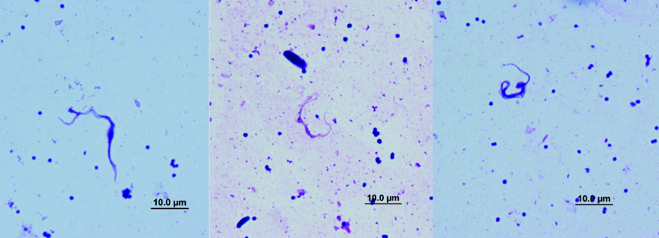
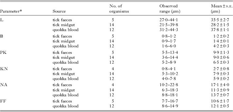
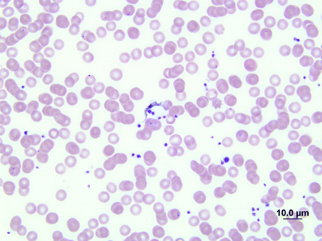
Of the 8 tick faecal smears examined from ticks collected from 8 individual quokkas and 1 Gilbert's potoroo, the detection of trypanosomes was observed in 1 wet smear of dried faeces isolated from ticks collected from quokka 2322 and, to the authors' knowledge, this is the first time that trypanosomes have been identified in tick faeces. Morphologically the trypanosomes were slender in shape with a narrow undulating membrane, a large PK value (defined in Table 1) and a free flagellum (Fig. 1). The mean measurements for each morphological parameter for the trypanosomes are presented in Table 1.
Light photomicrographs of trypanosomes in Modified Wright's stain detected in the faeces of the tick.
Mean dimensions and standard error (s.e.) of morphological features of Trypanosoma copemani isolated from tick faeces from quokka 2322, tick midgut and bloodmeal from quokka 2917 and bloodstream trypanosomes from quokka blood

* L, length of body measured along the mid-line including free flagellum (total length); B, maximum breadth measured at the level of the nucleus (including undulating membrane); PK, distance between the posterior end and the kinetoplast; KN, distance between the kinetoplast and posterior edge of the nucleus; NA, distance between the anterior edge of the nucleus and the anterior end of the body; FF, length of the free flagellum.
Trypanosomes present in tick
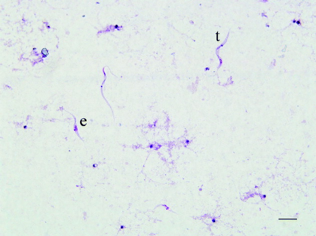
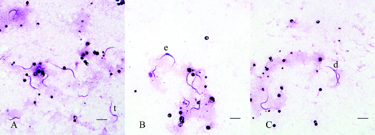
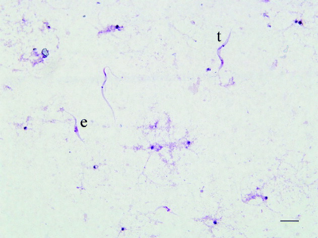
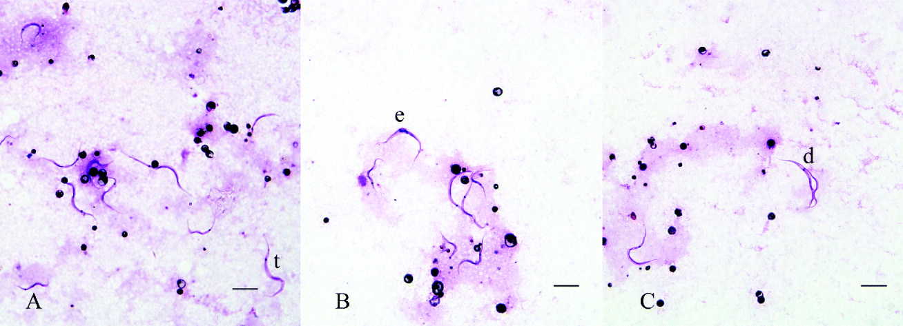
Motile trypanosomes were detected in 2 ticks of the 10 ticks collected from animals from Bald Island that were dissected and examined. Trypanosomes were observed in pieces of midgut dissected from Ixodes australiensis collected from quokkas 2322 and 2917. Numerous motile trypanosomes were also detected in the haemolymph (Fig. 2), as well as in the midgut and bloodmeal from 1 tick collected from a Gilbert's potoroo. Morphologically, the trypanosome tick isolates resembled the slender pleomorphic trypomastigote forms that are present in in vitro cultures isolated from the blood of the Gilbert's potoroo as previously described (Austen et al. Reference Austen, Jefferies, Friend, Ryan, Adams and Reid2009). However, the free flagellum of the trypanosomes within the midgut and bloodmeal of the tick isolated from quokka 2917 were very short or absent (Fig. 3). The mean measurements for each morphological parameter from trypanosomes isolated from within the tick collected from quokka 2917 are presented in Table 1. Attempts to culture the trypanosome tick isolate in MSEM were unsuccessful.
Light photomicrographs of Trypanosoma copemani in Modified Wright's stain detected in the haemolymph of the tick collected from the Gilbert's potoroo F170. Trypomastigote form (t), epimastigote form (e). Scale bar represents 10 μm.
Light photomicrographs of trypanosomes in Modified Wright's stain from the gut region and bloodmeal of a tick collected from quokka 2917. (A) Trypomastigote form. (B) Dividing epimastigote form (e). (C) Dividing trypomastigote form (d). Scale bar represents 10 μm.
Comparative analysis of morphological measurements of trypanosomes from quokka blood and from ticks
Comparison of the morphological measurements taken from the trypanosomes found in tick faeces and midgut, with measurements taken from the bloodstream forms of T. copemani in quokkas (Table 1), showed that trypanosomes from ticks were narrower (P<0·001) and their PK values were greater (P<0·001) than trypanosomes from quokkas. The length of the trypanosomes both from the tick faeces, and host bloodstream were similar, but a significant difference was noted in the length of the midgut trypanosomes which were shorter (P<0·001). The length of the KN parameter was highly variable; however, differences between trypanosomes from each of the 3 sources were significant (P<0·001). NA was only different between trypanosomes from the midgut and tick faeces (P<0·001). Comparison of the free flagellar length revealed no significant differences (P>0·329) between trypanosomes from the blood of the quokka and the faeces of the tick. The midgut trypanosomes did not possess a free flagellum.
Characterization of Trypanosoma copemani isolated from blood and ticks
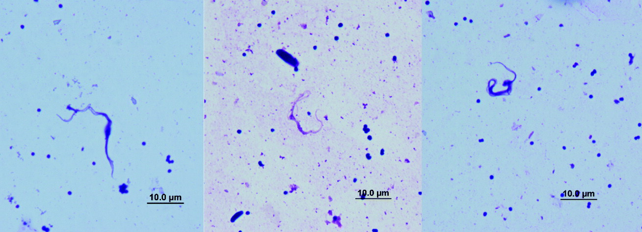
Two of the 3 quokkas (Q4489 and Q4539) from Bald Island were positive for trypanosomes through microscopical examination (Fig. 4) and amplification of the 18S rRNA gene. A tick from one of the same quokkas (Q4489) as well as a tick from another quokka from Bald Island (Q2322) and a tick isolate collected from a Gilbert's potoroo was also positive by PCR. The 18S rRNA sequences from the quokka blood (Q4489) and the Gilbert's potoroo tick isolate had 100% homology to each other and 100% homology to T. copemani (genotype A) and 99·9% homology to the T. copemani wombat AII isolate. The 18S rRNA sequences from the 2 Bald Island tick isolates were also confirmed as T. copemani. They had 100% homology to each other and just 1 bp difference compared to the trypanosome isolated from the quokka blood (Q4489) and the Gilbert's potoroo tick isolate. No amplification was detected from DNA extracted from the tick salivary glands and the faeces.
Light photomicrographs of Trypanosoma copemani in Modified Wright's stain detected in the blood of quokka 4489 captured from Bald Island.

DISCUSSION
Ticks are obligate ectoparasites of vertebrates that are capable of transmitting pathogens (viruses, bacteria, rickettsiae, helminths and protozoa) of veterinary and medical importance (Roberts, Reference Roberts1970). In Australia, various tick species of the family Ixodidae are responsible for transmitting zoonotic diseases, such as tick typhus, and Lyme disease (Roberts, Reference Roberts1970). This is the first time ectoparasites from quokkas on Bald Island have been examined. Ixodes myrmecobii and I. australiensis have previously been found occurring in various regions of the South West of Western Australia and also within regions of Tasmania. To the best of our knowledge, I. hirsti has previously only been located in the eastern states of Australia in NSW, Victoria and Tasmania (Roberts, Reference Roberts1970). This is therefore the first report of I. hirsti in Western Australia.
The trypanosomes found in the midgut, haemolymph and faeces of the ticks had similar morphology (long and slender) to trypanosomes detected in tick nymphs (Ixodes holocyclus) removed from bandicoots (Mackerras, 1959). The long slender trypanosomes found in the faeces are likely to be the metacyclic form of T. copemani, as suggested by Austen et al. (Reference Austen, Jefferies, Friend, Ryan, Adams and Reid2009). In that study, slender trypanosomes in in vitro cultures of blood from infected potoroos were identified and named as T. copemani (Austen et al. Reference Austen, Jefferies, Friend, Ryan, Adams and Reid2009). This parasite is now known to infect several species of marsupials including quokkas, Gilbert's potoroos, koalas and common wombats (Noyes et al. Reference Noyes, Stevens, Teixeira, Phelan and Holz1999; Austen et al. Reference Austen, Jefferies, Friend, Ryan, Adams and Reid2009; McInnes et al. Reference McInnes, Gillett, Ryan, Austen, Campbell, Hanger and Reid2009). Cultured trypanosomes from the blood of the potoroos were maintained at around 25°C, which is suitable for the growth of the vector stages of trypanosomes (Vickerman, Reference Vickerman1985). Morphological comparison of the trypanosomes within the vector and those from infected hosts shows that ticks harbour pleomorphic trypanosomes with some tick trypanosome life cycle stages found to represent the multiplication forms. The difference in PK and KN values could be due to kinetoplast migration which occurs when trypanosomes are in their multiplication and division stage (Hoare, Reference Hoare1972).
In the present study, motile trypanosomes were observed in the midgut of ticks dissected 49 and 117 days after removal of the ticks from the host. Motile trypanosomes were also detected in the haemolymph 117 days after removal. When xenodiagnosis is used for the detection of low levels of T. cruzi infection in humans or animals, first-instar Triatoma nymphs are allowed to feed on the suspect individual and after 30 days the bugs are examined for the presence of trypanosomes (Machado et al. Reference Machado, Fernandes, Murta, Vitor, Camilo, Pinheiro, Lopes, Adad, Romanha and Pinto Dias2001). It is unlikely that the trypanosomes observed in this study originated from the bloodmeal of the ticks examined because of the extended period between the last meal and detection of trypanosomes (49 days and 117 days). This is longer than the 30 days used in the xenodiagnostic test. It is interesting that live T. cruzi can not be detected in the small intestines of reduviid bugs at the fifth instar 60 days after the bug's last bloodmeal (Kollien and Schaub, Reference Kollien and Schaub1998). The detection of motile trypanosomes at day 117 within the tick midgut, suggests that T. copemani may multiply in the tick. Indeed, the observation of dividing trypanosomes in stained smears from the gut and gut contents of the tick and the detection of slender trypanosomes in the tick haemolymph suggest that the ticks were systemically infected with T. copemani. This further supports the conclusion that I. australiensis is naturally infected and is the vector of T. copemani.
Trypanosomes have previously been detected in the haemolymph and gut of a variety of tick species (Mackerras, 1959; Shastri and Deshpande, Reference Shastri and Deshpande1981; El Kady, Reference El Kady1998; Latif et al. Reference Latif, Bakheit, Mohamed and Zweygarth2004; Thekisoe et al. Reference Thekisoe, Honda, Fujita, Battsetseg, Hatta, Fujisaki, Sugimoto and Inoue2007). However, there is insufficient information to fully determine the mode of natural trypanosome transmission via ticks. The detection of intact trypanosomes and trypanosome DNA in smears of dried tick faeces and the detection of DNA in the midgut but not from salivary glands suggest that transmission is likely to be contaminative via tick faeces. The faecal oral route of transmission that occurs with T. lewisi when rats eat infected fleas and/or their droppings containing metatrypanosomes (Hoare, Reference Hoare1972) could be a similar process of transmission that could be occurring between T. copemani and its marsupial host. Ticks located on quokkas are generally found around the ears and the tail while on the Gilbert's potoroo they are more widely distributed ranging from ears, tail, rim of pouch in females and around the scrotum in males as well as amongst the fur on the body which are body parts generally accessible to grooming. The stercorarian nature of T. copemani, as previously suggested by Austen et al. (Reference Austen, Jefferies, Friend, Ryan, Adams and Reid2009) is consistent with a faecal route of trypanosome transmission. However, it is not possible to rule out the salivary route of transmission because only a limited number of salivary glands were analysed. Furthermore, there is evidence that similar trypanosomes such as T. rangeli are transmitted via the contaminative and inoculative routes (Guhl and Vallejo, Reference Guhl and Vallejo2003).
Sequence analysis of the amplified 18S rRNA gene from the DNA extracted from the gut showed that ticks on Bald Island and Two Peoples Bay are carrying a strain of T. copemani (genotype A) which has previously been isolated from a quokka and a Gilbert's potoroo and is similar to T. copemani isolates from wombats in Victoria, Australia (Noyes et al. Reference Noyes, Stevens, Teixeira, Phelan and Holz1999). The single base-pair change may have been due to an error introduced by the polymerase. The wide geographical distribution of T. copemani across Australia may be accounted for by the presence of Ixodes tick species, which have been found in most states of Australia. Ticks show varying degrees of host specificity (Roberts, Reference Roberts1970). Ixodes ticks are generally not host-specific. Ixodes australiensis has been recorded from numerous hosts including small marsupials, such as the bettongs, (Bettongia lesueuri, and B. penicillata), potoroos (Potorous tridactylus and P. gilbertii), the quokka (Setonix brachyurus), as well as kangaroos, dogs, humans and cows (Roberts, Reference Roberts1970; Austen et al. Reference Austen, Jefferies, Friend, Ryan, Adams and Reid2009). Leeches have been identified as vectors of trypanosomes in Australia (Hamilton et al. Reference Hamilton, Stevens, Gidley, Holz and Gibson2005) and several authors have suggested various ectoparasites as potential vectors for native Australian trypanosomes (fleas, biting flies, and ticks) (Noyes et al. Reference Noyes, Stevens, Teixeira, Phelan and Holz1999; Jakes et al. Reference Jakes, O'donoghue and Adlard2001).
Australian trypanosomes have been found in a wide range of marsupials from mainland Australia (Mackerras, 1959; Noyes et al. Reference Noyes, Stevens, Teixeira, Phelan and Holz1999; Jakes et al. Reference Jakes, O'donoghue and Adlard2001; Hamilton et al. Reference Hamilton, Stevens, Gidley, Holz and Gibson2005; Austen et al. Reference Austen, Jefferies, Friend, Ryan, Adams and Reid2009; McInnes et al. Reference McInnes, Gillett, Ryan, Austen, Campbell, Hanger and Reid2009) as well as offshore islands (Bettiol et al. Reference Bettiol, Jakes, Le, Goldsmid and Hocking1998). A recent survey of the infection status of the quokkas on Rottnest Island off the coast of Western Australia revealed that these animals are not infected with T. copemani (J. Austen, unpublished data) whereas quokkas on Bald Island and at Two Peoples Bay are infected. Interestingly, no ticks or fleas were detected on the quokkas from Rottnest and the only ectoparasite found on quokkas caught on the island was a biting louse (Heterodoxus quadriseriatus). This is in contrast to the quokka populations at Two Peoples Bay and on Bald Island, which are usually infested with ticks (J.A. Friend, unpublished observation).
The pathogenicity of T. copemani in naïve animals is not known and therefore the identification of the vector for this parasite is important for elucidation of the complete life cycle of T. copemani. Knowledge of the life cycle will aid in management decision making when re-introduction of naïve animals occurs into areas where the parasite is known to be present in the same or other species of marsupials. This is particularly important when a population of animals from the same species are genetically and morphologically different, as occurs with quokkas on Rottnest in comparison to mainland and Bald island quokka populations (Sinclair, Reference Sinclair1998, Reference Sinclair2001).
ACKNOWLEDGEMENTS
We would like to thank Rottnest Island Authority who funded activities on Rottnest Is. and Shane Kearney the environmental coordinator from Rottnest Island. The technical assistance of Russ Hobbs, Aileen Elliot, Simon Fleetwood and Gordon Thomson is also acknowledged.