In a manuscript published in the current issue of the journal, Prabhu and his colleagues suggest a new categorisation for anomalies of the right aortic arch.
Reference Prabhu, Mehra and Kasturi1
As the authors rightly emphasise, the anomalies of the right aortic arch, once it has crossed the right bronchus, can all be explained on the basis of the hypothetical double arch as formulated by Edwards.
Reference Edwards2
They further explain that the concept, as put forward by Edwards, is itself derived from the understanding of the remodelling of the arteries of the pharyngeal arches, with these changes usually being interpreted on the basis of the diagram prepared long since by Rathke.
Reference Rathke3
Of the several important lessons to be learned from their analysis, the first is that it is much better to describe the findings, rather than to create yet another complex alphanumeric code. For example, at one point in their manuscript, they describe how the differentiation between the situation when the left subclavian artery is isolated from the arch, and the arterial duct is closed, represented by the arterial ligament, as opposed to the situation when the duct and ligament are absent, can only be made in the operating room. The significance of this difference is immediately obvious. Since they have identified at least seven variants, which can exist with or without an arterial duct or ligament, it might have been tempting for them to create a system of classification such that these findings could be expressed as “the differentiation between type 4C versus type 6 and between type 5C versus type 7 can only be made intra-operatively”. Fortunately, they have resisted this temptation. Indeed, although their descriptions are often somewhat lengthy, they are a model of clarity. They serve to emphasise the intrinsic deficiencies of all alphanumeric classifications. Our goal should be to achieve uniformity in understanding. As is now shown in their report, this is best achieved by avoiding unnecessary codifications.
There is, however, a much more significant second lesson to be taken from their investigation. As already indicated, the diagrams in which they show the different patterns are based on the hypothetical arrangements that can be predicted on the basis of the model of the perfect double arch proposed by Edwards.
Reference Edwards2
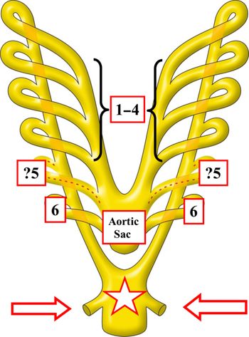
And, again as already stated, these findings are based on the knowledge of the remodelling, during cardiac development, of the arteries that percolate through the mesenchyme of the pharyngeal arches. These developmental changes are usually depicted on the basis of the Rathke diagram. As is shown by Prabhu and his colleagues, it is now necessary to redraw the Rathke diagram. This is because the “classical” diagram has, as its starting point, the presence of six pairs of arteries. These vessels extend from the aortic sac in a symmetrical fashion to reach the dorsal aorta (Fig 1). In the drawing provided by Prabhu and his colleagues, the fifth of these pairs of arteries, as in our own diagram, is shown by dotted lines. This is because the so-called “fifth arch arteries” have long been controversial. As most, if not all, paediatric cardiologists are well aware, various congenital cardiac malformations have been interpreted on the basis of their persistence in postnatal life. In the past, one of us was an enthusiastic supporter of the notion that these entities can be recognised when approached on the basis of the “great pretender”.
Reference Gerlis, Ho, Anderson and Da Costa4
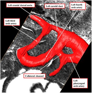
We have subsequently investigated large numbers of developing murine embryos using the technique of episcopic microscopy. In none of these, developing mice were we able to find evidence either of a fifth aggregation of pharyngeal mesenchyme to form an “arch”, nor of any additional arterial channels extending in parallel and symmetrical fashion from the aortic sac between the arteries of the fourth arches and those of the so-called “sixth” arches. In half of the embryos examined, nonetheless, we did find bilateral collateral channels at the terminations of the fourth and pulmonary arch arteries as they are inserted into the descending aorta (Fig 2).
Reference Bamforth, Chaudhry and Bennett5
We did also, however, claim collectively to have discovered the only example of the persistence of a “fifth arch artery” in a developing human embryo.
Reference Bamforth, Chaudhry and Bennett5
We now take the stance that the vessel is better considered as a collateral channel, which is enclosed in its own segment of the mesenchyme of the ultimate left pharyngeal arch. On the basis of our initial interpretation, we had conducted an extensive literature review of the lesions that, in our opinion, could properly be interpreted as representing the persistence of the enigmatic fifth arch arteries.
Reference Gupta, Bamforth and Anderson6,Reference Gupta, Gulati and Anderson7
We must now revise these interpretations. This is because, as is now explained by Prabhu and colleagues, strong evidence has emerged showing that, in reality, there is never any formation of the purported fifth pharyngeal arch. By extension, there is never any formation of “fifth arch arteries”. This is also why we have now reinterpreted our original finding in a human embryo
Reference Bamforth, Chaudhry and Bennett5
as representing a collateral channel. The developmental evidence in question was provided by Graham and his colleagues.
Reference Graham, Poopalasundaram, Shone and Kiecker8
We are now in possession of additional evidence endorsing in full all of their conclusions.
Figure 1.The drawing shows the “classical” diagram offered by Rathke to explain the development of the arteries formed within the pharyngeal arches. The so-called “fifth arches” are shown with a dotted line, since their existence has long been questioned by embryologists. The diagram shows the descending aortas coming together to form the solitary midline structure shown by the white star with red borders. The white arrows with red borders show the seventh cervical intersegmental arteries, which will remodel to become the subclavian arteries.
Figure 2.The image shows the left side of the arteries percolating through the pharyngeal mesenchyme in a developing mouse embryo, reconstructed from an episcopic microscopic dataset. Comparable collateral channels to the one shown between the terminal parts of the arteries extending through the fourth and pulmonary arches were found in half of all the datasets examined
Over and above the examination of murine embryos, as described above, we have now been fortunate also to be granted access to a series of reconstructions made by Hikspoors and Lamers, working at the University of Maastricht in the Netherlands, from serially sectioned human embryos between Carnegie stages 10 and 23. This material is as yet unpublished. We are also in the process of collating our own evidence regarding the remodelling of the arteries of the murine pharyngeal arches. Suffice it to say that our findings in both the human and murine embryos support the conclusions made by Graham and his associates.
Reference Graham, Poopalasundaram, Shone and Kiecker8
There are no arteries of the fifth arches, nor indeed any fifth pharyngeal arches. Prabhu and his colleagues, therefore, are to be congratulated for grasping the nettle of describing the arteries in an appropriate fashion as they undergo remodelling to form, on the one hand, the normal systemic and pulmonary arterial pathways, and on the other hand, the different patterns they have observed in the setting of the right aortic arch.
These revelations, of course, are of particular significance to the fashion in which we will need to describe not only the malformations afflicting the right aortic arch, but all those lesions previously interpreted on the basis of persistence on the non-existent arteries of the alleged fifth pharyngeal arches.
Reference Gupta, Bamforth and Anderson6,Reference Gupta, Gulati and Anderson7
Even more significantly, they call into question our very description of “sixth arch arteries”. It is an inescapable conclusion of the study reported by Graham and colleagues,
Reference Graham, Poopalasundaram, Shone and Kiecker8
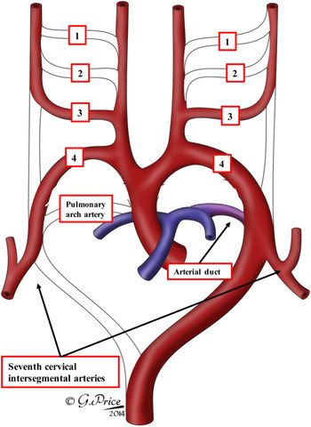
and endorsed by our own investigations, that the arterial duct cannot be a derivative of the “sixth arch artery”, since during normal mammalian development there are only five sets of arteries formed within the pharyngeal mesenchyme. For paediatric cardiologists, however, it would make no sense simply to rename the “sixth arch arteries” as “fifth arch arteries”, despite the fact that they are truly the fifth set of bilaterally symmetrical arteries extending through the pharyngeal mesenchyme. The solution of this enigma offered by Graham and his associates was, indeed, to rename the sixth arches as fifth arches. This is a suitable solution for basic scientists, and the logic is inescapable. But as emphasised above, for those dealing with congenital cardiac malformations, this would create nothing but confusion. Instead, as is suggested by Prabhu and associates, it makes more sense to consider the ultimate arteries as coursing through the pulmonary arches. This approach would also be suitable, surely, for embryologists? This is because, unlike the first through the fourth pharyngeal arches, the pulmonary arches contain only the arterial vessels destined to become the arterial duct, or its ligamentous remnant. Since it is the first and second sets of arteries that do disappear almost completely during the course of remodelling, it also makes sense to redraw the Rathke diagram, removing the channels representing the non-existent fifth arch arteries (Fig 3). The redrawn Rathke diagram will still remain as the basis for the creation of the Edwards’ hypothetical double arch,
Reference Edwards2
which as Prabhu and associates also show, remains the “gold standard” for interpreting the multiple patterns of vascular rings.
Figure 3.The drawing indicates how the Rathke diagram as shown in Fig 1 can now be redrawn so as to recognise the fact that the so-called “fifth arches” do not exist. The ultimate pharyngeal arch, however, is best designated as the pulmonary arch. It persists on the left side to become the arterial duct. The components of the double arch system that do not persist in normal development are shown as open channels.






 You have
Access
In a manuscript published in the current issue of the journal, Prabhu and his colleagues suggest a new categorisation for anomalies of the right aortic arch. Reference Prabhu, Mehra and Kasturi1 As the authors rightly emphasise, the anomalies of the right aortic arch, once it has crossed the right bronchus, can all be explained on the basis of the hypothetical double arch as formulated by Edwards. Reference Edwards2 They further explain that the concept, as put forward by Edwards, is itself derived from the understanding of the remodelling of the arteries of the pharyngeal arches, with these changes usually being interpreted on the basis of the diagram prepared long since by Rathke. Reference Rathke3
Of the several important lessons to be learned from their analysis, the first is that it is much better to describe the findings, rather than to create yet another complex alphanumeric code. For example, at one point in their manuscript, they describe how the differentiation between the situation when the left subclavian artery is isolated from the arch, and the arterial duct is closed, represented by the arterial ligament, as opposed to the situation when the duct and ligament are absent, can only be made in the operating room. The significance of this difference is immediately obvious. Since they have identified at least seven variants, which can exist with or without an arterial duct or ligament, it might have been tempting for them to create a system of classification such that these findings could be expressed as “the differentiation between type 4C versus type 6 and between type 5C versus type 7 can only be made intra-operatively”. Fortunately, they have resisted this temptation. Indeed, although their descriptions are often somewhat lengthy, they are a model of clarity. They serve to emphasise the intrinsic deficiencies of all alphanumeric classifications. Our goal should be to achieve uniformity in understanding. As is now shown in their report, this is best achieved by avoiding unnecessary codifications.
There is, however, a much more significant second lesson to be taken from their investigation. As already indicated, the diagrams in which they show the different patterns are based on the hypothetical arrangements that can be predicted on the basis of the model of the perfect double arch proposed by Edwards. Reference Edwards2 And, again as already stated, these findings are based on the knowledge of the remodelling, during cardiac development, of the arteries that percolate through the mesenchyme of the pharyngeal arches. These developmental changes are usually depicted on the basis of the Rathke diagram. As is shown by Prabhu and his colleagues, it is now necessary to redraw the Rathke diagram. This is because the “classical” diagram has, as its starting point, the presence of six pairs of arteries. These vessels extend from the aortic sac in a symmetrical fashion to reach the dorsal aorta (Fig 1). In the drawing provided by Prabhu and his colleagues, the fifth of these pairs of arteries, as in our own diagram, is shown by dotted lines. This is because the so-called “fifth arch arteries” have long been controversial. As most, if not all, paediatric cardiologists are well aware, various congenital cardiac malformations have been interpreted on the basis of their persistence in postnatal life. In the past, one of us was an enthusiastic supporter of the notion that these entities can be recognised when approached on the basis of the “great pretender”. Reference Gerlis, Ho, Anderson and Da Costa4 We have subsequently investigated large numbers of developing murine embryos using the technique of episcopic microscopy. In none of these, developing mice were we able to find evidence either of a fifth aggregation of pharyngeal mesenchyme to form an “arch”, nor of any additional arterial channels extending in parallel and symmetrical fashion from the aortic sac between the arteries of the fourth arches and those of the so-called “sixth” arches. In half of the embryos examined, nonetheless, we did find bilateral collateral channels at the terminations of the fourth and pulmonary arch arteries as they are inserted into the descending aorta (Fig 2). Reference Bamforth, Chaudhry and Bennett5 We did also, however, claim collectively to have discovered the only example of the persistence of a “fifth arch artery” in a developing human embryo. Reference Bamforth, Chaudhry and Bennett5 We now take the stance that the vessel is better considered as a collateral channel, which is enclosed in its own segment of the mesenchyme of the ultimate left pharyngeal arch. On the basis of our initial interpretation, we had conducted an extensive literature review of the lesions that, in our opinion, could properly be interpreted as representing the persistence of the enigmatic fifth arch arteries. Reference Gupta, Bamforth and Anderson6,Reference Gupta, Gulati and Anderson7 We must now revise these interpretations. This is because, as is now explained by Prabhu and colleagues, strong evidence has emerged showing that, in reality, there is never any formation of the purported fifth pharyngeal arch. By extension, there is never any formation of “fifth arch arteries”. This is also why we have now reinterpreted our original finding in a human embryo Reference Bamforth, Chaudhry and Bennett5 as representing a collateral channel. The developmental evidence in question was provided by Graham and his colleagues. Reference Graham, Poopalasundaram, Shone and Kiecker8 We are now in possession of additional evidence endorsing in full all of their conclusions.
The drawing shows the “classical” diagram offered by Rathke to explain the development of the arteries formed within the pharyngeal arches. The so-called “fifth arches” are shown with a dotted line, since their existence has long been questioned by embryologists. The diagram shows the descending aortas coming together to form the solitary midline structure shown by the white star with red borders. The white arrows with red borders show the seventh cervical intersegmental arteries, which will remodel to become the subclavian arteries.
The image shows the left side of the arteries percolating through the pharyngeal mesenchyme in a developing mouse embryo, reconstructed from an episcopic microscopic dataset. Comparable collateral channels to the one shown between the terminal parts of the arteries extending through the fourth and pulmonary arches were found in half of all the datasets examined
Over and above the examination of murine embryos, as described above, we have now been fortunate also to be granted access to a series of reconstructions made by Hikspoors and Lamers, working at the University of Maastricht in the Netherlands, from serially sectioned human embryos between Carnegie stages 10 and 23. This material is as yet unpublished. We are also in the process of collating our own evidence regarding the remodelling of the arteries of the murine pharyngeal arches. Suffice it to say that our findings in both the human and murine embryos support the conclusions made by Graham and his associates. Reference Graham, Poopalasundaram, Shone and Kiecker8 There are no arteries of the fifth arches, nor indeed any fifth pharyngeal arches. Prabhu and his colleagues, therefore, are to be congratulated for grasping the nettle of describing the arteries in an appropriate fashion as they undergo remodelling to form, on the one hand, the normal systemic and pulmonary arterial pathways, and on the other hand, the different patterns they have observed in the setting of the right aortic arch.
These revelations, of course, are of particular significance to the fashion in which we will need to describe not only the malformations afflicting the right aortic arch, but all those lesions previously interpreted on the basis of persistence on the non-existent arteries of the alleged fifth pharyngeal arches. Reference Gupta, Bamforth and Anderson6,Reference Gupta, Gulati and Anderson7 Even more significantly, they call into question our very description of “sixth arch arteries”. It is an inescapable conclusion of the study reported by Graham and colleagues, Reference Graham, Poopalasundaram, Shone and Kiecker8 and endorsed by our own investigations, that the arterial duct cannot be a derivative of the “sixth arch artery”, since during normal mammalian development there are only five sets of arteries formed within the pharyngeal mesenchyme. For paediatric cardiologists, however, it would make no sense simply to rename the “sixth arch arteries” as “fifth arch arteries”, despite the fact that they are truly the fifth set of bilaterally symmetrical arteries extending through the pharyngeal mesenchyme. The solution of this enigma offered by Graham and his associates was, indeed, to rename the sixth arches as fifth arches. This is a suitable solution for basic scientists, and the logic is inescapable. But as emphasised above, for those dealing with congenital cardiac malformations, this would create nothing but confusion. Instead, as is suggested by Prabhu and associates, it makes more sense to consider the ultimate arteries as coursing through the pulmonary arches. This approach would also be suitable, surely, for embryologists? This is because, unlike the first through the fourth pharyngeal arches, the pulmonary arches contain only the arterial vessels destined to become the arterial duct, or its ligamentous remnant. Since it is the first and second sets of arteries that do disappear almost completely during the course of remodelling, it also makes sense to redraw the Rathke diagram, removing the channels representing the non-existent fifth arch arteries (Fig 3). The redrawn Rathke diagram will still remain as the basis for the creation of the Edwards’ hypothetical double arch, Reference Edwards2 which as Prabhu and associates also show, remains the “gold standard” for interpreting the multiple patterns of vascular rings.
The drawing indicates how the Rathke diagram as shown in Fig 1 can now be redrawn so as to recognise the fact that the so-called “fifth arches” do not exist. The ultimate pharyngeal arch, however, is best designated as the pulmonary arch. It persists on the left side to become the arterial duct. The components of the double arch system that do not persist in normal development are shown as open channels.
Financial support
No funding was received to produce this commentary.
Conflicts of interest
None