Introduction
Fascioloides magna is a liver trematode, commonly found in several areas in North America and Europe (Erhardová-Kotrlá, Reference Erhardová-Kotrlá1971; Foreyt, Reference Foreyt1996). In Europe, F. magna was first detected in 1875 in Italy (Malek, Reference Malek1980) and then during the next century in: the Czech Republic (Ullrich, 1930), Germany (Salomon, Reference Salomon1932), Poland (Slusarski, Reference Slusarski1955), Austria (Pfeiffer, Reference Pfeiffer1983), Slovakia (Rajsky et al., Reference Rajsky, Patus and Bukovjan1994) and Hungary (Majoros & Sztojkov, Reference Majoros and Sztojkov1994). The presence of F. magna in Croatia was confirmed in 2000, although the fluke was probably present in that area years before (Marinculić et al., Reference Marinculić, Džakula, Janicki, Hardy, Lučinger and Živičnjak2002). Fascioloides magna enter uninfected habitats by natural migration of cervids and by translocation of animals (Pybus, Reference Pybus1990). The fluke probably entered Croatia by natural migration of cervids along the Danube River from neighbouring Hungary (Marinculić et al., Reference Marinculić, Džakula, Janicki, Hardy, Lučinger and Živičnjak2002) and by migration of parasite life cycles flooded by water from the Danube (Slavica et al., Reference Slavica, Florijančić, Janicki, Konjević, Severin, Marinculić and Pintur2006). According to Králová-Hromadová et al. (Reference Králová-Hromadová, Bazsalovicsová, Štefka, Špakulová, Vávrová, Szemes, Tkach, Trudgett and Pybus2011), presently there are two stable foci of F. magna in Europe: (1) the central and southern parts of the Czech Republic; and (2) Danube floodplain forests (territories of Austria, Slovakia, Hungary and Croatia).
Red deer (Cervus elaphus), white-tailed deer (Odocoileus virginianus) and fallow deer (Dama dama) are considered to be normal definitive hosts for F. magna in Europe (Erhardová-Kotrlá, Reference Erhardová-Kotrlá1971) Although F. magna infection in North American natural definitive hosts is usually asymptomatic (Foreyt & Todd, Reference Foreyt and Todd1976; Pybus, Reference Pybus1990), in the Croatian red deer population moderately poor body condition was noticed (Marinculić et al., Reference Marinculić, Džakula, Janicki, Hardy, Lučinger and Živičnjak2002). Rajković-Janje et al. (Reference Rajković-Janje, Bosnić, Rimac and Gojmerac2008) investigated a Croatian red deer population and found F. magna eggs in 34.53% of faeces samples and F. magna adults in 28.57% of liver samples. A high rate of red deer invasion with F. magna in Croatia has been confirmed by several authors (Janicki et al., Reference Janicki, Konjević and Severin2005; Slavica et al., Reference Slavica, Florijančić, Janicki, Konjević, Severin, Marinculić and Pintur2006; Rajković-Janje et al., Reference Rajković-Janje, Bosnić, Rimac and Gojmerac2008), with great variation in the rate of invasion between the eastern part (high prevalence) and other regions (only sporadic) of Croatia (Rajković-Janje et al., Reference Rajković-Janje, Bosnić, Rimac and Gojmerac2008).
The high prevalence of fascioloidosis in Croatia makes it an economically important disease in wild ruminants, and different diagnostic methods are being investigated in order to improve the health status of the infected populations (Severin et al., Reference Severin, Konjević, Janicki, Slavica, Marinculić and Cvrković2007). Serum biochemistry could be used as valuable diagnostic information in the evaluation of wild animal health and disease (Mašek et al., Reference Mašek, Konjević, Severin, Janicki, Grubešić, Krapinec, Bojanc, Mikulec and Slavica2009). The specific rise in liver enzymes and changes in other serum metabolites after infection with the common liver fluke (Fasciola hepatica) have been investigated extensively in domestic ruminants (Simesen & Nansen, Reference Simesen and Nansen1974; Anderson et al., Reference Anderson, Berrett, Brush, Hebert, Parfitt and Patterson1977, Reference Anderson, Berrett and Patterson1978, Reference Anderson, Matthews, Berrett, Brush and Patterson1981; Rowlands & Clampitt, Reference Rowlands and Clampitt1979; Sykes et al., Reference Sykes, Coop and Robinson1980; Bulgin et al., Reference Bulgin, Anderson, Hall and Lang1984; Doy & Hughes, Reference Doy and Hughes1984; Hawkins, Reference Hawkins1984; Wyckoff & Bradley, Reference Wyckoff and Bradley1985; Ferre et al., Reference Ferre, López, Gonzalo-Orden, Julian, Rojo-Vázquez and González-Gallego1995; Yang et al., Reference Yang, Mao, Ferre, Bayón, Mao and González-Gallego1998; Bossaert et al., Reference Bossaert, Lonneux, Godeau, Peeters and Losson1999; Vengušt et al., Reference Vengušt, Klinkon, Bidovec and Vengušt2003; Matanović et al., Reference Matanović, Severin, Martinković, Šimpraga, Janicki and Barišić2007). In contrast, there are only a few published papers on some aspects of serum biochemistry in domestic and wild animals infected with F. magna (Foreyt & Todd, Reference Foreyt and Todd1979; Presidente et al., Reference Presidente, McCraw and Lumsden1980; Stromberg et al., Reference Stromberg, Conboy, Hayden and Schlotthauer1985; Conboy & Stromberg, Reference Conboy and Stromberg1991) and, to our knowledge, none on C. elaphus. Moreover, there is a significant difference in the lesions produced in the liver by F. magna and F. hepatica because F. hepatica adults live in the bile ducts (Bulgin et al., Reference Bulgin, Anderson, Hall and Lang1984), while F. magna adults live within fibrous hepatic capsules in the hepatic parenchyma, which open to the biliary system (Foreyt & Todd, Reference Foreyt and Todd1979). Therefore, at least some differences in blood biochemistry between fasciolosis and fascioloidosis could be expected.
The present study was undertaken to investigate the serum biochemistry response to F. magna infection in the naturally infected red deer population in Croatia, as well as to better understand the pathogenesis of F. magna as a less known parasite in red deer. The relationship between serum biochemistry values and patho-anatomical changes in the liver was also examined.
Materials and methods
Parasitological procedures
The trial was performed on 47 red deer shot during routine culls in hunting grounds of eastern Croatia during the winters of 2006/2007 and 2007/2008. Faecal samples were collected directly from the rectum of each deer and examined by faecal sedimentation technique (Zajac, Reference Zajac, Sloss, Kemp and Zajac1994) for detection and quantification of F. magna fluke egg count. Eggs were counted in Petri dishes under a microscope (magnification × 40) and an estimate was made of eggs per gram of faeces. After they were shot, the deer were submitted to the Department of Veterinary Pathology (Faculty of Veterinary Medicine, Zagreb, Croatia) and standard necropsy was performed. All internal organs were examined grossly for F. magna and lesions compatible with fluke migration. The liver from each animal was sliced perpendicularly into 1.0–2.0 cm slices, and the slices were carefully examined for grossly visible flukes, migratory lesions, fibrous capsules, haematin pigment accumulation and other liver fluke lesions. All recovered flukes were counted, measured and classified as immature or mature according to Behm & Sangster (Reference Behm, Sangster and Dalton1999). According to the presence or absence of migratory lesions, F. magna infected deer were divided into the FC +M group (fibrous capsules and migratory lesions found in the liver) and the FC group (only fibrous capsules found in the liver).
Serology
Blood samples were collected from the heart into a blood tube without additives (BD Vacutainer; Becton Dickinson, Plymouth, Devon, UK). The blood was left in tubes to clot at room temperature and then centrifuged at 3500 rpm for 10 min within 1 h after sampling. The sera were stored frozen at − 20°C for a maximum of 30 days until analysed. Blood serum total protein, albumin, glucose, urea, creatinine, triglycerides, cholesterol, aspartate aminotransferase (AST), alanine aminotransferase (ALT), alkaline phosphatase (ALP), γ-glutamyltransferase (GGT), lactate dehydrogenase (LDH) and glutamate dehydrogenase (GLDH) were assayed by a Technicon RA-1000 (Technicon Istruments, Tarrytown, New York, USA) automatic analyser by using commercial kits of reagents (Randox Laboratories, Crumlin, County Antrim, UK). The concentrations for globulin were calculated.
Data analyses
We calculated mean value and standard deviations for each blood characteristic. The two groups were considered significantly different if the P value was lower than 0.05. The data were subjected to ANOVA procedure and post-hoc Tukey–Kramer test. Pearson's correlation coefficient was applied to evaluate the strength of association between serum biochemistry values and the number of immature flukes, mature flukes, migratory lesions and fibrous capsules. Statistica Version 9, 2010 (StatSoft Inc., Tulsa, Oklahoma, USA) was used for all data analysis.
Results
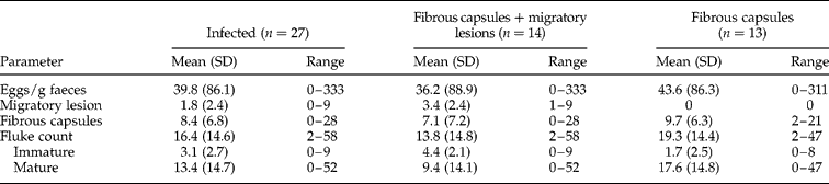
At necropsy, F. magna infection was confirmed in 27 animals (57.4%). Two to 58 flukes (mean ± standard deviation, 16.4 ± 14.6) were recovered from the infected animals. In the liver, flukes were found in fibrous capsules or migrating through the parenchyma (table 1). Formed fibrous capsules with mature flukes were observed in 14 livers simultaneously with migratory lesions, suggesting re-infection with F. magna. The other 13 infected animals had only fibrous capsules without the presence of migratory lesions, and there were no animals with only migratory lesions found in the liver. In five infected animals (18.5%) only immature flukes were found and in 9 animals only mature flukes were found (33.3%) while in most animals (13, 48.1%) both immature and mature flukes were found.
Liver necropsy findings in F. magna infected deer.
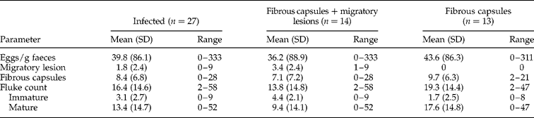
SD, standard deviation.
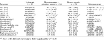
The differences in blood biochemistry values between F. magna infected and uninfected deer are presented in table 2. Both groups of infected deer had significantly higher values for globulin (P < 0.05) and significantly (P < 0.01) lower values for albumin/globulin ratio and glucose (P < 0.01) in comparison to the uninfected deer. Values for LDH and GLDH were significantly higher (P < 0.01) only for the FC + M group compared to the uninfected animals and ALT value was significantly higher (P < 0.05) only for the FC group in comparison to the uninfected animals.
Mean values and standard deviation (in parentheses) of serum biochemistry values in F. magna infected and non-infected red deer.

AST, aspartate aminotransferase; ALT, alanine aminotransferase; ALP, alkaline phosphatase; GGT, γ-glutamyltransferase; LDH, lactate dehydrogenase; GLDH, glutamate dehydrogenase.
a,b Rows with different superscripts differ significantly, *P < 0.05.
1 Reference range (Goddard et al., Reference Goddard, Keay and Grigor1997; Marco & Lavin, Reference Marco and Lavín1999; Padilla et al., Reference Padilla, Bouda, Quiroz-Rocha, Davalos and Sanchez2000; Rosef et al., Reference Rosef, Nystøyl, Solenes and Arnemo2004).
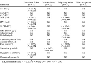
Statistically significant and positive correlations were observed between the number of immature flukes and AST (r = 0.570; P = 0.033), GGT (r = 0.612; P = 0.020), LDH (r = 0.721; P = 0.004), GLDH (r = 0.604; P = 0.022), urea (r = 0.599; P = 0.024) and triglyceride (r = 0.846; P = 0.033) values and between the number of migratory lesions and GGT (r = 0.668; P = 0.009), GLDH (r = 0.768; P = 0.001), globulin (r = 0.556; P = 0.039) and urea (r = 0.595; P = 0.025) values (table 3). No statistically significant correlations were determined between blood biochemistry values and the number of mature flukes and fibrous capsules with the exception of the creatinine values which were positively correlated to the number of mature flukes (r = 0.673; P = 0.008).
Pearson's correlation coefficient between biochemistry values and number of immature flukes, mature flukes, migratory lesions and fibrous capsules.

AST, aspartate aminotransferase; ALT, alanine aminotransferase; ALP, alkaline phosphatase; GGT, γ-glutamyltransferase; LDH, lactate dehydrogenase; GLDH, glutamate dehydrogenase.
NS, not significant; P>0.10; 1P < 0.10; *P < 0.05; **P < 0.01.
Discussion
There is an evident similarity between the pathophysiological effects of F. magna and F. hepatica in the initial phase of invasion when young, immature flukes migrate through the hepatic parenchyma. At a later stage of invasion, F. magna remains encapsulated in the hepatic parenchyma while F. hepatica adults penetrate into the bile ducts, where they reach sexual maturity (Foreyt & Todd, Reference Foreyt and Todd1979). Therefore, F. magna is responsible for liver damage mainly by the mechanical migration of immature flukes in the acute phase and by the creation of fibrous capsules around the mature flukes in the chronic phase of the infection.
There are only a few published reports on the influence of F. magna infection on serum biochemistry. Conboy & Stromberg (Reference Conboy and Stromberg1991) found only a minor increase in GGT and sorbitol dehydrogenase (SDH) values in F. magna infected calves and a significant increase in AST values in F. magna infected guinea pigs. Similarly, Stromberg et al. (Reference Stromberg, Conboy, Hayden and Schlotthauer1985) did not find significant changes in liver enzymes in F. magna infected sheep. In contrast, our experiment showed significant differences in ALT, GLDH and LDH values, as well as in globulin and glucose values. Nevertheless, it is important to notice that these experiments were performed on different aberrant hosts. Foreyt & Todd (Reference Foreyt and Todd1979) performed research on the definitive host (white-tailed deer) and found a significant increase in total protein and globulin values without significant differences in liver enzyme values. In F. hepatica infected animals, different liver enzymes were confirmed as useful indicators of the different stages of infection (Yang et al., Reference Yang, Mao, Ferre, Bayón, Mao and González-Gallego1998). Clinically, the most important enzymes are AST and GLDH, which provide information on the passage of immature flukes through the liver parenchyma, and GGT, which indicates penetration into the bile ducts and the chronic stage of infection (Anderson et al., Reference Anderson, Berrett, Brush, Hebert, Parfitt and Patterson1977; Sykes et al., Reference Sykes, Coop and Robinson1980; Bulgin et al., Reference Bulgin, Anderson, Hall and Lang1984; Yang et al., Reference Yang, Mao, Ferre, Bayón, Mao and González-Gallego1998).
Serum ALT activity was significantly higher in F. magna infected animals without migratory lesions (FC group) than in uninfected animals, while that increase did not reach a significant difference in F. magna infected animals with migratory lesions (FC+M group) in comparison to uninfected animals. An increase in serum ALT activity was also noticed by Vengušt et al. (Reference Vengušt, Klinkon, Bidovec and Vengušt2003) in F. hepatica infected fallow deer. Nevertheless, ALT activity is not hepatic specific for cattle (Hoffman & Solter, Reference Hoffman, Solter, Kaneko, Harvey and Bruss2008) and it could be significantly influenced by other factors, such as the method of capture (Marco & Lavin, Reference Marco and Lavín1999; Topal et al., Reference Topal, Gul and Yanik2010).
Total serum LDH activity in both F. magna infected groups was higher than the reference range observed by other authors for red deer (Goddard et al., Reference Goddard, Keay and Grigor1997; Marco & Lavin, Reference Marco and Lavín1999; Rosef et al., Reference Rosef, Nystøyl, Solenes and Arnemo2004) and LDH activity was significantly higher in F. magna infected deer with migratory lesions than in uninfected deer. Nevertheless, the diagnostic value of that parameter is questionable because of the distribution of LDH isoenzymes within red deer tissues. In red deer, LDH-1 activity is higher in the cardiac muscle, liver and kidneys than in the lungs, spleen and skeletal muscles, while LDH-5 activity is higher in skeletal muscles than in other tissues (Goddard et al., Reference Goddard, Keay and Grigor1997).
Serum GLDH activity was the highest in F. magna infected deer with migratory lesions present in the liver and the mean GLDH value for both F. magna infected groups was higher than the reference value proposed by Rosef et al. (Reference Rosef, Nystøyl, Solenes and Arnemo2004). Additionally, a positive correlation was observed between GLDH activity and the number of immature flukes and migratory lesions. These results were also noticed in F. hepatica infected animals, where serum GLDH activity was increased during the parenchymal phase of infection, while in the chronic phase GLDH activity was normal (Sykes et al., Reference Sykes, Coop and Robinson1980; Yang et al., Reference Yang, Mao, Ferre, Bayón, Mao and González-Gallego1998). Normalization of GLDH activity is probably caused by liver tissue regeneration in the later stage of infection. Unlike AST or LDH, GLDH activity can be considered to be specific for liver parenchyma (Hoffman & Solter, Reference Hoffman, Solter, Kaneko, Harvey and Bruss2008), which gives important diagnostic value to this enzyme in fascioloidosis.
During our trial GGT activity was only numerically higher in F. magna infected deer. In F. hepatica infected animals, GGT values are usually increased because that parasite penetrates into the bile ducts with subsequent damage to the biliary epithelium (Bulgin et al., Reference Bulgin, Anderson, Hall and Lang1984) where GGT is concentrated (Naftalin et al., Reference Naftalin, Child and Morley1969). In contrast, F. magna adult flukes live within fibrous hepatic capsules in the hepatic parenchyma (Foreyt & Todd, Reference Foreyt and Todd1976). Nevertheless, besides numerically higher GGT values in F. magna infected deer, we also found a significant correlation between GGT values and the number of immature flukes and migratory lesions. These findings could mean that F. magna causes damage to the biliary epithelium during the parenchymal phase of infection.
An increased value of globulin and the subsequent change in the albumin/globulin ratio was also noticed in F. magna infected white-tailed deer (Foreyt & Todd, Reference Foreyt and Todd1979) and in F. hepatica infected animals (Matanović et al., Reference Matanović, Severin, Martinković, Šimpraga, Janicki and Barišić2007). This increase is probably due to an immune response to F. magna (Foreyt & Todd, Reference Foreyt and Todd1979).
It could be concluded that the liver enzyme response to F. magna infection in red deer did not completely correspond to that of F. hepatica infection. The most probable explanations are the differences in pathogenesis between these two diseases in the chronic phase, with F. magna forming fibrous capsules and F. hepatica penetrating into the bile ducts. In our investigation the most relevant enzyme diagnostically was GLDH, although further investigations in controlled experiments are needed to clarify the influence of F. magna on blood biochemistry in different stages of infection. Nevertheless, in our opinion, it is obvious that these findings would be extremely hard to interpret in naturally infected animals because of the different stages of infection, common superinfections and the high variability of blood parameters in free-ranging animals.
Acknowledgements
This study was supported by the Ministry of Science, Education and Sport of the Republic of Croatia, in the projects: No. 053-0532400-2399 and No. 053-0532265-2244. This research complies with current laws of the Republic of Croatia.