Introduction
Schistosomosis caused by Schistosoma spindale and Schistosoma indicum is one of the major economically important trematode infections of ruminants in India. It is speculated that at least 165 million cattle are infected with schistosomosis worldwide, while another 530 million live in endemic areas (De Bont & Vercruysse, Reference De Bont and Vercruysse1997). Although abattoir surveys revealed a high prevalence of schistosome infection in bovines of southern India (Sumanth et al., Reference Sumanth, D'Souza and Jagannath2004; Ravindran et al., Reference Ravindran, Lakshmanan, Ravishankar and Subramanian2007; Lakshmanan et al., Reference Lakshmanan, Rauoof, Fawaz and Subramanian2011), the disease was underestimated in live animals, probably due to the low sensitivity of conventional coprodiagnostic approaches (Pardo et al., Reference Pardo, Carranza, Turrientes, Arellano, Velez, Ramajo and Muro2004). Immunodiagnostic assays using different antigens of Schistosoma spp. have been standardized (Sumanth et al., Reference Sumanth, D'Souza and Jagannath2003; Singh et al., Reference Singh, Singh and Chaudhri2004; Mahfouz et al., Reference Mahfouz, Mahana, Rabee and El-Amir2011) in spite of the potential problems associated with immunological detection of schistosomes as reviewed by Doenhoff et al. (Reference Doenhoff, Chiodini and Hamilton2004). The 18S and 28S rDNA sequences have been identified as good genetic markers for identification and phylogenetic studies of the genus Schistosoma (Johnston et al., Reference Johnston, Kane and Rollinson1993; Barker & Blair, Reference Barker and Blair1996; Lockyer et al., Reference Lockyer, Olson, Ostergaard, Rollinson, Johnston, Attwood, Southgate, Horak, Snyder, Le, Agatsuma, McManus, Carmichael, Naem and Littlewood2003; Webster et al., Reference Webster, Southgate and Littlewood2006). Detection of Schistosoma spp. DNA in faeces has been accomplished by several workers using molecular tools (Pontes et al., Reference Pontes, Dias-Neto and Rabello2002; Gobert et al.; Reference Gobert, Chai, Duke and McManus2005, Oliveira et al., Reference Oliveira, Santos, Goncalves, Barreto and Peralta2010). Amplification of Schistosoma mansoni DNA from faecal material, by targeting a previously reported 121 bp highly repeated sequence (Hamburger et al., Reference Hamburger, Turetski, Kapeller and Deresiewicz1991) was successful (Pontes et al., Reference Pontes, Dias-Neto and Rabello2002). Hamburger et al. (2004) used the Dra1 repeat to detect Schistosoma haematobium infection in snails. Specific amplification of Schistosoma spp. 28S rRNA from human urine samples (Sandoval et al., Reference Sandoval, Siles-Lucas, Peres-Arellano, Carranza, Puente, Lopez-Aban and Muro2006) and Schistosoma japonicum mitochondrial DNA from faeces of infected mammalian hosts (Gobert et al., Reference Gobert, Chai, Duke and McManus2005) are also reported. But tools for molecular detection of bovine schistosome species in India have not been investigated comprehensively. In the context of the high prevalence of infection among slaughtered cattle in the State and the low sensitivity of routine coprodiagnostic approaches, early and specific detection of bovine schistosomosis is imperative for timely detection and treatment. Hence, we report a new copro-polymerase chain reaction (copro-PCR) protocol for sensitive and specific detection of Indian species of schistosomes affecting bovines. To evaluate the specificity of the protocol, we also analysed the comparative efficacy of the designed primers with faecal DNA samples positive for other helminths which were prevalent among bovines in this region.
Materials and methods
Collection and examination of adult worms and faecal samples
Adult schistosome worms residing in the mesenteric veins were recovered from the mesenteries of cattle (n= 50) slaughtered in the local abattoir of Thrissur district, following a previously described method (Murthy et al., Reference Murthy, D'Souza and Isloor2013). The worms recovered from each mesentery were washed several times in sterile phosphate-buffered saline (PBS, pH 7.4), identified morphologically based on the tubercles, eggs and number of testes (Kumar, Reference Kumar1999) and stored separately at − 20°C until DNA extraction. The adult schistosomes recovered were S. spindale and S. indicum. Based on the number of worms recovered from each mesentery, the intensity of infection was graded as high (more than 100 worms), moderate (50–100 worms), low (10–20 worms) and very low (fewer than 10 worms). In addition, amphistomes were also recovered from the rumen and identified as Gastrothylax crumenifer and Fischoederius elongatus (Kumar, Reference Kumar1999). Faecal samples (10 g) were collected from slaughtered animals (n= 30), as well as from live animals clinically suspected to have schistosomosis (n= 30). The faecal samples were processed for microscopic detection of eggs as well as for DNA extraction.
Faecal samples were analysed microscopically for the presence of parasite eggs by flotation, sedimentation (Hendrix & Robinson, Reference Hendrix and Robinson2006) and the potassium hydroxide digestion method (Marshall et al., Reference Marshall, Morrison and Nyrinden1989). Amphistome, strongyle and S. spindale eggs could be detected in different faecal samples.
Molecular analysis
Total DNA was extracted from pooled samples of each fluke species using QIAGEN DNeasy Blood and Tissue Kit (Qiagen, Hilden, Germany). The protocol specified in the kit was adopted with relevant modifications. Approximately 25 mg of each species of fluke were mixed with 180 μl of buffer ATL and 20 μl of proteinase K, vortexed thoroughly for 15 s and incubated at 56°C overnight until the tissues were lysed completely. In the case of amphistomes it was necessary to add 40 μl of proteinase K for optimal DNA yield.
The faecal samples (1 g each) were processed for DNA extraction using QIAGEN QIA amp DNA stool Mini Kit, following manufacturer's instructions for large-volume samples. The DNA content and purity of the final eluates were estimated using a nanospectrophotometer (Nano drop 200 C, Thermoscientific, Pittsburgh, Pennsylvania, USA). The concentration was determined at 260 nm and purity by the 260:280 nm ratio. Samples that yielded a ratio between 1.7 and 1.9 were selected for analysis.
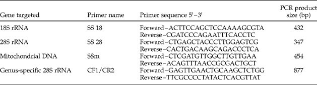
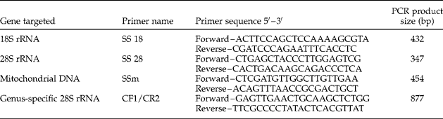
Species-specific diagnostic primers were not available for the fluke species under study and hence we decided to design the primers. The gene sequences of S. spindale corresponding to complete 18S rRNA (Z11979), 28S rRNA (Z46505) and mitochondrial genes (DQ 157223) available in GenBank (www.ncbi.nlm.nih.gov) were used for designing the primers. The primers were designed using Primer 3 Web Version 4.0.0 (gmdd.shgmo.org/primer 3/?sequid=47), their specificity confirmed using BLASTn (National Center for Biotechnology Information) and suitability confirmed using Sequence Manipulation Suite Version 2 (www.bioinformatics.org/sms2/pcr_primer_stats.html). The sequence of primers designed in this study, along with the previously published (Sandoval et al., Reference Sandoval, Siles-Lucas, Peres-Arellano, Carranza, Puente, Lopez-Aban and Muro2006) genus-specific primers targeting 28S rRNA of Schistosoma sp. (CF1, CR2) are given in table 1.
The number of primers used in the present study together with sequences, gene targets and the size of PCR products (base pairs, bp).
Gradient PCR was performed using SS 18, SS 28, SSm and CF1/CR2 primers to standardize the protocol with DNA extracted from adult S. spindale and S. indicum collected from the mesenteries. These primers were also tested with DNA extracted from F. elongatus and G. crumenifer, the most commonly occurring amphistome species in Kerala, to check the specificity. The standardized protocol was applied to faecal DNA from samples positive for eggs of schistosomes, amphistomes and strongyles.
The PCR was performed in a 25 μl reaction volume containing 2.5 μl of buffer (10 × ) without MgCl2, 200 μm of each deoxynucleoside triphosphate (dNTP), 25 pmol each of forward and reverse primers, 1.5 mm MgCl2, 1 U of Taq DNA polymerase and 5.0 μl of template DNA. All the reagents were obtained from Sigma Aldrich (St. Louis, Missouri, USA). A gradient thermal cycling programme (MJ Mini, Biorad, California, USA) with initial denaturation at 94°C for 5 min followed by 35 cycles of denaturation (94°C, 1 min), annealing (55–67°C, 30 s) and extension (72°C, 1 min), and a final extension at 72°C for 7 min was adopted. The amplicons were electrophoresed in 2% agarose gel and sizes were resolved using a 100 bp ladder (Thermoscientific).The amplicons were purified using silica gel purification columns (GeneJET, Thermoscientific), sequenced using Sangers dideoxy chain termination method and the sequences aligned using Sequencher Version 5.0 (Sci Genom Labs Pvt Ltd, Cochin, India).
The sequences obtained in the study using SS 18, SS 28 and SSm primers were submitted to GenBank and assigned the following accession numbers: KF 317666, KF 317669, KF 317667, KF 317668, KF 425713, KF 425714, KF 830697, KF 830698, KF 830696 and KF 850451.
Results
Gradient PCR with annealing temperatures varying from 55°C to 67°C using SS 18 and SS 28 primers, amplified a 432 bp and 347 bp product, respectively, at all temperatures, when template DNAs of S. spindale, S. indicum, F. elongatus and G. crumenifer were used. Previously published primer sets, namely CFI/CR2, also yielded a product of about 877 bp using DNA of all the four parasite species studied.
In the absence of specificity for ribosomal gene markers, mitochondrial primers (SSm) were designed for amplification of mitochondrial DNA of Schistosoma spp., targeting the partial rrnL (16S rRNA), tCys (transfer RNA for cysteine) and partial rrnS (12S rRNA) sequences. Gradient PCR confirmed the specificity of SSm primers, with an optimum annealing temperature of 60°C. A specific 454 bp product was obtained with the protocol using Schistomoma spp. template DNA. No amplification product was detected with the SSm primers when F. elongatus and G. crumenifer DNAs were used as templates.
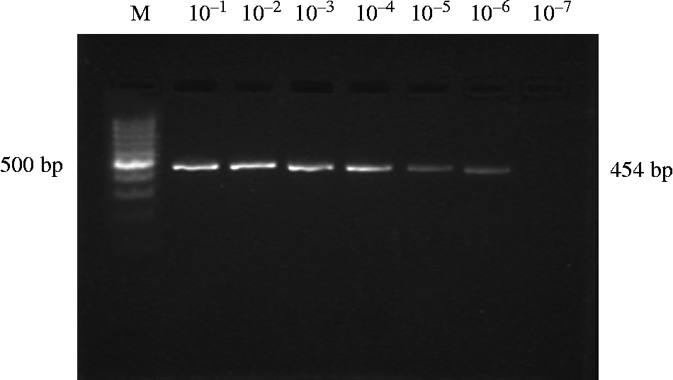
To ascertain the PCR detection limit of the primers, tenfold serial dilutions of template DNA of S. spindale, with an initial concentration of 45 ng/μl, were prepared and subjected to PCR to identify the lowest concentration of DNA that can be amplified by SSm using the standardized protocol. Although the brightness of the bands was gradually reduced with increasing dilution, amplicons could be detected up to 10− 6 dilution. Thus the detection limit of SSm primers was determined to be 0.225 pg of template DNA (fig. 1).
PCR reactions with SSm primers carried out with serial dilutions of adult S. spindale worm DNA. M, DNA ladder; lane headings denote serial dilutions of template S. spindale DNA.

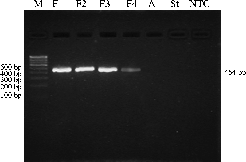
Microscopic analysis of faecal samples of cattle suffering from diarrhoea and emaciation revealed amphistome eggs in 12 samples, S. spindale eggs in 10 samples and strongyle eggs in 1 sample. There was no mixed parasitic infection. None of the samples were positive for S. indicum eggs. Faecal DNA extracted from samples positive for Schistosome and amphistome eggs yielded positive PCR signals with SS 18, SS 28 and CF1/CR2 primer sets. However, faecal DNA sample from the animal with a heavy load of strongyle eggs (330 eggs/g (epg)), failed to amplify with these primers. A distinct product of 454 bp was obtained using SSm primers with template DNA extracted from faecal samples positive for S. spindale eggs. PCR with the same primer pairs did not result in a detectable amplification product when faecal DNA samples with heavy loads of amphistome and strongyle eggs were used as templates (fig, 2), thus confirming the diagnostic specificity of the copro-PCR protocol employing SSm primers. The detection limit of the copro-PCR protocol in our study was 40 epg, as indicated by positive PCR signals from samples containing ≥ 40 epg and the absence of amplification in two samples containing 15 and 10 epg, respectively.
PCR products from faecal material containing schistosome, amphistome and strongyle eggs, using SSm primers. Lanes: M, DNA ladder; F1–F4, positive faecal DNA for schistosome, A, amphistome DNA; St, strongyle DNA; NTC, no template control.
The sensitivity of the PCR protocol was also determined using post-mortem samples, by assuming that samples from worm-positive slaughtered cattle are true positives. The faecal samples from animals that harboured a high intensity (n= 4), moderate intensity (n= 6) and low intensity (n= 15) of schistosome infections yielded a positive PCR signal. It is noteworthy that five animals that failed to yield positive PCR signals harboured very low intensity infection (fewer than 10 worms per mesentery). The copro-PCR protocol had a sensitivity of 83.3% when compared with post-mortem diagnosis of schistosome infection. The prevalence of S. indicum infection was low during the abattoir survey. Moreover, none of the faecal samples revealed S. indicum eggs during microscopic examination. Hence, further analysis was focused on S. spindale and amphistomes.
Sequence analysis
Nucleotide BLAST analysis of sequence data of S. spindale DNA amplified with SS 18 and SS 28 primers revealed 98–100% identity with those of several published schistosome species. The complete sequence of Gastrothylax sp. and Fischoederius sp. were not available in the databank. Hence, the nucleotide sequences of pouched amphistomes obtained in the study were further analysed and compared with the partial gene sequences of amphistomes and schistosomes, which revealed significant similarity with the sequences of several Schistosoma spp. and with Paramphistomidae sp.
The mitochondrial DNA sequence of S. spindale amplified using SSm primers, comprising partial 16S rRNA, complete tCys and partial 12S rRNA, exhibited 99% identity with that of S. spindale (DQ 157223). The results of sequence analysis highlight the urgent need to sequence the complete ribosomal and mitochondrial DNA of the common amphistome isolates of India.
Discussion
Schistosomosis and amphistomosis are among the most economically important snail-borne trematode infections of bovines in the country. The economic importance of bovine schistosomosis is mainly attributed not only to morbidity, mortality, liver condemnation, reduced productivity and poor subsequent reproductive performance (McCauley et al., Reference McCauley, Majid and Tayeb1984), but also to increased susceptibility to other diseases (Dargie, Reference Dargie1980). Acute infections are symptomatically similar, and routine coprodiagnostic approaches for egg detection often suffer from low sensitivity. The present study focused on development and evaluation of a PCR protocol for specific detection of schistosome infection in faecal samples of bovines. Amphistomes and strongyles are the most frequently identified parasite aetiology of diarrhoea among bovines of the State. Gastrothylax crumenifer and F. elongatus were the amphistomes recovered from the rumen of slaughtered cattle, and hence were selected to study the possibility of non-specific amplification. In addition, faecal samples positive for amphistome and strongyle eggs were also subjected to copro-PCR to ascertain the specificity of the protocol.
Schistosoma spindale 18S and 28S rRNA genes were initially targeted for primer design, owing to the fact that these sequences were effective genetic markers to identify schistosomes (Johnston et al., Reference Johnston, Kane and Rollinson1993; Chen et al., Reference Chen, Li, Sugiyama, Weng, Zou, Lin, Yuan, Song, Zhu and Zhao2011). Further, Sandoval et al. (Reference Sandoval, Siles-Lucas, Peres-Arellano, Carranza, Puente, Lopez-Aban and Muro2006) had also developed a PCR protocol of high specificity with CF1/CR2 primers, which amplified a 877 bp product of the 28S rRNA region specific to the members of the genus Schistosoma. The specificity of CF1/CR2 primers had been checked with DNA from Fasciola hepatica, Echinococcus multilocularis, and various other nematodes and protozoa infecting human beings, but not with any of the amphistome species.
In this study, primers designed from 18S and 28S ribosomal RNA gene sequences, as well as the previously published primers targeting 28S rRNA primers, failed to differentiate between schistosomes and amphistomes in cattle. Concurrent results were obtained with copro-PCR as well. In a tropical country such as India, with special emphasis on the waterlogged marshy plains of Kerala State, which were once used for paddy cultivation, snail-borne trematode infections, such as amphistomes and schistosomes, are prevalent among grazing cross-bred cattle. Hence, it is imperative to distinguish between these parasitic infections. No published records exist of cross-amplification of schistosomes and amphistomes with 18S and 28S rRNA gene markers.
Further investigation into nucleotide sequence similarity with Fischoederius sp. and Gastrothylax sp. was futile, since complete ribosomal RNA gene sequence data of these flukes were unavailable in databases. However, significant similarity that exists between the ribosomal RNA gene sequences of Paramphistomidae sp. – a closely related amphistome – F. elongatus, G. crumenifer and S. spindale, observed during BLASTn analyses, enabled cross-amplification.
The mitochondrial DNA sequences, which are present in multiple copies within cells, were suggested as ideal targets for PCR diagnosis of schistosome eggs in human stool samples (Gordon et al., Reference Gordon, Gray, Gobert and McManus2011). The complete sequencing of the mitochondrial genome of S. spindale (Littlewood et al., Reference Littlewood, Lockyer, Webster, Johnston and Le2006) facilitated the design of SSm primers, which proved to be specific and sensitive for detection of the egg stages of Schistosoma spp. in naturally infected bovine hosts. The detection limit of primers was measured by several workers, as the minimum quantity of detectable DNA in PCR using those primers. The level of detection of S. mansoni adult worm DNA by PCR was reported to be 10 fg (Hamburger et al., 1998; Suzuki et al., Reference Suzuki, Osada, Kumagai, Hamada, Okuzawa and Kanazawa2006) and 0.98 pg (Sandoval et al., Reference Sandoval, Siles-Lucas, Peres-Arellano, Carranza, Puente, Lopez-Aban and Muro2006),while that of egg-derived DNA was 1.0 fg (Pontes et al., Reference Pontes, Dias-Neto and Rabello2002) and 3.0 fg (Gomes et al., Reference Gomes, Marques, Enk, Coelho and Rabello2009). Webster et al. (Reference Webster, Rollinson, Stohard and Littlewood2010) developed multiplex PCR utilizing primers targeting the cytochrome oxidase subunit 1 gene of mitochondrial DNA, with a sensitivity of 0.78 ng of DNA derived from adult S. haematobium and Schistosoma bovis. The sensitivity cut-off for copro-PCR to detect S. mansoni and S. japonicum mitochondrial DNA was ≥ 60 epg during experimental infections in mice (Gobert et al., Reference Gobert, Chai, Duke and McManus2005). In our study, the newly designed SSm primers detected a minimal DNA concentration of 0.23 pg in ethidium bromide stained agarose gels. The sensitivity cut-off of the protocol was determined to be ≥ 40 epg during natural infections in definitive hosts. The worm-free DNA in the samples could also have contributed to the specific amplification in copro-PCR. Samples with lower epg yielded false-negative PCR signals, probably due to the lack of uniform distribution of eggs in faecal samples or due to the presence of PCR inhibitors in the sample (Oliveira et al., Reference Oliveira, Santos, Goncalves, Barreto and Peralta2010). When compared with post-mortem worm detection, copro-PCR failed to detect infection in five true-positive samples which harboured very low-intensity infections. These samples could have been from chronically infected cattle, which might have developed immunity against schistosomes, thus leading to reduced, as well as fluctuating, egg excretion in the faeces (Aradaib et al., Reference Aradaib, Omer, Abbas, Bushara, Elmalik, Saad, Osburn and Taylor1995). This study proved conclusively, for the first time, the high specificity of mitochondrial DNA markers for detection of natural infections by S. spindale and S. indicum among bovines. These markers distinguished efficiently schistosome, amphistome and strongyle infections in bovines. The results also suggested a high degree of homology between partial nucleotide sequences of 18S and 28S ribosomal RNA genes of S. spindale, S. indicum, G. crumenifer and F. elongatus isolates from southern India. Further studies on molecular detection of developing stages of schistosomes in snails, by developing species-specific probes, will be potentially useful to trace endemic pockets of infection in the country. Investigations should also be directed towards sequencing complete 18S and 28S ribosomal RNA, as well as mitochondrial genes, of pouched amphistomes of the region, and to provide a better understanding of the molecular epidemiology of snail-borne trematode infections.
Acknowledgements
The authors are grateful to Kerala Veterinary and Animal Sciences University for the facilities provided to conduct the study.
Financial support
This work was financially supported by Kerala Veterinary and Animal Sciences University, Pookode, Wayanad, Kerala, India.
Conflict of interest
None.