Implications
Melatonin is a potential therapeutic to overcome negative effects of nutrient restriction during pregnancy because of its roles in regulating blood flow and gastrointestinal and pancreatic function. We hypothesized that melatonin supplementation would positively impact fetal pancreatic function in nutrient restricted ewes. Results indicate nutrient restriction during gestation had greater impacts on digestive enzyme concentrations in ewes than fetuses and that nutrient restriction decreased pancreatic insulin concentrations. Melatonin supplementation did not influence fetal pancreatic function suggesting that its role as a therapeutic for overcoming negative effects of undernutrition during pregnancy on fetal pancreatic development is limited.
Introduction
Although nutrient restriction can result in reduced performance of both the dam and fetus, the development of proper intervention techniques may mitigate harmful consequences. Understanding the influence of maternal nutrition on fetal development provides the potential to design supplementation strategies that may help overcome these negative impacts. Melatonin, most widely known for its role in regulating circadian rhythms, has been shown to increase utero-placental blood flow when supplemented to pregnant ewes which has the potential to reduce or offset the negative effects of nutrient restriction on maternal blood flow and return utero-placental nutrient transfer to normal levels (Lemley et al., Reference Lemley, Meyer, Camacho, Neville, Newman, Caton and Vonnahme2012). Melatonin can be synthesized by many tissues of the body with the largest source being produced in the gastrointestinal tract (GIT; Jaworek et al., Reference Jaworek, Nawrot-Porabka, Leja-Szpak, Bonior, Szklarczyk, Kot, Konturek and Pawlik2007 and Reference Jaworek, Szklarczyk, Jaworek, Nawrot-Porąbka, Leja-Szpak, Bonior and Kot2012). Melatonin secretion increases with the ingestion of food and can influence GIT function by causing a reduction in the speed of intestinal peristalsis thereby slowing food transit time and allowing for greater nutrient absorption (Bubenik, Reference Bubenik2008). Melatonin receptors have also been found on the pancreas and can influence pancreatic insulin secretion, glucose homeostasis and provide protective mechanisms against oxidative stress, which can selectively destroy β-cells (Sartori et al., Reference Sartori, Dessen, Mathieu, Monney, Bloch, Nicod, Scherrer and Duplain2009; Li et al., Reference Li, Hou, Lu and Kang2011). Because of its potential influence on nutrient supply and pancreatic function, melatonin supplementation may mitigate negative impacts of maternal nutrient restriction on fetal growth and pancreatic development. We hypothesized that melatonin supplementation may have positive effects on the functional morphology of the pancreas in nutrient-restricted pregnant ewes and their fetuses. Therefore, the objectives were to determine the effect of dietary melatonin supplementation on pancreatic digestive enzyme activity, morphology and plasma insulin levels in undernourished pregnant ewes and their fetuses.
Material and methods
This experiment was approved by the North Dakota State University Institutional Animal Care and Use Committee.
Animals and dietary treatments
Animal care and dietary treatments were described previously (Lemley et al., Reference Lemley, Meyer, Camacho, Neville, Newman, Caton and Vonnahme2012). Briefly, 32 nulliparous ewes carrying singletons were selected for the study; however, one ewe was removed due to a perforated esophagus, which existed before dietary treatment. Ewes were transported on day 28 of gestation to a facility that provided a temperature controlled environment at 14°C and a 12 : 12 light–dark cycle with lights on at 0700 h and off at 1900 h each day. On day 50 of gestation, the ewes began their dietary treatments. Ewes were individually penned and provided 100% (ADQ; adequate diet) or 60% (RES; restricted diet) of nutrient recommendations for a pregnant ewe lamb in mid- to late-gestation (NRC, 2007) and diets were supplemented with either 5 mg of melatonin (MEL; Spectrum Chemical Mfg. Corp., Gardena, CA, USA) or no melatonin (CON) in a 2×2 factorial arrangement of treatments. The four treatment groups consisted of CON : ADQ (n=7), MEL : ADQ (n=8), CON : RES (n=8) or MEL : RES (n=8). The diet was pelleted and contained beet pulp, alfalfa meal, corn, soybean meal and wheat middlings. All melatonin supplemented ewes were fed melatonin-enriched pellets at 1400 h (5 h before the start of the dark cycle at 1900 h). The pellets were prepared using melatonin powder dissolved in 95% ethanol at a concentration of 5 mg/ml. The day before feeding, 100 g of the control pellet was placed into a small plastic bag and top dressed with 1 ml of melatonin solution. The ethanol was allowed to evaporate overnight at room temperature in complete darkness before feeding (Lemley et al., Reference Lemley, Meyer, Camacho, Neville, Newman, Caton and Vonnahme2012). The amount of feed offered to individual ewes was adjusted weekly based on BW and change in BW to ensure the desired average daily gains were achieved (Reed et al., Reference Reed, Ward, Vonnahme, Neville, Julius, Borowicz, Taylor, Redmer, Grazul-Bilska, Reynolds and Caton2007). Ewes were maintained on their given dietary treatments until 130 days of gestation.
Sample collection
On day 130 of gestation, dams were weighed. Maternal and fetal blood plasma collection was conducted as described by Lemley et al. (Reference Lemley, Camacho, Meyer, Kapphahn, Caton and Vonnahme2013). Briefly, ewes were anesthetized with 3 mg/kg of BW sodium pentobarbital (Fort Dodge Animal Health, Overland Park, KS, USA) and a catheter was placed into the maternal saphenous artery. Surgery was conducted to expose the uterus via a midventral laparotomy and simultaneous blood samples were collected from the catheterized maternal saphenous artery and the gravid uterine vein. Immediately following collection of maternal blood, the gravid uterine horn was dissected. The umbilical cord was located and simultaneous blood samples were collected from the umbilical artery and umbilical vein. Maternal and fetal blood samples were placed into tubes containing EDTA, placed on ice, and plasma collected after centrifugation at 2000×g for 20 min. Plasma samples were stored at −20°C until further analysis. Following surgery, ewes were euthanized with sodium pentobarbital as described by Lemley et al. (Reference Lemley, Camacho, Meyer, Kapphahn, Caton and Vonnahme2013). Pancreases were immediately removed from ewes and fetuses and trimmed of mesentery and fat. Pancreases were then weighed and a sample of tissue was flash-frozen in liquid nitrogen super-cooled with isopentane (Swanson et al., Reference Swanson, Kelly, Salim, Wang, Holligan, Fan and McBride2008) until analyses for protein concentration and α-amylase, trypsin and lipase activities. A subsample of pancreatic tissue was also fixed in 10% formalin and later embedded in paraffin blocks to be used in immunohistochemistry analyses.
Pancreatic protein, α-amylase, trypsin and lipase activity analyses
Pancreatic tissue (0.25 g) was homogenized in 0.9% NaCl (2.25 ml) using a polytron (Brinkmann Instruments Inc., Westbury, NY, USA). Protein concentration was measured using the bicinchoninic acid procedure with bovine serum albumin as the standard (Smith et al., Reference Smith, Krohn, Hermanson, Mallia, Gartner, Provenzano, Fujimoto, Goeke, Olson and Klenk1985). Activity of α-amylase was determined using the procedure of Wallenfels et al. (Reference Wallenfels, Fold, Niermann, Bender and Linder1978) utilizing a kit from Teco Diagnostics (Anaheim, CA, USA). Trypsin activity was assayed using the methods described by Geiger and Fritz (Reference Geiger and Fritz1986) after activation with 100 U/l enterokinase (Swanson et al., Reference Swanson, Kelly, Salim, Wang, Holligan, Fan and McBride2008). Lipase activity was determined using a colorimetric reagent kit from Point Scientific Inc. (Canton, MI, USA) and the methods of Imamura and Misaki (Reference Imamura and Misaki1984). Analyses were adapted for use on a microplate spectrophotometer (SpectraMax 340; Molecular Devices, Sunnyvale, CA, USA). One unit (U) of enzyme activity equals 1 µmole product produced per min. Enzyme activity data are expressed as U/g wet tissue, U/g protein, kU/pancreas and U/kg BW.
Determination of insulin-positive tissue area in pancreas
Maternal and fetal pancreatic tissue collected from a representative area of the pancreas was immersion-fixed in formalin and embedded in paraffin blocks with proper and persistent orientation as described previously (Borowicz et al., Reference Borowicz, Arnold, Johnson, Grazul-Bilska, Redmer and Reynolds2007). From each sample, 5-μm tissue sections were obtained from the blocks and mounted on slides. Tissue sections on the slides were later deparaffinized and rehydrated. Slides were incubated in staining enhancer citrate buffer heated to 95°C for 5 min then cooled to room temperature. Slides were covered in a solution of 10% donkey serum and 1% bovine serum albumin diluted in tris-buffered saline for a period of 20 min then incubated with 1 : 100 mouse anti-pig insulin (Santa Cruz Biotech, Dallas, TX, USA) primary antibody in 1% BSA and tris-buffered saline at 4°C on a plate rocker for 24 h. Next, the slides were washed with tris-buffered saline with added tween 20 (TBST) and incubated with 1 : 100 CFTM633 Goat Anti-Mouse IgG secondary antibody (Biotium, Inc., Hayward, CA, USA) for 1 h in complete darkness. A final wash with TBST was performed and coverslips were applied using mounting medium containing 4',6-diamidino-2-phenylindole in order to visualize all pancreatic cell nuclei. Negative control slides were prepared by omitting primary antibody.
Histological analysis
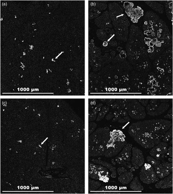
Photomicrographs were taken with a Zeiss Imager M2 epifluorescence microscope (Carl Zeiss Microscopy, Jena, Germany) using a 10× objective and AxioCam HR camera with a Zeiss piezo automated stage. To describe morphology of the pancreas, the mosaic image of a large tissue area of ~144 pictures (12×12 pictures) on the slide was taken using the MosaiX module of Zeiss AxioVision software (Carl Zeiss Microscopy). This method allowed creation of a single image covering all the pancreatic tissue present on the histological slide. The MosaiX images were then analyzed using the ImagePro Premier software (ImagePro Premier 9.0; Media Cybernetics, Silver Spring, MD, USA) for the insulin-positive tissue area per section of tissue and size of insulin-containing clusters. The images were analyzed by creating regions of interest corresponding to the whole area of pancreatic tissue visible on the picture. Within the regions of interest, images were segmented based on positive insulin staining and measurements of total positive insulin staining within pancreatic tissue expressed as percent as well as of individual insulin-containing clusters (Figure 1). Insulin-containing clusters were further classified based on size in order to perform population density measurements of small clusters (1 to 2 000 μm2), medium clusters (2 001 to 32 000 μm2), large clusters (32 001 to 512 000 μm2) and giant clusters (⩾512 000 μm2). Insulin-positive cell clusters do not correspond to islets of Langerhans because not every cell in the islet is insulin positive.
Influence of nutrient restriction and melatonin supplementation on insulin-positive staining. Sections represent the maternal and corresponding fetal pancreatic tissues of representative ewes fed 5 mg/day melatonin and receiving 100% of nutrient requirements (a, b) and 60% of nutrient requirements (c, d). The white arrows indicate insulin-containing clusters. Magnification 10×.

Insulin analysis of blood plasma
Plasma insulin concentrations were measured using an insulin RIA kit (MP Biomedicals, LLC, Santa Ana, CA, USA). The intra-assay CV was 6.99%.
Statistical analysis
Data were analyzed as a completely randomized design with a 2×2 factorial arrangement of treatments using the MIXED procedure of SAS (SAS software version 9.2; SAS Institute Inc., Cary, NC, USA). The model statement included: melatonin treatment, plane of nutrition and their interaction. The model statement also initially included fetal sex and breeding date that ranged from early September to late December but were later removed from the model as they were found not to influence (P>0.25) the measured variables. To determine if there were differences in enzyme activity and islet morphology between ewes and fetuses, data were analyzed using the MIXED procedure of SAS and physiological stage (maternal or fetal), melatonin treatment, plane of nutrition and the interactions between all factors were included in the model. Statistical significance was declared at P<0.05. Tendencies were declared when 0.05⩾P⩽0.10.
Results
There were no nutrient restriction×melatonin supplementation interactions in maternal or fetal data (P⩾0.05). Consequently, the main effects of nutrient restriction and melatonin supplementation are discussed.
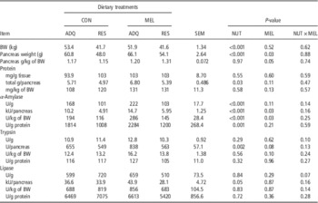
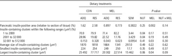
As expected, and previously reported (Lemley et al., Reference Lemley, Meyer, Camacho, Neville, Newman, Caton and Vonnahme2012), nutrient restriction decreased (P<0.001) maternal BW (Table 1). Nutrient restriction decreased (P⩽0.001) maternal pancreas weight (g) and melatonin supplementation increased (P=0.03) maternal pancreas weight (g) with a tendency (P=0.05) for an increase in pancreas weight when expressed as g/kg BW. Total protein content (g) in the maternal pancreas was decreased (P⩽0.03) by nutrient restriction and unaltered by melatonin. Maternal pancreatic α-amylase activity (U/g, U/g protein, kU/pancreas and U/kg of BW) was decreased (P⩽0.001) by nutrient restriction and increased (kU/pancreas and U/kg BW; P⩽0.03) by melatonin supplementation. Nutrient restriction decreased (P=0.002) maternal pancreatic trypsin activity (U/pancreas), while melatonin tended (P⩽0.10) to increase maternal pancreatic trypsin activity when expressed as U/pancreas and U/kg BW. No differences were observed in maternal pancreatic lipase activity in response to nutrient restriction or melatonin supplementation; however, nutrient restriction tended (P=0.05) to decrease lipase activity when expressed as kU/pancreas. Melatonin supplementation decreased (P=0.002) pancreatic insulin-positive tissue area (relative to section of tissue: Table 2). Ewes consuming 60% of requirements had an increased percentage of small insulin-containing cell clusters (1 to 2 000 µm2) while those receiving adequate nutrition had a greater percentage medium insulin-containing cell clusters (2 000 to 32 000 µm2). The percentage of large insulin-containing cell clusters was reduced (P=0.03) in ewes supplemented with melatonin. Restricted ewes had a tendency for the largest insulin-containing cell cluster (P=0.06) while supplementation with melatonin reduced the size of the largest insulin-containing cell cluster (µm2; P=0.04). There was a tendency (P=0.09) for an interaction between intake and melatonin supplementation for the largest insulin-containing cell cluster.
Influence of nutrient restriction and melatonin supplementation on maternal BW, pancreas weight and pancreatic enzymes at 130 days of gestationFootnote 1

CON=control; MEL=melatonin; ADQ=adequate; RES=restricted; NUT=nutrition.
1 Data are presented as least square means per treatment±SEM, n=15 or 16.
Influence of nutrient restriction and melatonin supplementation on insulin concentration and measurements of insulin-containing clusters in maternal pancreas at 130 days of gestationFootnote 1

CON=control; MEL=melatonin; ADQ=adequate; RES=restricted; NUT=nutrition.
1 Data are presented as least square means per treatment±SEM, n=15 or 16.
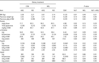
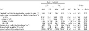
As previously described, fetal BW decreased (P<0.01) with nutrient restriction but was not influenced by melatonin supplementation (Lemley et al., Reference Lemley, Meyer, Camacho, Neville, Newman, Caton and Vonnahme2012; Table 3). Fetal pancreatic weight, protein concentration, and α-amylase, trypsin and lipase activity were not influenced by nutrient restriction or melatonin supplementation. Nutrient restriction decreased (P⩽0.04) fetal pancreatic insulin-positive area (relative to section of tissue), along with the percentage of large and giant insulin-containing clusters (Table 4). The average size of the cell clusters was reduced (P⩽0.04) in restricted animals and the size of the largest insulin-containing cell cluster was smaller.
Influence of nutrient restriction and melatonin supplementation on fetal BW, pancreas weight and pancreatic enzymes at 130 days of gestationFootnote 1

CON=control; MEL=melatonin; ADQ=adequate; RES=restricted; NUT=nutrition.
1 Data are presented as least square means per treatment±SEM, n=15 or 16.
Influence of nutrient restriction and melatonin supplementation on insulin concentration and measurements of insulin-containing clusters in fetal pancreas at 130 days of gestationFootnote 1

CON=control; MEL=melatonin; ADQ=adequate; RES=restricted; NUT=nutrition.
1 Data are presented as least square means per treatment±SEM, n=15 or 16.
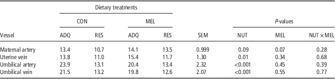
Insulin concentrations in the maternal artery at day 130 of gestation tended (P=0.09) to decrease with nutrient restriction and increased (P=0.07) with melatonin supplementation (uIU/ml; Table 5). Plasma insulin concentration decreased (P=0.01) in the uterine vein of nutrient restricted ewes. Likewise, insulin concentrations in the umbilical artery and vein also decreased (P<0.001) with nutrient restriction and was unaltered by melatonin supplementation.
Influence of nutrient restriction and melatonin supplementation on plasma insulin concentrations (uIU/ml) at 130 days of gestationFootnote 1

CON=control; MEL=melatonin; ADQ=adequate; RES=restricted; NUT=nutrition.
1 Data are presented as least square means per treatment±SEM, n=15 or 16.
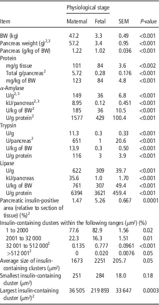
Interactions existed between physiological stage (maternal v. fetal) and intake (P<0.05) for pancreas weight, protein content (total g/pancreas), α-amylase concentration (U/g and U/g protein), α-amylase content (kU/pancreas and U/kg BW) and trypsin content (U/pancreas). Physiological stage×melatonin interactions were also observed (P<0.05) for pancreas weight, and α-amylase concentration (U/g) and content (kU/pancreas). However, interactions occurred primarily because of differences in magnitude between maternal and fetal pancreas with intake or melatonin group and not because of re-ranking between maternal and fetal measurements. Consequently, only the main effects associated with maternal and fetal pancreatic outcomes are presented (Table 6). Pancreatic mass and all measurements of enzyme activity were greater (P<0.02) in maternal than fetal pancreas at day 130 of gestation (Table 6). Fetal pancreas had a larger (P<0.001) insulin-positive area (relative to section of tissue) and a greater percentage of small insulin-containing cell clusters (P=0.02). Maternal pancreas had a greater number of medium insulin-containing clusters (P=0.01) while the fetal pancreas had a greater percent of large insulin-containing cell clusters (P<0.001). A tendency (P=0.05) was observed for fetal pancreas to have a higher percent of giant insulin-containing cell clusters and for the average size of cell clusters to be larger. However, due to the high number of small cell clusters, the average size among fetal and maternal pancreases was not significantly different despite the fetal pancreas exhibiting the largest insulin-containing cell clusters (µm2; P<0.001).
Influence of physiological stage (maternal v. fetal) on digestive enzyme activity and ratio of insulin-positive staining and islet size at 130 days of gestationFootnote 1

1 Data are presented as least square means±SEM, n=31.
2 Physiological stage×intake (P<0.05).
3 Physiological stage×melatonin (P<0.05).
P-values are representative of significance due to physiologic stage.
Discussion
Nutrient restriction from mid- to late-pregnancy decreased both maternal and fetal BW as reported previously (Lemley et al., Reference Lemley, Meyer, Camacho, Neville, Newman, Caton and Vonnahme2012) as well as maternal pancreatic mass (g). However, fetal pancreatic mass was not influenced by nutrient restriction, contradicting past research (Reed et al., Reference Reed, Ward, Vonnahme, Neville, Julius, Borowicz, Taylor, Redmer, Grazul-Bilska, Reynolds and Caton2007), which reported that fetal pancreatic mass from undernourished ewes was decreased compared with those from ewes adequately fed. Moreover, the melatonin supplementation strategy did not rescue fetal growth restriction brought about by maternal nutrient restriction from mid- to late-pregnancy (Lemley et al., Reference Lemley, Meyer, Camacho, Neville, Newman, Caton and Vonnahme2012).
In the growing ruminant, an increase in nutrient intake generally increases pancreatic content of or secretion of exocrine digestive enzymes (Corring, Reference Corring1980; Harmon, Reference Harmon1992; Wang et al., Reference Wang, Ogawa, Suda, Taniguchi, Uike, Kumagai and Mitani1998; Swanson and Harmon, Reference Swanson and Harmon2002). The ewes in this study on the ADQ treatment had increased pancreatic α-amylase and trypsin activities compared with ewes on the RES treatment indicating that changes in feed intake also impact pancreatic exocrine function in mature pregnant ewes.
A study conducted by Jaworek et al. (Reference Jaworek, Katarzyna, Konturek, Leja-Szpak, Thor and Pawlik2004) reported that, in anaesthetized rats, exogenous melatonin or its precursor l-tryptophan resulted in a dose-dependent increase in pancreatic α-amylase secretion. These increases were believed to be manifested through the stimulation of cholecystokinin and the triggering of vagal sensory nerves (Jaworek et al., Reference Jaworek, Katarzyna, Konturek, Leja-Szpak, Thor and Pawlik2004). We also observed this effect of melatonin on maternal pancreatic α-amylase content relative to BW but did not observe effects of melatonin on fetal pancreatic weight or enzyme activity. This, therefore, disagrees with our hypothesis as melatonin supplementation had limited effects on the fetal development of the exocrine pancreas in our study.
Past work has suggested that diets containing lower protein concentration than those with adequate nutrition resulted in decreased pancreatic insulin concentration (Dahri et al., Reference Dahri, Reusen, Remacle and Hoet1995). Other studies have suggested that the β-cell proportional area (relative to total cellular area) and islet number was decreased in rats undergoing nutrient restriction for a period of 4 weeks when compared with a control group fed for ad libitum intake (Chen et al., Reference Chen, Yang, Zheng, Hu, Kong and Zhang2010). Our results suggested no effect of global nutrient restriction on insulin-positive area (relative to section of tissue) in ewes. As expected, however, the insulin-positive area and size of insulin-containing clusters were reduced in fetuses from restricted ewes. Developmental changes in islet size and number potentially could have effects on development in regards to energetics and function of β-cells. When investigating islet size of the endocrine pancreas, pups borne from mothers fed low protein diets were found to have islet cell proliferation reduced by 12% (Dahri et al., Reference Dahri, Reusen, Remacle and Hoet1995).
Plasma insulin concentrations were greater in ADQ ewes and fetuses. This was not surprising as insulin is released in response to nutrient intake (Takahashi et al., Reference Takahashi, Kurose, Kobayashi, Sugino, Kojima, Kangawa, Hasegawa and Terashima2006). Because these animals were not subjected to nutrient restriction they would have been secreting insulin at a higher rate to accommodate the rise in blood glucose and to promote protein synthesis. Although no significant differences were observed, there were numerically greater insulin concentrations in the maternal artery in ewes supplemented with melatonin. This trend is unexpected as melatonin supplementation resulted in a decrease in pancreatic insulin-positive area (relative to section of tissue). Interestingly, Lemley et al. (Reference Lemley, Meyer, Camacho, Neville, Newman, Caton and Vonnahme2012) reported an interaction between nutrient restriction and melatonin supplementation on umbilical artery glucose concentrations suggesting that glucose concentrations were influenced differently compared with plasma insulin concentration and islet size measurements. Melatonin, on the other hand, did not influence the insulin-positive area (relative to section of tissue) or insulin-containing cluster morphology in the fetal pancreas, suggesting that supplemental melatonin at the dose and timing relative to the light–dark cycle that we provided had minimal effects on pancreatic fetal endocrine function.
The concentration of pancreatic digestive enzymes was much lower in the fetal than the maternal pancreas. However, when observing the size of insulin containing clusters, fetuses had a greater number of large islets compared with those of their respective dams amongst all treatments (Table 6). Similar results have also been observed when comparing calves to adult cattle (Merkwitz et al., Reference Merkwitz, Lochhead, Böttger, Matz-Soja, Sakurai, Gebhardt and Ricken2012). In the examination of the pancreatic tissue of calves, islets of two differing sizes were discovered. The larger islets (perilobular giant islets) were found in a much smaller quantity while smaller islets (interlobular islets) appeared more frequently. These interlobular islets were shown to persist into adulthood while the perilobular giant islets underwent regression. It has also been shown that mass related metabolic rate tended to decrease with increasing body size of the organism (Singer, Reference Singer2006). Therefore, these differences in fetal islet size and number may be associated with differences in function of the endocrine pancreas during development and later in life. Further investigation is needed in this area.
In conclusion, nutrient restriction during gestation has more of an effect on pancreatic protein and digestive enzyme concentrations in the dam than in the fetus. This may be because the dam is adapting to protect the fetus from changes in nutritional status or because of the much lower concentrations of enzymes produced in the fetal compared with the maternal pancreas. In contrast to our hypothesis, melatonin supplementation did not impact the effect of nutrient restriction on maternal and fetal pancreatic function and therefore may not be an appropriate supplementation option to help therapeutically mitigate the effects of maternal nutrient restriction on pancreatic development.
Acknowledgments
The authors would like to thank the employees of the NDSU Animal Nutrition and Physiology Center and students and staff who assisted with this project. In addition, the authors would like to thank Ellen Kuemper for assistance with sample collection and analysis. This project was partially supported by a Postdoctoral Basic Research Fellowship grant from the Lalor Foundation, Inc. and USDA Postdoctoral Fellowship grant no. 2011-67012-30683 from the National Institute of Food and Agriculture to C. O. Lemley.