Introduction
Much of the early history of fossil therapsids (the lineage that includes mammals) is recorded in the rocks of the Karoo Basin, South Africa (Rubidge, Reference Rubidge1995; Rubidge and Sidor, Reference Rubidge and Sidor2001; Rubidge, Reference Rubidge2005). The continuous record of therapsids in Middle Permian through Middle Triassic strata has allowed the establishment of eight faunal assemblage zones in the Beaufort Group (Karoo Supergroup), six of them representing Permian and two Triassic assemblages. This succession presents one of few available windows into the transition between Permian and Triassic terrestrial ecosystems. This transition is of enormous importance because it documents the worst catastrophe in earth history: the end-Permian extinction, a global biotic crisis characterized by significant biodiversity loss and reduced ecosystem complexity in both marine and terrestrial systems (Ward et al., Reference Ward, Botha, Buick, Dekock, Erwin, Garrison, Kirschvink and Smith2005; Smith and Botha, Reference Smith and Botha2005; Botha and Smith, Reference Botha and Smith2006; Smith and Botha-Brink, Reference Smith and Botha-Brink2014).
The nonmammalian therapsids were a major component of Permo-Triassic terrestrial communities, represented by a variety of lineages that together constituted much of amniote diversity during this time. Among these lineages, the therocephalians, a speciose and ecomorphologically varied group, first diversified during the Middle Permian as with most other major therapsid clades (Abdala et al., Reference Abdala, Rubidge and van den Heever2008). This group has traditionally been recognized as monophyletic by a variety of authors (e.g. Romer, Reference Romer1969; Hopson and Barghusen, Reference Hopson and Barghusen1986; van den Heever, Reference Heever1994), although non-cladistic anatomical investigations and more recent cladistically informed studies have suggested that cynodonts may share a sister group relationship with the whaitsiid Theriognathus, the subject of the present study (Kemp, Reference Kemp1972a; Abdala, Reference Abdala2007; Botha et al., Reference Botha, Abdala and Smith2007). The latter hypothesis is significant because it suggests that cynodonts represent a subgroup of a paraphyletic Therocephalia, and that the origins of cynodont morphological structure may therefore be found among the early Therocephalia. However, new phylogenetic investigations by Huttenlocker and colleagues including large numbers of therocephalian specimens have offered robust morphological support for the traditional view of a monophyletic Therocephalia (Huttenlocker, Reference Huttenlocker2009; Huttenlocker et al, Reference Huttenlocker, Sidor and Smith2011; Sigurdsen et al., Reference Sigurdsen, Huttenlocker, Modesto, Rowe and Damiani2012; Huttenlocker, Reference Huttenlocker2013). Detailed anatomical reassessments and investigations of morphological variation in well-represented taxa such as Theriognathus are therefore of great importance in understanding functional homology and the order of character evolution in mammal-line amniotes.
Theriognathus was a large, predatory therocephalian and was one of the most abundant representatives of the group in Late Permian faunas of South Africa. Its first occurrence is in the Cistecephalus Assemblage Zone (AZ) of the western Karoo Basin in South Africa, although it was more common in the lower portion of the Dicynodon AZ and may have been a victim of the end-Permian extinction (Smith and Botha, Reference Smith and Botha2005; Botha and Smith, Reference Botha and Smith2006). As in the case of other abundant therapsid taxa from Late Permian faunas of South Africa (e.g., the cynodont Procynosuchus), Theriognathus is also known from faunas outside the Karoo, such as those of Tanzania (von Huene, Reference Huene1950; Kemp, Reference Kemp1972a,Reference Kempb; Maisch, Reference Maisch2002; Weide et al., Reference Weide, Sidor, Angielczyk and Smith2009; Sidor et al., Reference Sidor, Angielczyk, Weide, Smith, Nesbitt and Tsuji2010) and Zambia (Sidor et al., Reference Sidor, Vilhena, Angielczyk, Huttenlocker, Nesbitt, Peecook, Steyer, Smith and Tsuji2013). Given its abundance and widespread occurrence in southern Africa (Fig. 1), Theriognathus may be a useful biostratigraphic marker for Late Permian faunal assemblages, and such studies would benefit from a redescription and understanding of its morphological variation, species composition, and review of its known occurrences.
Stratigraphic and geographic distribution of Theriognathus in southern Africa. Principal Theriognathus-bearing localities are arranged along strike between the Late Permian uppermost Cistecephalus (light gray) and Dicynodon (medium gray) Assemblage Zones (AZs) in the Karoo Basin, and are found in equivalent deposits in Tanzania and Zambia. Abbreviations: Chx=Changhsingian, Cisteceph AZ=Cistecephalus AZ, Cyn sub A=Cynognathus AZ subzone A, Dicyno AZ=Dicynodon AZ, Lystro AZ=Lystrosaurus AZ, Ma=million years ago, Olen=Olenekian, Prist AZ=Pristerognathus AZ, Trop AZ=Tropidostoma AZ.

The first discovery of a whaitsiid specimen was based on a partial skull missing the anterior portion of the snout (NHMUK 47065; Fig. 2.1) from the Sneeuberge range near Graaff-Reinet, South Africa, and donated to the Natural History Museum in London by A. G. Bain. The specimen was described and illustrated in Owen’s (Reference Owen1876) “Catalogue of the Fossil Reptiles of South Africa in the Collections of the British Museum” where it was assigned to a new genus and species, Theriognathus microps, although it was originally identified as an anomodont due in part to its lack of postcanine teeth. The specimen was later recognized as a therocephalian by Broom (Reference Broom1910) although new specimens refining its affinities within Therocephalia would not be discovered until several years later.
Photographic plates of selected type specimens. (1) Theriognathus microps Owen (NHMUK 47065) in ventral view. (2) Whaitsia platyceps Haughton (SAM-PK-4006) in ventral (left) and lateral (right) views. (3) Whaitsia majora Broom (NHMUK R5694) in ventrolateral view, showing lower mandibles in articulation. (4) Moschorhynchus latirostris Broom (TM 264) in ventral (left) and dorsal (right) views. Abbreviations: cr. cho.=crista choanalis, ept=epipterygoid, f.-n. cr.=median frontonasal ridge/crest, m.-v. br.=maxillo-vomerine bridge, pc. r.=postcanine ridge, pt. tr. pr.=transverse process of pterygoid, V=exit for trigeminal nerve.
A prolific period of expansion of whaitsiid taxonomy followed the description of Whaitsia platyceps (Fig. 2.2) by Haughton (Reference Haughton1918), and especially after several contributions by Broom (Reference Broom1920, Reference Broom1925, Reference Broom1931, Reference Broom1932, Reference Broom1935, Reference Broom1936; Broom and Robinson, Reference Broom and Robinson1948). Haughton (Reference Haughton1918) erected the family name Whaitsiidae for the new taxon. Broom (Reference Broom1920) named Alopecopsis atavus, based on a partial skull that also lacked postcanines. He recognized the new taxon as a therocephalian, but did not discuss its precise relationships to other therocephalians. In the same contribution, Broom also discussed and illustrated the palate of the earlier described Whaitsia platyceps, which he also considered a therocephalian. Broom (Reference Broom1925) later gave a brief description (without illustration) of the skull of Notosollasia laticeps, which he proposed to be “nearly related to Whaitsia platyceps” (Broom, Reference Broom1925: p. 322). Additional specimens were described in the decade that followed (Broom, Reference Broom1930). In another short description without illustration, Broom (Reference Broom1931) erected the new species Whaitsia major (sic) (Fig. 2.3) based on five skulls, including a particularly large specimen. Broom distinguished W. major from other whaitsiids based on very few proportional differences, noting “the dentary is less curved and rather stronger than in W. platyceps, and the back part differently shaped” (Broom, Reference Broom1932: p. 86). Boonstra (Reference Boonstra1934) provided an important account of specimens within the Natural History Museum that included full preparation and redescription of the holotype and only specimen of Theriognathus microps. Comparisons to similar taxa represented in the collection that lacked postcanine teeth, including Notosollasia laticeps and Whaitsia majora, facilitated Boonstra’s recognition of their familial affinities. Broom (Reference Broom1935) described and named a partial skull of another whaitsiid having a nearly complete lower jaw as Hyenosaurus platyceps, as well as three new forms the following year (Broom, Reference Broom1936): Moschorhynchus latirostris (a skull lacking the mandible; Fig. 2.4), Notosollasia luckhoffi (represented by the anterior portion of the skull; Fig. 3.1), and Notaelurops paucidens (a skull with a poorly preserved mandible; Fig. 3.2). Broom and Robinson (Reference Broom and Robinson1948) erected two new whaitsiid taxa based on the relatively small sizes of the specimens: Whaitsia pricei (Fig. 3.3) and Aneugomphius ictidoceps, the latter specimen representing the smallest known whaitsiid discovered to date (Figs. 3.4, 4.1). Watson and Romer (Reference Watson and Romer1956) suggested that the latter specimen represented a juvenile of the former. However, in his redescription of the skull, Brink stated that Aneugomphius “is not a juvenile specimen” (1956: p. 111) and that it instead represented a distinct therocephalian taxon of unknown affinities.
Photographic plates of selected type specimens (continued). (1) Notosollasia luckhoffi Broom (RC 108/L2) in ventral view. (2) Notaelurops paucidens Broom (TM 280) in dorsal (left) and lateral (right) views. (3) Whaitsia pricei Broom and Robinson (BP/1/164) in dorsal (left) and lateral (right) views. (4) Aneugomphius ictidoceps Broom and Robinson (BP/1/182) in ventral (left) and lateral (right) views. Abbreviations: cr. cho.=crista choanalis, ept=epipterygoid, f.-n. cr.=median frontonasal ridge/crest, m.-v. br.=maxillo-vomerine bridge, pc. r.=postcanine ridge, pt. b.=pterygoid boss, pt. tr. pr.=transverse process of pterygoid.
Huene (Reference Huene1950) provided a major contribution when he documented the occurrence of whaitsiids outside of South Africa for the first time, based on two specimens he identified as Notosollasia and Notaelurops from Permian-aged rocks of the Ruhuhu Basin of Tanzania. Proalopecopsis haughtoni was described by Brink and Kitching (Reference Brink and Kitching1951) based on an almost complete skull and dentition, including the mandible, from South Africa. Though presently not considered a whaitsiid, the skull morphology of this species bore superficial resemblance to Alopecopsis atavus from the perspective of the original authors, despite the fact that P. haughtoni featured six to seven “apparently degenerate molars” (Brink and Kitching: Reference Brink and Kitching1951: p. 1222). Brink and Kitching (Reference Brink and Kitching1951) speculated that postcanine presence could be an ontogenetic feature of young individuals, which subsequently lost their teeth with age. Brink (Reference Brink1954) built on this earlier work naming several new whaitsiids, including Moschorhynchus brachyrhinus, M. gracilis, Notosollasia boonstrai, and Noto. longiceps. At this stage and as in the case of P. haughtoni, Brink (Reference Brink1954) also included within Whaitsiidae taxa having postcanine teeth that are now placed amongst Akidnognathidae (e.g., Promoschorhynchus and Moschorhinus Broom).
Mendrez (Reference Mendrez1974) removed taxa such as Proalopecopsis, Promoschorhynchus, and Moschorhinus from Whaitsiidae, recognizing akidnognathids and whaitsiids as two distinct groups (the latter group being monotypic, consisting only of T. microps Owen). Although Mendrez’s revision was not formally published, she proposed extensive synonymies between T. microps and all African whaitsiids lacking postcanines and having an incipient secondary palate, including the genera Whaitsia, Alopecopsis, Notosollasia, Hyenosaurus, Notaelurops, Aneugomphius, and Moschorhynchus. In a reversal of his previous interpretation of Aneugomphius, Brink (Reference Brink1980) adopted this revision, but recognized four morphotypes within the genus Theriognathus (named as ‘Whaitsia,’ ‘Notosollasia,’ ‘Moschorhynchus,’ and ‘Aneugomphius’ morphotypes). He suggested that these types differed quantitatively in their overall size and proportions, and grouped them into four distinct species: T. platyceps, T. microps, T. latirostris, and T. pricei. More recent reports and applications to biostratigraphic studies have not treated Theriognathus to the species level, with records often documented simply as Theriognathus or Theriognathus sp. (e.g., Rubidge, Reference Rubidge1995; Ward et al., Reference Ward, Botha, Buick, Dekock, Erwin, Garrison, Kirschvink and Smith2005).
Here, we present a taxonomic revision of Theriognathus from the uppermost Cistecephalus and Dicynodon AZs of South Africa, including a detailed description based on nearly all specimens in existing collections. We also present a quantitative analysis of cranial allometry in the genus using traditional morphometrics with the purpose of corroborating the species composition of Theriognathus, as well as size-related trends in the skull of this and other eutheriodont therapsids (i.e., therocephalians and cynodonts). We suggest that the studied materials represent a single allometric series referable to Theriognathus microps Owen, making this early-described taxon one of the most abundant theriodont predators of the upper Cistecephalus and Dicynodon AZs, and highlighting its applications both for biostratigraphy and for understanding cranial development and evolution (particularly of the jaw suspensorium) in Late Permian theriodonts.
Materials and methods
To assess qualitative and allometric cranial variation in African whaitsiids referred to Theriognathus, we examined more than 70 partial and complete skulls across a variety of sizes (basal skull length 67 to 336 mm; Fig. 4). To our knowledge, these included all available skulls representing the genus (Appendix).
Skull size ranges (1) and measurements (2–4) used in the allometric analysis. Values for (1) are the basal skull lengths (BSL) of the largest (BP/1/4008, left) and smallest (BP/1/182, right) specimens. Skull drawings are presented in ventral (2), dorsal (3), and left lateral (4) views. Abbreviations: BCW=bicanine width, IOB=interorbital breadth, BSL=basal skull length, NAL=nasal length, SOB=breadth of skull at suborbital bar, MZB=maximum breadth at zygoma, PCL=parietal crest length, CCL=crista choanalis length, PAB=palatal breadth (measured at the level of the postcanine ridge), MRH=maximal rostral height (measured at level of canine), OL=orbit length, TL=temporal fenestra maximum length (measured on the diagonal), EPH=epipterygoid height, EPDL=epipterygoid maximum length of dorsal region, EPML=epipterygoid length at midpoint constriction, DEL=dentary length, RLL=reflected lamina length, ML=mandible total length.
Allometric analysis
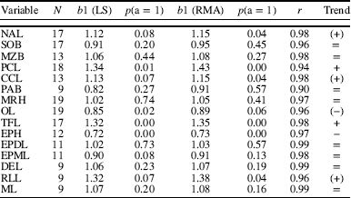
Complete skulls representing a wide range of ontogenetic stages and exhibiting satisfactory preservation for precise cranial measurements were chosen for the allometric analysis (Supplemental Data). These included 19 specimens out of our total studied sample (see Supplemental Data). A series of measurements representing length, width and height of the skull components were considered for a bivariate analysis of allometry (Fig. 4; Table 1). Measurements were taken to the nearest millimeter using Mitutoyo digital calipers. Of the 17 dependent variables originally measured we selected those having a minimum of nine values and also represented in the smallest specimen of the sample (i.e., Aneugomphius ictidoceps holotype; BP/1/182). The total variables represented following these requisites were 15 (see Supplemental Data). Basal skull length was selected as an estimator of overall size (Emerson and Bramble, Reference Emerson and Bramble1993). Estimation of variation in size of each skull measurement was made through linear (log10) transformation of the exponential growth equation [y=x b1b 0e], where y is the variable of interest, b 0 the y-intercept, x the basal skull length, b 1 the coefficient of allometry, and e the error term (Simpson et al., Reference Simpson, Roe and Lewontin2003). Two models were used to obtain the coefficients of allometry: 1) least squares (LS) and 2) reduced major axis (RMA) regressions (Abdala and Giannini, Reference Abdala and Giannini2000; Flores et al., Reference Flores, Abdala and Giannini2010). RMA assumes error in both the dependent and independent variables, a more realistic assumption as all the variables are measured with error. Departures from isometry (b 1=1) were estimated by two-tailed t-tests. If b 1 was significantly less than 1.0 for both models, then a negative allometric trend was assumed with respect to skull length. If b 1 was significantly greater than 1.0, then positive allometry was assumed. Trends were considered weakly supported if they were only significant for one of the models. The ‘model/linear’ menu from the program PAST (Palaeontological Statistics) version 2.12 (Hammer et al., Reference Hammer, Harper and Ryan2001; Hammer and Harper, Reference Hammer and Harper2005) was used to produce the bivariate results. The results of these tests are presented in Table 1.
Bivariate analysis of Theriognathus microps by least square and reduced major axis methods
See Figure 4 for variable acronyms.
b1, coeffeicient of allometry; +, positive allometry, −, negative allometry;=, isometry
Trend in parentheses is significant under one method but not significant under the other.
Institutional abbreviations
AM, Albany Museum, Grahamstown; AMNH, American Museum of Natural History, New York; BP, Evolutionary Studies Institute (former Bernard Price Institute for Palaeontological Research), Johannesburg; CG, Council for Geoscience, Pretoria; GPIT, paleontological collection of Tübingen University; NHCC, National Heritage Conservation Commission collections, Lusaka; NHMUK, Natural History Museum, London; NMQR, National Museum, Bloemfontein; NMT, National Museum of Tanzania, Dar es Salaam; RC, Rubidge Collection, Wellwood; SAM, Iziko: South African Museum, Cape Town; TM, Ditsong National Museum of Natural History (former Transvaal Museum), Pretoria; UCM, University of Colorado Museum of Natural History, Boulder; UCMP, University of California Museum of Paleontology, Berkeley; UMZC, University Museum of Zoology, Cambridge; USNM, National Museum of Natural History, Washington, D.C.
Systematic paleontology
Therapsida Broom, 1905
Eutheriodontia Hopson and Barghusen, Reference Hopson and Barghusen1986
Therocephalia Broom, 1903
Family Whaitsiidae Haughton, Reference Haughton1918
Genus Theriognathus Owen, Reference Owen1876
Theriognathus microps Owen, Reference Owen1876
1876 Theriognathus microps Owen
1890 ‘Endothiodon’ microps Lydekker
1918 Whaitsia platyceps Haughton
1920 Alopecopsis atavus Broom
1925 Notosollasia laticeps Broom
1931 Whaitsia major Broom
1932 Whaitsia majora (Broom)
1935 Hyenosaurus platyceps Broom
1936 Moschorhynchus latirostris Broom
1936 Notosollasia luckhoffi Broom
1936 Notaelurops paucidens Broom
1948 Whaitsia pricei Broom and Robinson
1948 Aneugomphius ictidoceps Broom and Robinson
1954 Moschorhynchus brachyrhinus Brink
1954 Moschorhynchus gracilis Brink
1954 Notosollasia boonstrai Brink
1954 Notosollasia longiceps Brink
Holotype
NHMUK 47065 (Fig. 2.1), partial skull and lower jaw from Stylkrans, Nieu Bethesda (Graaff-Reinet District), Eastern Cape Province, Republic of South Africa (RSA); original stratigraphic provenance documented as “Cistecephalus beds” of the Karoo Basin, although Kitching (Reference Kitching1977: pp. 67, 83) reported exposures of both Cistecephalus and Dicynodon AZs at this locality with whaitsiid occurrences restricted to the latter unit.
Diagnosis (revised)
Medium to large therocephalian with five upper and four lower incisors, a single upper and lower canine (a single deciduous precanine may be present in subadults), and complete absence of upper and lower postcanines; postcanine alveolar margin replaced by prominent maxillary ridge (or ‘postcanine crest’) located lateral to and parallel with crista choanalis; ventral maxillary groove present on postcanine crest immediately posterior to canine and confluent with canine alveolus; suborbital vacuities extremely reduced or absent in large individuals; interpterygoid vacuity extremely narrow or reduced in large individuals; pineal foramen extremely reduced or absent in large individuals; expanded epipterygoid with well-ossified, up-turned posterior apophysis contributing to a distinct trigeminal foramen superficially resembling that of cynodonts; anterior border of pterygo-paroccipital foramen formed almost exclusively by squamosal.
Occurrence
Occurs in both uppermost Cistecephalus AZ and lower to middle levels of the Dicynodon AZ (Upper Permian, late Wuchiapingian) throughout the Graaff-Reinet (Eastern Cape Province) and Murraysburg (Western Cape Province) districts and, less frequently, in the Free State and Northern Cape provinces, Karoo Basin, RSA. Occurrences outside the Karoo Basin have been documented in the Usili Formation (Ruhuhu Basin, Tanzania) and upper Madumabisa Mudstone Formation (Luangwa Basin, Zambia), and are interpreted as Cistecephalus AZ-equivalent (von Huene, Reference Huene1950; Kemp, Reference Kemp1972a, Reference Kempb; Maisch, Reference Maisch2002; Weide et al., Reference Weide, Sidor, Angielczyk and Smith2009; Sidor et al., Reference Sidor, Angielczyk, Weide, Smith, Nesbitt and Tsuji2010, Reference Sidor, Vilhena, Angielczyk, Huttenlocker, Nesbitt, Peecook, Steyer, Smith and Tsuji2013). See Figure 1 and Appendix 1 for detailed provenance data.
Description
Skull roof
The premaxilla is a robust, tripartite element with distinct maxillary, vomerine, and dorsal processes. The dorsal process is tall with a long posterior arch that wedges strongly between the nasals and anterolaterally facing external nares. The rostral surface bears one or two small (paired) anterior premaxillary foramina (Fig. 7.1), and a small nervous foramen that is located on the suture between the premaxilla and maxilla. On the palatal surface, five alveoli are present for the deeply rooted incisors. Immediately posterior to the first incisor alveolus is another small (paired) posterior premaxillary foramen and a median palatal foramen located on the midline suture of the paired vomerine processes. The vomerine process is long and forms much of the medial wall of the anterior portion of the internal naris (i.e., the lower canine fossa). The vomerine process does not extend posteriorly to the maxillary palatal process (unlike some derived baurioids), allowing the vomer to participate in the posteromedial border of the internal naris.
The septomaxilla has a broad facial exposure anterolaterally (similar to akidnognathids), but with a restricted footplate having a straight (horizontal) suture with the premaxilla on the ventral margin of the naris. The septomaxilla is completely preserved with well-delineated sutures in RC 232, where it preserves an extensive facial exposure and somewhat concave medial process within the naris (Fig. 7.1). There is often a shallow oval fossa present on the posterolateral surface (between the septomaxilla and dorsal lamina of the maxilla) and a foramen situated between the narial portion and the maxilla (‘septomaxillary foramen’).
The maxilla is a robust element with a tall dorsal lamina, strong canine buttress, and a tapering jugal process that extends posteriorly beneath the orbit. The outer surface of the dorsal lamina bears fine pitting with some larger nervous foramina located near the canine buttress, a condition similar to that of other therocephalians. Posterior to the canine buttress is a marked medial constriction of the snout, which rolls gently onto the palatal surface. The smoothly sloping maxilla forms a palatal platform that ends in a marked ridge that extends from behind the canine to near the base of the pterygoid process. This structure allows a substantial portion of the dorsal lamina to remain visible in ventral view, and gives the snout a strongly circular cross-section (a configuration that is shared with other whaitsiids and hofmeyriids).
The canine alveolus is large and usually located anteriorly, although it may be located closer to the transverse midline of the maxilla in smaller (presumably subadult) specimens. The alveolus frequently incorporates spongy alveolar bone that would have surrounded the root of the canine. The alveolus tapers posteriorly, forming a teardrop shape, and continues along the ridged alveolar margin as the postcanine ‘groove.’ There are no postcanine alveoli in the upper jaw. Medially, a sharp crista choanalis is present and extends anteriorly from the level of the canine and continues far posteriorly along the palatine. A small maxillopalatine foramen is present on the maxilla-palatine suture between the postcanine ridge and the crista choanalis. A stout medial process is present anterior to this foramen and medial to the canine alveolus, forming a maxillo-vomerine bridge. This specialized, finger-like process bears a strong suture with distinctive lateral processes of the vomer, separating the internal naris into anterior (lower canine fossa) and posterior (choanal) compartments.
The lacrimal is a delicate element that is positioned on the anterior wall of the orbit. It is interrupted anterodorsally by the tall dorsal lamina of the maxilla and the long anterior process of the prefrontal, and therefore does not contact the nasal on the snout (as in all therocephalians except lycideopid baurioids). The lacrimal canal is located on the posteroventral border of the bone, just inside the anterior wall of the orbit (rather than outside the orbit as it is in some hofmeyriids).
The nasal is a long, quadrangular element that is bordered anteriorly by the premaxilla and external naris, anterolaterally by the septomaxilla, laterally by the dorsal lamina of the maxilla, posterolaterally by the prefrontal, and posteriorly by the frontal. In dorsal view, the element is very slightly waisted near its center, just behind the canine buttress of the maxilla. The midline suture is relatively straight and there is always a shallow buttress located posteriorly on the suture (i.e., the median frontonasal ridge) as in akidnognathids. The frontonasal suture is strongly transverse and highly interdigitated.
The prefrontal is a relatively large element, having a long anterior process bearing broad contact with the nasal (comparable to that which is seen in the akidnognathids Promoschorhynchus and Olivierosuchus). It forms the majority of the anteromedial border of the orbit with an equally long posterior process that nearly contacts the postorbital and leaves only a very narrow contribution of the frontal to the dorsomedial wall of the orbit.
The paired frontals form a broad, pentagonal element that separates the orbits on the skull table. Anteriorly, the frontal forms a strongly interdigitating suture with the nasal and bears the posterior extent of the median frontonasal ridge along the midline. Posteriorly, the frontal tapers to its pinched contact with the parietal where it also forms a narrow interdigitating suture. Laterally, it is extremely restricted in its contribution to the dorsomedial wall of the orbit, due to the well-developed posterior and anterior processes of the prefrontal and postorbital, respectively. As stated above, this condition resembles some hofmeyriids (e.g., Hofmeyria and Mirotenthes) and, to a lesser degree, some early nonmammaliaform cynodonts. However, unlike hofmeyriids and cynodonts, these processes do not come into direct contact to form the dorsomedial wall of the orbit, allowing a small portion of the frontal to be exposed on the orbital rim and continuing ventrally to form the dorsomedial wall of the orbit down to its contact with the orbitosphenoid.
The postorbital is a thin element that borders the posteromedial portion of the orbit. The jugal process, which contributes to the postorbital bar, is fairly robust in adults having a tapering descending branch that forms a scarf joint along the posterior side of the jugal. Posteriorly, the postorbital forms a thin sheet of bone that broadly overlaps the parietal and contributes to the anteromedial border of the temporal fenestra. This short process extends about as far as the level of the pineal enclosure and is widely separated from the squamosal by the parietal.
The jugal is a robust, tripartite bone that frames the suborbital region and cheek. It extends anteriorly just beyond the anterior border of the orbit and pinches out just ventral to the lacrimal. This region is very deep, particularly in large adults, forming a strong suborbital bar. Two very small but distinctive nutrient foramina are present just ventral to the orbit, one contained completely within the jugal and another anterior foramen often situated on or near the jugal-maxilla suture. In adults, the postorbital process is relatively thick at its contact with the postorbital, but may be weaker in smaller subadult specimens (e.g., BP/1/182). Posteriorly, the subtemporal bar is robust and bears a long, arching branch that underlaps the squamosal on the cheek. The subtemporal bar is fairly horizontal but becomes oriented ventrally at its posterior margin contributed by the squamosal.
The squamosal is a complex element forming much of the posterolateral portion of the temporal region, and bracing the suspensorium. A tall dorsomedial lamina broadly overlaps the parietal and contributes to the lambdoidal crest, which borders the occiput. A wide flaring of the posterolateral rim of the temporal fossa forms a strongly triangular skull in dorsal profile. The temporal opening is the largest known in a therocephalian and its wide posterior breadth resembles that of cynognathian cynodonts. Ventrally, a finger-like otic process of the squamosal originates from near the quadrate recess and extends anteromedially to its contact with the prootic (Figs. 7.3, 8). This structure bounds the pterygo-paroccipital foramen and is formed almost exclusively by the squamosal (i.e., lacking any lateral contribution from the prootic, unlike other therocephalians). Posterolaterally, the squamosal accommodates the suspensorium. Its anterior surface bears the quadrate recess, which loosely surrounds the quadrate-quadratojugal complex, and a laterally located ‘squamosal slit’ observable in lateral view. The posterior surface of the squamosal bears a broad, shallow sulcus (sometimes termed ‘external auditory meatus’) just lateral to its contact with the paraoccipital process of the opisthotic.
The parietal is a large sheet-like bone that forms the dorsomedial border of the temporal fenestra and contributes to the dorsolateral walls of the braincase. An anterior midline suture may be found in some specimens, but disappears where the paired left and right parietals fuse along the sagittal crest. The crest is long (at least one-half to two-thirds the length of the temporal fenestra) and high in large specimens. Although an externally expressed parietal foramen is absent in large specimens, a tall, bulbous pineal enclosure marks the position of the pineal gland, approximately one-third of the distance from the anterior to posterior margin of the sagittal crest. This enclosure is visible in ventral view in some specimens preserving the braincase (e.g., SAM-PK-K5512), exposing a tall cavity in which the dorsal pineal body was situated. This enclosure is absent in the Permo-Triassic baurioid Tetracynodon, the ventral surface of the parietal instead being relatively flat and smooth (Sigurdsen et al., Reference Sigurdsen, Huttenlocker, Modesto, Rowe and Damiani2012). The appearance of a small parietal foramen encountered in some large Theriognathus specimens is usually linked to erosion of the dorsal part of the skull roof after fossilization. A small parietal foramen may only be naturally preserved in specimens that were likely immature (e.g., BP/1/182) and the opening is usually medially compressed or oval in shape. The rear portion of the parietal contributes to the lambdoidal crest and the dorsal portion of the occiput. A very extensive occipital exposure of the parietal is preserved in SAM-PK-K5512 (Fig. 8.1, 8.2) as described in Regisaurus (Mendrez, Reference Mendrez1972) and Promoschorhynchus (Huttenlocker et al., Reference Huttenlocker, Sidor and Smith2011), indicating that this character is more widespread in therocephalians than previously recognized.
Braincase and occiput
Elements of the sphenethmoid complex are present in a few of the studied specimens, including BP/1/182, BP/1/655 (preserved in sagittal-section), and UMZC T902. The lateral walls of the complex are divided into a median ventral ossification (‘presphenoid’ of Crompton, Reference Crompton1955; ‘interorbital septum’ of Kemp, Reference Kemp1972a) and an overlying U-shaped orbitosphenoid, which braces directly against the ventral surface of the frontal. These elements are clearly separated by an anterodorsally-oriented fissure in the smallest specimen (BP/1/182), and therefore represent separate ossifications of the neurocranium. A similar U-shaped orbitosphenoid atop an interorbital septum was also recently described in the bauriid Microgomphodon (Abdala et al., Reference Abdala, Jashashvili, Rubidge and van den Heever2014a), whereas a single Y-shaped element was revealed in Tetracynodon by micro-CT imaging (Sigurdsen et al., Reference Sigurdsen, Huttenlocker, Modesto, Rowe and Damiani2012) and was interpreted as forming a unique bone. It is possible that the sphenethmoid of Tetracynodon was similarly divided into two distinct ossifications, but that the micro-CT images available in that study did not provide sufficient resolution to delineate them. Further phylogenetic surveys of the neurocranial elements in therocephalians are needed to resolve this issue.
The anterior (prootic) and posterior (opisthotic) ossifications of the otic capsules are well formed, documenting unique morphology in Theriognathus. The prootic is situated anteromedially to the squamosal and ventral to the interparietal and supraoccipital (Fig. 8.3, 8.4). It has a tall ascending process that is directed anteriorly where it suturally contacts a dorsal flange of the epipterygoid above the trigeminal foramen. The lateral wall of the prootic provides contact for the otic process of the squamosal, but, unlike other therocephalians, does not form lateral processes of its own. As a result, the outer borders of the pterygo-paroccipital foramen are formed exclusively by the squamosal in Theriognathus. The opisthotic is best exposed on the occipital surface where it is seen bordering the post-temporal fenestra. Dorsally, a large flange roofs the post-temporal fenestra and shares a strongly-interdigitating suture with the squamosal. Ventrally, the stout paroccipital process of the opisthotic bears a strongly transverse orientation toward its lateral contact with the squamosal, and bears a shallow depression on its dorsal surface in the floor of the post-temporal fenestra (as in other eutherocephalians).
The para-basisphenoid complex is fairly wide in ventral view where is it situated between the quadrate processes of the pterygoid. Anteriorly it is strongly buttressed against the flanges of the para-basisphenoid process of the pterygoid. A thin sutural connection is present between these two elements, and between them are the paired internal carotid foramina sometimes visible on the ventral surface. A strong ventral-median keel is also present as in other therocephalians. A marked ventral projection, the basal tuber, is present at the posterolateral contact between the para-basisphenoid and basioccipital, a common condition in therocephalians.
The supraoccipital appears as a very shallow and broad sheet-like element on the occipital surface of the skull. Here, it forms the dorsal border of the foramen magnum (Fig. 8.1, 8.2). A portion of the nuchal line is preserved along the midline (continuing dorsally onto the interparietal) and the sutural connections with the tabular and interparietal are extremely sinuous and interdigitating. Anteriorly, the supraoccipital sends an ascending process along the lateral walls of the braincase where it wedges between the interparietal (above) and prootic (below) and sutures anteriorly to the dorsal most portion of the ascending process of the epipterygoid (Fig. 8.3, 8.4).
The interparietal (sometimes termed ‘postparietal’) is a thin, unpaired wing-shaped element that forms much of the dorsal occipital surface. As in other therocephalians, the element is relatively shallow being wider than it is tall. It is bordered dorsally by the parietal, laterally by the paired tabulars, and ventrally by the supraoccipital. Its external surface frequently bears a shallow vertical ridge on the midline that continues from the supraoccipital: the nuchal line. An anterior flange of the interparietal shares a long sutural connection to the underside of the parietal, contributing to the posterolateral walls of the braincase (Fig. 8.1, 8.2).
The paired tabulars rest posterolaterally to the squamosal and bear an overlapping sutural connection with the latter element. Along with the parietal and interparietal, the tabular contributes broadly to the occipital surface. The element is bordered dorsomedially by the parietal, medially by the interparietal, ventrally by the supraoccipital, and laterally by the squamosal. It appears to bear a tall dorsal contribution to the lambdoidal crest (shared with the squamosal), but is limited ventrally, lacking a ventral process that extends around or below the post-temporal fenestra. As a result, the tabular is well retreated from contact with the post-temporal fenestra (Fig. 8.1, 8.2).
The lateral borders of the foramen magnum are formed by the exoccipitals. The paired exoccipitals appear as tripartite or anvil-shaped with an expanded dorsal process and a ventral contribution to the occipital condyle. Ventrolaterally they are pierced by a small hypoglossal foramen.
The basioccipital forms the central (or median) portion of the tripartite occipital condyle (a single, convex rolling surface formed by three separate ossifications in contact with the atlas [C1]). This entire structure is formed from the floor of the neurocranium. Ventrally, it is wide with moderately-sized paired basal tubera.
Palate, splanchnocranium, and suspensorium
The vomer, best observed in palatal view, is an elongated yet broad element with a long, nondenticulated interchoanal process that expands anteriorly as in other therocephalians. However, the point of greatest expansion is located not at its anterior connection to the premaxilla vomerine process, but at the contact with the maxillae at the level of the canine (the maxillo-vomerine bridge; Figs. 5.2, 6.2). Posteriorly, the vomer is keeled, bearing a ventral median ridge that extends onto the main body of the vomer from the interchoanal process as in other scylacosaurian therocephalians. At its posteriormost extent, however, it is usually fairly flat. The vomer is almost completely fused although a small midline suture may sometimes be seen on the posteriormost portion of the vomer.
The palatine is a broad, nondenticulated bone on the palate that borders the choana along with the maxilla. It bears the posterior portion of the crista choanalis, which is sharp and well raised anteriorly and becomes flatter toward its posterior contact with the ectopterygoid. An additional ridge is often present on the palatal surface just lateral to the crista choanalis, representing the posterior extension of the postcanine ridge. There is no posterior emargination for the suborbital vacuity in most specimens as this feature is usually lacking in both subadults and adults. Small juveniles may sometimes bear a small, scalloped fenestra in this region, however, which may be present on both sides of the skull or only one side. Huene (Reference Huene1950: fig. 44) figured paired vacuities on the palate of a small specimen he referred to Notosollasia (GPIT/RE/07143).
The ectopterygoid is a small, nondenticulated, crescent-shaped bone located on the anterior border of the subtemporal fossa. Its concave posterior margin forms the anterior rim of the subtemporal fossa in the vicinity of the transverse flanges. It is bordered laterally by the jugal and maxilla, anteromedially by the palatine, and posteromedially by the pterygoid.
The pterygoid is a large, radiating bone with sufficient ontogenetic variation to be described in some detail. In individuals of all sizes, the element is complex, bearing an anterior process with several raised bosses, transverse flanges, parasagittal para-basisphenoid processes, and posteriorly diverging quadrate rami (Fig. 6.2). The ventral surface of the anterior process, bound by the ectopterygoid, palatine, and vomer, bears paired ventral intermediate crests that are confluent with the pterygoid boss, and a median tubercle or crest that is present in all therocephalians. The pterygoid boss bears a denticle pair in the smallest individual (BP/1/182) and may also be present in some medium-sized subadult specimens (e.g., BP/1/724, NMQR 3375), although they were ultimately lost (Figs. 5.2, 6.2). Given their frequent absence in specimens (particularly larger specimens), the pterygoid boss teeth were likely only present during a very short window of ontogeny. Similarly, the interpterygoid vacuity between the paired parasagittal ridges of the para-basisphenoid processes also exhibits some size-related variation. Smaller specimens typically retain a small interpterygoid vacuity, whereas larger adults show no vacuity at all (Figs. 5.2, 6.2). The reduction of the interpterygoid vacuity and elaboration of the basicranial girder appears to have been a common trait in whaitsiids as in cynodonts (Huttenlocker et al., Reference Huttenlocker, Sidor and Smith2011; Sigurdsen et al., Reference Sigurdsen, Huttenlocker, Modesto, Rowe and Damiani2012; Huttenlocker, Reference Huttenlocker2013; Abdala et al., Reference Abdala, Jashashvili, Rubidge and van den Heever2014a).
The epipterygoid is a tall and anteroposteriorly lengthened element that forms a well-ossified lateral wall of the braincase (more so than in any other therocephalians). The ventral footplate shares a long connection to the quadrate ramus of the pterygoid. The processus ascendens shows some waisting toward its center, but then becomes extremely anteroposteriorly elongated at its dorsal sutural connection to the parietal, giving an overall anvil-shaped appearance in lateral view (Fig. 8.3, 8.4). This lengthening has resulted in an epipterygoid that is at least as long as it is tall (differing from the primitively slender, columnar epipterygoid of earlier therocephalians). The posterior and dorsal most portions share sutural connections to the interparietal and supraoccipital. An additional posterior process connects to the prootic above the posterior apophysis. The latter process is well ossified and up-turned, creating a specialized housing for the exit of the trigeminal nerve (cranial nerve V) from the braincase (Kemp, Reference Kemp1972a), which is regarded here as apomorphic in Theriognathus (Fig. 8.3, 8.4).
The functional morphology of the quadrate-quadratojugal complex in Theriognathus was explained in detail by Kemp (Reference Kemp1972a), and is described only briefly here. The quadratojugal is extremely reduced and sutured tightly to the quadrate, separated only by a small posterolaterally-oriented foramen that pierces the structure. Together, this complex is situated loosely on the anterior face of the squamosal in the quadrate recess. Posterior to this recess is an open notch (‘quadrate notch’) so that the condylar surface of the quadrate is well exposed on the skull in posterolateral view. This notch is confluent with the shallow squamosal sulcus on the posterior surface of the squamosal. More laterally, the quadratojugal is slightly exposed in the vicinity of the ‘squamosal slit.’ The quadrate-quadratojugal complex is freely mobile within this housing such that it is often disarticulated or missing from specimens. This loose articulation, coupled with the stresses imposed on the jaw by the slight medial declination of the condylar surface of the quadrate, suggests an obligatory mobile (or streptostylic) function of the suspensorium in whaitsiids like Theriognathus (Kemp, Reference Kemp1969) and probably in many early eutheriodonts (but see Laurin, Reference Laurin1998).
The stapes is preserved in situ in few specimens (e.g., BP/1/4093, NMQR 3375, SAM-PK-K5512). It is a relatively straight, dumbbell-shaped element with a strongly transverse orientation (Fig. 7.4). The distal portion, which contacts the quadrate, is more expanded anteroposteriorly than the proximal portion. As in other therocephalians, it is imperforate and lacks a dorsal process.
Lower jaw
The lower jaw is a fairly distinctive element with a long, bowed dentary ramus that tapers anteriorly toward the symphysis. The degree of bowing is fairly pronounced in larger individuals, which may also bear a conspicuous dentary angle. The lateral surface of the dentary is fairly smooth, lacking a lateral groove or trough as in hofmeyriids (which also share a relatively bowed dentary). The coronoid process is well developed, but is not as high as in hofmeyriids, often terminating just below the level of the midpoint of the orbit. The posterior margin borders a narrow mandibular fenestra as in other eutherocephalians and also in basal cynodonts, where it is a tiny opening. Medially, the dentary houses a long, longitudinal trough that accommodates the prearticular and splenial just below and deep to the coronoid. The splenial is relatively thin and restricted in its extent, obscured completely in lateral view by the robust dentary. In ventral view, the left and right dentaries and splenials meet at a shallow angle near the symphysis and are often preserved disarticulated or shifted from their resting positions. The symphysis was therefore fairly loose in life and may have been somewhat mobile or compliant (rather than rigid), accommodating the streptostylic jaw suspension. This high mobility between the hemi-mandibles may also reflect that prey items could be grasped between bone ridges of the maxilla and dentary during occlusion.
The angular is a long, thin element located medially and posteriorly to the dentary. Its ventral margin is displaced from that of the dentary, as in other eutheriodonts, and borders the mandibular fenestra (Figs. 5.3, 6.3). The reflected lamina is large, flat and elongated posteriorly taking on a spade-like shape. It is ornamented by fine, radiating ridges and grooves as in other therocephalians and bears a deep dorsal notch that is positioned halfway between the dentary and the articular. It is bordered dorsally by the surangular.
The surangular is a flat element positioned dorsally and medially to the articular. Its lateral and medial surfaces are smooth and, in lateral view, it takes on a strong arching pattern with a convex dorsal rim that parallels that of the arching temporal bar (Figs. 5.3, 6.3).
The articular is a compact element located posterior to the angular and surangular, and participating in the jaw suspension by joining the lower jaw to the quadrate-quadratojugal complex and squamosal. It bears a smooth, transversely oriented condylar surface that articulates with the condyles of the quadrate. At its posterior extent, a thin, short retroarticular process is sometimes preserved wrapping around the condyle posteriorly.
Dentition
In the premaxilla, five upper incisors are frequently preserved, just anterior to the maxillary canine (Fig. 7.1) which may or may not be accompanied by one alternating replacement canine in small individuals such as BP/1/182. There are no postcanine teeth in either the upper or lower jaw. All incisors bear fine, longitudinal striations or fluting on the surface of the enamel. These striae differ in texture and coarseness from the flat facets described in some derived akidnognathids (e.g., Promoschorhynchus, Olivierosuchus, Cerdosuchoides), although some form of surface texture on the incisors was apparently common and probably plesiomorphic for early eutherocephalians (Hopson and Barghusen, Reference Hopson and Barghusen1986; Huttenlocker, Reference Huttenlocker2009, Reference Huttenlocker2013; Huttenlocker et al., Reference Huttenlocker, Sidor and Smith2011). The incisors and canines are circular in cross-section (rather than compressed) and lack any serrations or carinae in contrast to more basal therocephalians (e.g., lycosuchids, scylacosaurids, chthonosaurids, and akidnognathids).
Sclerotic ring
Although frequently absent or badly crushed, the orbits of AMNH FR 8226 and BP/1/717 preserve well the sclerotic ring. Although the ventrolateral portion of the ring is obscured in BP/1/717 (the larger of the two specimens), 12 individual chevron-shaped plates that loosely overlap are exposed with room for about six more completing the ring. In this specimen, the sclerotic ring measures 20.3 mm in outer diameter, approximately 57% the diameter of the orbit. This ratio is consistent with that of mesopic predators among therapsids and extant tetrapods (e.g., crepuscular or active in dense vegetation or other low-light conditions; Angielczyk and Schmitz, Reference Angielczyk and Schmitz2014).
Quantitative results: cranial allometry in Theriognathus
All studied morphometric variables in the allometric analysis showed a strong correlation with basal skull length (0.90 < R > 0.99, Table 1). Only three variables, however, showed statistically significant deviation from isometry for both models. Two of them, parietal crest length (PCL) and temporal fenestra length (TFL) are positively allometric, whereas the third, epipterygoid height, is negatively allometric with respect to skull length. To this, we can add four trends that we interpret as marginally significant (in that they were found to be significant in one of the two methods). Weakly positive trends included nasal length (NAL) (Fig. 9), crista choanalis length (CCL), and the length of the reflected lamina of the angular (RLL), whereas the only marginally negative trend was orbital length (OL). Several of these features represent variations expected during individual development (i.e., larger anchoring sites for jaw musculature, elongated rostrum, and smaller relative brain and sensory capsule sizes in adults).
Two of the measured variables related to the brain and sensory capsules appear to show negative allometry in Theriognathus, whereas four that may be functionally associated with the splanchnocranium and suspensorium are positively allometric. The positively allometric trend in the length of the reflected lamina of the angular is noteworthy. This feature, in conjunction with the lengthening of the temporal fenestra, has the highest coefficient of allometry (LS=1.32; RMA=1.38). Two different functional interpretations of the reflected lamina in early theriodonts (i.e., Gorgonopsia and Therocephalia) have been proposed. Some workers have suggested an early sensory function, arguing that the lamina was involved in encapsulating the acoustic chamber (Allin, Reference Allin1975), whereas a function related to occlusal muscular attachments has been suggested by others (Parrington, Reference Parrington1955; Crompton, Reference Crompton1963; Barghusen, Reference Barghusen1968; Kemp, Reference Kemp1969). It is therefore tempting to interpret that the reflected lamina of the angular in Theriognathus maintained its primitive function as a suspensorial structure, its growth being influenced by increased attachment of occlusal musculature. However, we interpret this cautiously as we only have nine samples for this variable in our regression. Therefore, improved sampling is necessary to reach sound conclusions about this particular trend.
Discussion
Taxonomic status of Theriognathus and cranial allometry in nonmammaliaform theriodonts
Revision of all the material attributed to different species of Whaitsiidae in African faunas implies a marked uniformity in features, and the variations identified are best interpreted as individual (likely ontogenetic) variation. The allometric analysis supports the existence of a single ontogenetic series represented in our sample corresponding to the type species Theriognathus microps Owen. The conclusions that can be drawn from the quantitative analysis include the following: (1) Most variation can be attributed to differences in body size, (2) allometric trends are consistent with predicted growth trajectories occurring during individual ontogeny (e.g., larger areas for attachment of jaw muscles in larger individuals), and (3) structures associated with the braincase and sensory capsules exhibit negative allometry, whereas those hypothesized to share a function in the suspensorium and jaw musculature exhibit positive allometry (and, by extension, offer possible functional explanations for the reflected lamina of the angular). Moreover, the observed trends provide a useful point of comparison to other theriodont therapsids in which growth series are known (e.g., other therocephalians and cynodonts), and consequently offer insights into cranial allometry in the eutheriodont forerunners of mammals.
As the relative size of the brain decreased with respect to skull length, and the jaw musculature became more extensive, the parietal crest correspondingly expanded in length and height in large, robust individuals. This process likely influenced the closure of the parietal foramen in subadults and adults, which left only a bulbous median enclosure for the pineal body in large specimens (with no external foramen). The dorsal housing for the pineal body is visible only on the ventral surface of the parietals in large individuals where it formed a hollowed vault. Incidentally, this feature is apparently absent in some small baurioids where the braincase has been described completely (e.g., Tetracynodon; Sigurdsen et al., Reference Sigurdsen, Huttenlocker, Modesto, Rowe and Damiani2012), the ventral surface of the parietal instead being smooth and flat. The ontogenetic closure of the parietal (pineal) foramen is a phenomenon that is represented in the ontogeny of the Middle Triassic traversodontid cynodont Massetognathus pascuali (Abdala and Giannini, Reference Abdala and Giannini2000). Interestingly, Massetognathus is one of the last cynodonts showing a pineal opening at any point during ontogeny, as geologically younger species of cynodonts lack this opening altogether. This is not the case in therocephalians, in which the pineal opening is widely represented in species from the latest Permian and even in some geologically younger representatives of the Middle Triassic (e.g., Microgomphodon oligocynus; Abdala et al., Reference Abdala, Jashashvili, Rubidge and van den Heever2014a).
Intertemporal to interorbital width ratio is about 0.68 in small (juvenile) specimens (e.g., BP/1/182, BSL=67 mm). In larger individuals, including specimens inferred to represent subadults and adults, the ratio is closer to 0.45, indicating that the rate of expansion of the interorbital region exceeds expansion of the posterior capsules of the brain at an early stage. This allometry could also be influenced by the smaller relative size of the orbits in adult animals.
Pterygoid teeth appear to have been retained over small-to-medium sizes (BP/1/724) but were lost in larger individuals. Loss of pterygoid teeth during ontogeny is a rare phenomenon in other tetrapods (e.g., non-therocephalian synapsids, archosaurs and other sauropsids), but its occurrence in Theriognathus represents an ontogenetically variable state that may be widespread in therocephalians and which should be considered in future phylogenetic analyses.
In small juveniles, the lower jaw is relatively slender with a low coronoid process that is restricted to the level of the ventral half of the orbit when preserved in articulation. Some of the largest individuals (e.g., BP/1/153) have relatively robust jaws, with a strongly bowed ramus, conspicuous angle, and a high coronoid process. However, small and large individuals alike preserve an anteriorly tapering mentum with a weak symphysis, indicating extensive mobility, unlike the strongly-joined mental protuberance of basal therocephalians (lycosuchids and scylacosaurids), akidnognathids, or gorgonopsian theriodonts, for example.
Discrete features of the dentition largely do not change with ontogeny, such as upper incisor number, which is always five. In BP/1/724 and BP/1/2243, replacement of the upper canine is occurring at both sides, with the new tooth erupting anteriorly to the old canine. However, BP/1/1367 and BP/1/870 has upper canine replacement occurring posteriorly to the functional one. The first two specimens have the canine replacement happening on both sides indicating that replacement of canines is a synchronized phenomenon as the new canines show similar degree of eruption on both sides of the skull. Evidence of new teeth erupting anterior to the old ones is also present in UMZC T904, a large specimen in which the functional canine is in front of an old, resorbing canine. There is only one case in which it is possible to observe the pattern of lower canine replacement (BP/1/2622). Although the pattern is not entirely clear, the posterior tooth seems to be the newly erupted one. Considering the evidence provided by the entire sample, Theriognathus shows an overall low rate of canine replacement, with most of the specimens showing only one canine in place. This contrasts with the rapid rate of canine replacement in lycosuchid therocephalians (Abdala et al., Reference Abdala, Kammerer, Day, Jirah and Rubidge2014b) and the almost continuous activity of canine replacement reported recently in the cynodont Thrinaxodon liorhinus (Abdala et al., Reference Abdala, Jasinoski and Fernandez2013).
Other taxa assigned to Whaitsiidae
Here, we follow the suggestion of Mendrez (Reference Mendrez1974) that Theriognathus is the senior synonym of several previously recognized southern African whaitsiids, including Whaitsia, Alopecopsis, Notosollasia, Hyenosaurus, Moschorhynchus, Notaelurops, and Aneugomphius. Theriognathus is therefore the best-represented whaitsiid genus, with abundant occurrences in South Africa and well-represented records in Tanzania and Zambia (Sidor et al., Reference Sidor, Vilhena, Angielczyk, Huttenlocker, Nesbitt, Peecook, Steyer, Smith and Tsuji2013). In addition to this taxon, two Russian forms previously placed in ‘Moschowhaitsiidae’ (Tatarinov, Reference Tatarinov1995; Ivakhnenko, Reference Ivakhnenko2011) can be added to the Whaitsiidae: Viatkosuchus from the Wuchiapingian Kotelnich locality and Moschowhaitsia from the Changhsingian Vyazniki-1 locality (Archosaurus AZ), Russia. The existence of closely-related Russian and South African forms mirrors the geographically widespread pattern seen in other therocephalian groups by the Wuchiapingian (e.g., akidnognathids). Other hitherto little-known South African forms may represent early-diverging members of this group, including, perhaps, Ictidochampsa Broom (Huttenlocker, Reference Huttenlocker2013), but this assignment merits more complete description in the future.
Historically, ‘Proalopecopsis,’ Promoschorhynchus, and Moschorhinus were also originally identified as whaitsiids (Brink, Reference Brink1954; Watson and Romer, Reference Watson and Romer1956), though all are currently recognized as belonging to a separate family: Akidnognathidae Nopsca, 1928. ‘Proalopecopsis’ is likely a junior synonym of the type genus Akidnognathus, given their shared dental formula with striated postcanines and characteristically procumbent lower incisors (Huttenlocker, Reference Huttenlocker2013). Although Hofmeyria and Mirotenthes were also previously included within the Whaitsiidae (Attridge, Reference Attridge1956; Brink, Reference Brink1956; Watson and Romer, Reference Watson and Romer1956), they are currently recognized as members of a separate though poorly-known group that is a close ally of the whaitsiids: Hofmeyriidae Hopson and Barghusen, Reference Hopson and Barghusen1986 (Huttenlocker, Reference Huttenlocker2009, Reference Huttenlocker2013; Huttenlocker et al., Reference Huttenlocker, Sidor and Smith2011).
Biostratigraphic utility of Theriognathus and implications for the end-Permian Extinction in the Karoo Basin of South Africa.—Our comprehensive revision supports the view that much of the morphological diversity of South African whaitsiids was oversplit, and that a single species, Theriognathus microps, represents one of the most abundant Late Permian therocephalians in southern Africa and a potentially useful biostratigraphic marker for the upper Cistecephalus-to-lower Dicynodon AZ transition (i.e., late Wuchiapingian) and equivalent beds. Records of Theriognathus are distributed broadly across the western part of the Karoo Basin, including Graaff-Reinet and Murraysburg districts, and in the Free State to a lesser degree. Kitching (Reference Kitching1977), who characterized several revised assemblage zones including the Cistecephalus AZ and the Daptocephalus AZ (=Dicynodon AZ, Rubidge, Reference Rubidge1995), reported a detailed account discriminating assemblage zones within localities of the Karoo Basin. Twenty-nine out of the 116 localities recognized as having levels of the Dicynodon AZ by Kitching (Reference Kitching1977) have remains of whaitsiids. Kitching identified all remains of whaitsiids as recovered from the Dicynodon AZ, although 17 of the 29 localities where these were found also have outcrops corresponding to the Cistecephalus AZ. From the outcrops where Theriognathus was recovered, only one, Oude Platz 103, was interpreted as Cistecephalus AZ-equivalent by Kitching (Reference Kitching1977). Additionally, however, a recent in situ record was discovered from the uppermost Cistecephalus AZ of Osfontein, Graaff-Reinet (R. Smith, pers. comm., 2013; Appendix 1). Though rare in this zone, confirmation of Theriognathus in at least the upper parts of the Cistecephalus AZ partially corroborates the proposed stratigraphic equivalency between this and the Wuchiapingian tetrapod faunas of the Ruhuhu and Luangwa basins (Tanzania and Zambia). In these basins, Theriognathus co-occurs with several Cistecephalus AZ dicynodonts including cistecephalids, Endothiodon, Kitchinganomodon, and Odontocyclops (Angielczyk et al., Reference Angielczyk, Steyer, Sidor, Smith, Whatley and Tolan2014). The stratigraphic range of Theriognathus is therefore relatively short, facilitating its utility as a biostratigraphic marker of the late Wuchiapingian of Gondwana.
Though more abundant at historic localities in the lower parts of the Dicynodon AZ, such as Doornplaats, Ferndale, and Hoeksplaas (Appendix 1), records from stratigraphically higher portions of the Dicynodon AZ are unconfirmed (e.g., upper portions of the Balfour Formation to the northeast of the basin) further corroborating its short stratigraphic range. It is possible that the genus was replaced by other theriodont predators leading up to the Permo-Triassic boundary, including the fast-growing akidnognathid Moschorhinus (Huttenlocker and Botha-Brink, Reference Huttenlocker and Botha-Brink2013, Reference Huttenlocker and Botha-Brink2014). The disparity in preserved sizes of Theriognathus (Fig. 9) and the multi-zonal histological structure of its limb bones strongly suggested that individuals spent much of their lives in an actively-growing, subadult phase (Huttenlocker and Botha-Brink, Reference Huttenlocker and Botha-Brink2014: figs. 2, 5, S6). This contrasts with the relatively rapid skeletal growth seen in Moschorhinus where no specimen has been found to be smaller than 50% the adult size (Huttenlocker and Botha-Brink, Reference Huttenlocker and Botha-Brink2013). A stepwise replacement of large theriodont predators like Theriognathus may reflect longer-term ecological turnover and a “stepped” extinction pattern recently proposed for terrestrial end-Permian faunas of South Africa (Smith and Botha-Brink, Reference Smith and Botha-Brink2014).
Restoration of subadult skull (‘Aneugomphius/Notosollasia’-type) in dorsal (1), ventral (2), and left lateral (3) views. Abbreviations: an=angular, art=articular, bo=basioccipital, can.=canine, cr. cho.=crista choanalis, d=dentary, ect=ectopterygoid, eo=exoccipital, ept=epipterygoid, f=frontal, ip=interparietal, ipt. vac.=interpterygoid vacuity, j=jugal, l=lacrimal, m=maxilla, m.-pal. f.=maxillo-palatine foramen, m.-v. br.=maxillo-vomerine bridge, n=nasal, ot. pr. sq.=otic process of squamosal, p=parietal, p. pr. op.=paroccipital process of opisthotic, pal=palatine, pbs=para-basisphenoid complex, pc. g.=postcanine groove, pc. r.=postcanine ridge, pmx=premaxilla, po=postorbital, prf=prefrontal, pro=prootic, pt=pterygoid, pt.-par. f.=pterygo-paroccipital foramen, q. pr. pt.=quadrate process of pterygoid, q-qj=quadrate-quadratojugal, q. r. sq.=quadrate recess of squamosal, r. can.=replacement canine, r. lam.=reflected lamina of angular, sa=surangular, smx=septomaxilla, smx. f.=septomaxillary foramen, so=supraoccipital, sq=squamosal, t=tabular, V=exit for trigeminal nerve, vo=vomer.
Restoration of adult skull (‘Whaitsia’-type) in dorsal (1), ventral (2), and left lateral (3) views. See Figure 5 for abbreviations.
Detailed photographs of skull anatomy. (1) Anterolateral view of snout of RC 232. (2) Anterior portion of braincase of UMZC T902 in left lateral view. (3) Ventral view of posterior portion of braincase of BP/1/100. (4) Ventral view of braincase and splanchnocranium (including stapes and hyoids) of BP/1/4093. Skulls are not to scale. Abbreviations: ba. tu.=basal tuber, bo=basioccipital, c1=first cervical vertebra (atlas), eo=exoccipital, hy=hyoid, is=interorbital septum, m=maxilla, me. k.=median keel of parasphenoid, n=nasal, os=orbitosphenoid, p. pr. op.=paroccipital process of opisthotic, pbs=para-basisphenoid, pm. f.=premaxillary foramina, pmx=premaxilla, pt=pterygoid, pt.-par. f.=pterygo-paroccipital foramen, q. pr. pt.=quadrate process of pterygoid, smx=septomaxilla, smx. f.=septomaxillary foramen, st=stapes.
Posterior skull and braincase of Theriognathus microps (based on SAM-PK-K5512) in occipital (1, 2) and left lateral (3, 4) views. Abbreviations: a. p. ept.=posterior apophysis of epipterygoid, bo=basioccipital, c1=first cervical vertebra (atlas), ept=epipterygoid, f. m.=foramen magnum, f. ptemp.=posttemporal fenestra, ip=interparietal, p=parietal, p. pr. op.=paroccipital process of opisthotic, pbs=para-basisphenoid, po=postorbital, pr. pbs. pt.=para-basisphenoid process of pterygoid, pro=prootic, q. pr. pt.=quadrate process of pterygoid, so=supraoccipital, sq=squamosal, t=tabular, V=exit for trigeminal nerve.
Bivariate plots showing selected trends (nasal length and temporal fenestra length versus log10 basal skull length). The dependent variable is slightly positively allometric with respect to skull length. Specimen measurements are provided in the online Supplemental Data and results of the complete bivariate analysis are shown in Table 1. Dashed circles indicate approximate ranges of Brink’s (Reference Brink1980) four morphotypes. Abbreviations: An=‘Aneugomphius’-type, BSL=basal skull length, Mo=‘Moschorhynchus’-type, NAL=nasal length, No=‘Notosollasia’-type, TFL=temporal fenestra length across the diagonal, Wh=‘Whaitsia’-type.
Acknowledgments
This research was supported by generous grants from the Society of Vertebrate Paleontology’s Estes Memorial Grant; Evolving Earth Foundation student grant program; Doris and Samuel P. Welles Research Fund (UCMP); University of Washington WRF-Hall fellowship; California State University’s Associated Students Incorporated travel grant and Sally Casanova Predoctoral Scholarship; and NSF Doctoral Dissertation Improvement Grant (#1209018) to AKH. For collections access, we thank J. Botha-Brink and E. Butler (NMQR), T. Culver and J. Eberle (UCM); P. Havlik (GPIT), E. De Kock and J. Neveling (CG), P. Holroyd and K. Padian (UCMP), S. Kaal and R. Smith (SAM), C. Mehling (AMNH), A. Milner (NHMUK), R. Rubidge (RC), and B. Zipfel and B. Rubidge (BP). C. Sidor provided access to new specimens from Tanzania and Zambia. Work in the Permian of Zambia was supported by the National Geographic Society (grant 8571-08 to S. Steyer and 8962-11 to C. Sidor) and the National Science Foundation (EAR-1337569 to C. Sidor). FA was funded by the National Research Foundation of South Africa.
Supplemental data
Supplemental data deposited in Dryad data package: http://dx.doi.org/10.5061/dryad.rs82m
Appendix
Catalog of Theriognathus specimens examined in this study and their provenance. Stratigraphic provenance data are updated from Kitching (Reference Kitching1977) or more recent museum collections records.
Upper Cistecephalus Assemblage Zone (CiAZ) localities or equivalent:
Dicynodon Assemblage Zone (DAZ) localities:
Specimens of uncertain stratigraphic provenance (Ci/DAZ?):