Surgical relief of pulmonary stenosis was one of the first successful congenital heart operations and was described independently by BrockReference Brock 1 and Sellors,Reference Sellors 2 with later studies confirming satisfactory results.Reference Mirowski, Shah, Neill and Taussig 3 , Reference Tandon, Nadas and Gross 4 This procedure was replaced by the less-invasive balloon valvuloplasty in the late 1980s.Reference Kan 5 , Reference Rao 6 The natural history of surgically relieved pulmonary stenosis is not well described, and some of these patients have been lost to follow-up.
In this population-based study, we describe a cohort of patients who were surgically treated for pulmonary stenosis in western Denmark (uptake area, three million). We report the long-term mortality, the frequency of re-intervention, and the functional level of these patients for up to 57 years post-operatively.
Materials and methods
Study population
We identified a cohort of patients who underwent an operation for pulmonary stenosis with or without atrial septal defect at Aarhus University Hospital by reviewing the protocols of all cardiothoracic surgeries performed during the period between 1 January, 1957 and 31 December, 2000. Patients could be identified by the national civil registration system, which identifies people by a 10-digit personal identification number. This system allows linking of data from different registries and the identification of medical records and electronic files in the hospital database.Reference Pedersen 7
A total of 81 patients underwent surgery, and one patient was excluded from the study due to missing records.
Baseline data
Baseline data including sex, age at surgery, ECG before surgery, NYHA classification, stenosis pathology, and surgical technique were obtained from each patient’s medical records.
Echocardiography
Cardiologists performed echocardiography according to the national guidelines. Descriptions of right ventricular dilatation, pulmonary regurgitation, tricuspid regurgitation, and left ventricular dysfunction were noted.
Functional capacity
The functional capacity according to the NYHA class (I–IV) was obtained from the hospital records.
Electrocardiogram
Standard 12-lead electrocardiograms were collected and analysed, and special attention was given to right bundle branch block, which was defined as incomplete if the QRS complex was <120 ms in leads I, II, III, aVL, and aVF, with an R-R' configuration – the R' amplitude is greater than R in leads V1 or V2 – and as complete when the QRS complex was >120 ms, with an RSR' configuration in leads V1 or V2.Reference Prineas, Crow and Blackburn 8 Supraventricular or ventricular arrhythmias were also noted.
Follow-up data
By August, 2014, the hospital records were assessed, and the available data were collected for each patient, including the time of latest contact with the Department of Cardiology as well as the ECG findings, NYHA classification, echocardiographic evaluation, and re-intervention time and type.
Statistical analyses
Descriptive tables of the main study variables are presented for the entire study population. Follow-up began on the date of surgical repair and continued to the date of death or to the end of the study on 1 August, 2014. A Kaplan–Meier survival curve was calculated to analyse mortality. Freedom from re-intervention was assessed using Fine & Grey’sReference Fine and Gray 9 competing risk regression analysis to compute the cumulative incidence of re-intervention, with time from the initial surgery as the underlying time scale and death as a competing risk. Analyses were carried out using STATA® software (version 12.1, STATA, College Station, Texas, United States of America).
This study was approved by the Danish Data Protection Agency (j.nr. 2013-41-2189) and by the Danish Health and Medicines Authority (j.nr. 3-3013-310/1/KWH).
Results
Pre-operative data
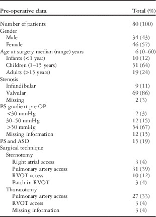
Table 1 shows the baseline data including age at surgery, pathology of the stenosis, pre-operative gradient of the stenosis, and the surgical technique used to relieve the pulmonary stenosis.
Pre-operative data of the study population.

ASD=atrial septum defect; OP=operation; PS=pulmonary stenosis; RVOT=right ventricular outflow tract
Valvular stenosis was the most common pathology (86%); 67% had a gradient across the valve of >50 mmHg, which is classified as severe pulmonary stenosis. The pulmonary gradient was measured by catheterisation for the majority of the population, as this was in the early days of echocardiography. In the majority of cases, the indication for intervention was a high gradient (>50 mmHg) across the pulmonary valve or the presence of symptoms. In some cases, the gradient was below 50 mmHg, and a concomitant atrial septal defect, partial abnormal pulmonary venous drainage, or right ventricular dilatation were the reasons for the intervention. For 72% of the patients, surgical access was through the pulmonary artery using cardiopulmonary bypass (n=43) or on the unsupported beating heart. See Table 1 for details regarding types of surgery. The stenosis was regarded as relieved if the jet felt at the pulmonary artery before repair disappeared. If the surgeons suspected elevated right ventricular pressure after relieving the stenosis, the per-operative direct pressure measurement was performed.
Follow-up data
Mortality
(Fig 1)
Kaplan–Meier survival curve for the 80 patients with surgically relieved pulmonary stenosis. Time is shown in years after surgery. The numbers of patients at risk at 0, 20, 40, and 60 years after surgery are shown.

Among all, two patients died within the first 24 hours of the operation due to circulatory insufficiency. The late mortality rate was 5% (4 of 80); one patient died at 40 years of age from sudden cardiac death, 31 years after repair; she had a pacemaker implanted due to congenital atrio-ventricular block and attended regular follow-up for this reason. The post-mortem report indicated dilated cardiomyopathy as the cause of death; however, no abnormalities were reported before her death. The second patient died at 91 years of age from pneumonia, 31 years after surgery. No echocardiography was performed before death. The third patient died at 60 years of age from colon cancer, 11 years after the operation. No echocardiography was performed before death. The time and cause of death are unknown for the last patient because the record could not be located.
Freedom from re-intervention
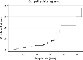
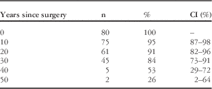
Freedom from re-intervention at 20 years after the initial surgery was 91%, and this decreased to 84, 52, and 26% after 30, 40, and 50 years, respectively (Fig 2, Table 2).
Cumulative incidence of re-intervention and death during the 57 years of follow-up. Analysis time is the time after the initial surgery (in years).

Freedom from re-intervention.

CI=confidence interval
n is the number of patients in the study who are free from re-intervention. At 10 years after repair, 95% are free from re-intervention, declining to 91, 84, 53, and 26% at 20, 30, 40, and 50 years after repair, respectively
Among all, 16 patients (20%) required re-intervention, with a median time to re-intervention of 30 years (with a range from 3 to 53 years); two-thirds of the re-interventions were pulmonary valve replacements, with indications of severe pulmonary regurgitation and/or dilatation of the right ventricle. Re-stenosis or elevated right ventricular pressure resulted in three surgical resections for re-stenosis. Balloon valvuloplasty was performed in three patients: one achieved successful reduction of the gradient, one did not achieve successful reduction and required surgical resection, and the last patient achieved successful dilatation of an implanted pulmonary valve prosthesis (Table 3).
Types of re-interventions.

PS=pulmonary stenosis
Distribution of re-interventions in the 16 patients who had at least one re-intervention
* After repair
Other re-interventions
Pacemakers were implanted in two patients: one due to congenital atrio-ventricular block and the other due to sick sinus syndrome.
Radiofrequency ablation was performed in two patients due to either atrial flutter or re-entry tachycardia. One patient had a coronary artery bypass in connection with a pulmonary valve replacement, and another patient had an atrial septal defect that was closed using catheter technique.
Functional capacity
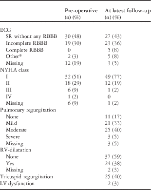
The functional NYHA class improved from before surgery to the time of the latest follow-up. A total of 63 patients were followed-up for >10 years; their results are presented in Table 4.
Follow-up (n=63, patients with >10 years follow-up).

AV=atrio-ventricular; LV=left ventricle; RBBB=right bundle branch block; RV=right ventricle; SR=sinus rhythm
* Other arrhythmias (atrial fibrillation, left bundle branch block, congenital AV-block)
Echocardiographic data
Standard echocardiography was performed at follow-up but only rarely at the time of surgery, because echocardiography was not the standard technique used in the 1970s and 1980s. Moderate or severe pulmonary regurgitation was found in 45% of the patients at the latest follow-up, and 38% had some degree of right ventricular dilatation without indication for re-intervention (Table 4). Correlation to type of surgery showed that moderate or severe pulmonary regurgitation was present in all patients with a patch in the right ventricular outflow tract, two out of three patients with access through the right atrium, and 7 out of 13 patients with access through the right ventricular outflow tract.
Electrocardiogram
At follow-up, incomplete and complete right bundle branch block increased from 30 to 36% and 0 to 8%, respectively (Table 4). Before the initial surgery, one patient had congenital complete atrio-ventricular block, and one patient had atrial fibrillation.
Discussion
Surgical treatment was the preferred procedure for pulmonary stenosis until the mid-1980s, when balloon valvuloplasty replaced surgery. Relieving the stenosis by splitting the valve leaflets is efficient but often causes insufficiency and subsequent right ventricular volume overload. During that era, volume overload was thought to be well tolerated by the right ventricle; therefore, regular follow-up was not the norm. Unaddressed right ventricular volume overload is now known to cause right ventricular failure with right ventricular dilatation and paradoxical septum movement, affecting left ventricular function and inducing arrhythmias.Reference Chaturvedi and Redington 10 Thus, these patients need regular follow-up.
The natural history of surgically repaired pulmonary stenosis has not been well described. In contrast to its diagnostic cousin tetralogy of Fallot, isolated pulmonary stenosis has not received much attention from the medical and scientific community.
From 1957 to 2000, 80 operations for pulmonary stenosis were performed at our institution. The long-term mortality was 5%, and one in five patients underwent re-intervention. The median follow-up period was 33 years.
Mortality
Additional cardiac details regarding patient deaths are important; however, in the instances reported in this study, only two – the patient with sudden cardiac death and the patient with missing information – could possibly be related to previous cardiac disease. Unfortunately, no information in the patient records provided insight regarding this issue. Our results, however, correspond to previous findings.Reference Morris and Menashe 11 Kopecky et alReference Kopecky, Gersh and McGoon 12 studied late survival in 191 patients 20–30 years after repair and found excellent survival and functional status; however, patients who had their initial repair in or after their third decade of life had a lower life expectancy compared with an age- and sex-matched control group.
Pulmonary regurgitation
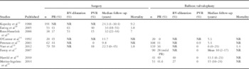
The most common finding in our population was the occurrence of pulmonary regurgitation; almost half the patients (45%) had moderate or severe pulmonary regurgitation that did not require re-intervention. This result is consistent with previous studies.Reference McNamara and Latson 13 Recent studies of both surgically treated and balloon-treated patients are shown in Table 5. For the surgically treated populations, the previous findings for pulmonary regurgitation are similar to ours.
Studies evaluating patients treated for pulmonary stenosis with either surgery or balloon valvuloplasty.

NR=not reported; PR=pulmonary regurgitation (moderate or severe); PVR=pulmonary valve replacement; RV=right ventricle
Results are shown as median follow-up in years (range) or per cent
In the three studiesReference O’Connor, Beekman, Lindauer and Rocchini 14 – Reference Voet, Rega and de Bruaene 16 that compared surgical versus balloon valvuloplasty relief of pulmonary stenosis, moderate or severe pulmonary regurgitation was most common in the surgically treated groups (45–59% compared with 0–36% in the balloon-treated groups). On the other hand, the balloon valvuloplasty groups were only followed-up for a median of 5.3–6 years, whereas the surgically treated groups had longer median follow-up periods of 9.8–22.5 years. Balloon valvuloplasty is obviously less invasive; however, the damage to the valve might be equally traumatic. Only time will tell whether pulmonary regurgitation in balloon valvuloplasty-treated patients will progress and ultimately require valve replacement. Recent studies of balloon valvuloplasty for pulmonary stenosis have shown the same incidences of pulmonary regurgitation, despite shorter follow-up periods (10–15 years mean/median)Reference Fawzy, Hassan and Fadel 17 – Reference Merino-Ingelmo, Santos-de Soto, Coserria-Sánchez, Descalzo-Señoran and Valverde-Pérez 19 (Table 5).
Haemodynamically unimportant tricuspid insufficiency was present in 40% of our population. This figure is somewhat larger than previously reported frequencies. Peterson et alReference Peterson, Schilthuis, Dodge-Khatami, Hitchcock, Meijboom and Bennink 15 reported mild or moderate tricuspid insufficiency in 22% of a surgically treated group and only in 12% of a balloon-treated group. Only one patient in the balloon-treated group required pulmonary valve replacement, and none of them required tricuspid valve repair.Reference Peterson, Schilthuis, Dodge-Khatami, Hitchcock, Meijboom and Bennink 15 The study by Peterson et al represents an intermediate “long-term” follow-up, and therefore may not reveal a need for pulmonary valve replacement. This intervention is more common in the third decade after the initial surgery, as shown by Earing et al.Reference Earing, Connolly, Dearani, Ammash, Grogan and Warnes 20
Right ventricular dilatation
Only two previous studies have described right ventricular dilatation in patients after treatment for pulmonary stenosis.Reference Earing, Connolly, Dearani, Ammash, Grogan and Warnes 20 , Reference Roos-Hesselink, Meijboom and Spitaels 21 Right ventricular dilatation is one of the parameters assessed when pulmonary valve replacement is considered. In the patients described by Earing et alReference Earing, Connolly, Dearani, Ammash, Grogan and Warnes 20 and Roos-Hesselink et al,Reference Roos-Hesselink, Meijboom and Spitaels 21 the frequency of right ventricular dilatation was reported to be 63 and 53%, respectively. Similar to our study, in which a rate of 38% was found, right ventricular dilatation was visually assessed using echo estimates. MRI is considered the gold standard for volume assessment but has not yet been used for patients with isolated pulmonary stenosis.
Freedom from re-intervention
In total, 20% of our patients underwent re-intervention, and pulmonary valve replacement was the most common re-intervention due to pulmonary regurgitation, right ventricular dilatation, and symptoms of right ventricular overload. Freedom from re-intervention rapidly declined >20 years after surgery. Previously reported re-intervention rates are 15Reference Roos-Hesselink, Meijboom and Spitaels 21 –40%,Reference Earing, Connolly, Dearani, Ammash, Grogan and Warnes 20 with a decline occurring >25 years after the initial surgery.Reference Earing, Connolly, Dearani, Ammash, Grogan and Warnes 20 Roos-Hesselink et alReference Roos-Hesselink, Meijboom and Spitaels 21 found that only two of six patients exhibited diminished right ventricular dimensions after valve replacement and only two patients showed improvements in their exercise capacity. These authors suggested that new techniques such as MRI and assessment of neurohormones could help stratify asymptomatic patients who will benefit from earlier pulmonary valve replacement for the right ventricle to maintain re-modelling capabilities.
In our population, none of the four patients with a patch in the right ventricular outflow tract required re-intervention, although this condition is a predictor of pulmonary regurgitation in patients with isolated pulmonary stenosis,Reference Roos-Hesselink, Meijboom and Spitaels 21 particularly in patients with tetralogy of Fallot.Reference Kirklin, Kirklin, Blackstone, Milano and Pacifico 22
Post-surgical pulmonary regurgitation in the setting of isolated pulmonary stenosis has been considered benign and does not require regular follow-up. As this and other studies have shown, pulmonary regurgitation can be well tolerated for a number of years; however, the need for re-operation rapidly increases after the second decade after repair. Guidelines from the American College of Cardiology/American Heart Association and European Society of Cardiology recommend periodic clinical follow-up for all patients after surgical or balloon pulmonary valvotomy with specific attention given to pulmonary regurgitation; right ventricular pressure, size, and function; and tricuspid regurgitation. The frequency of follow-up depends on the severity of the lesion; most patients will need a yearly visit, including evaluation in specialised centres for grown-up CHD. Patients with mild valvular or residual pulmonary stenosis should be seen at least every 5 years.Reference Warnes, Williams and Bashore 23 , Reference Baumgartner, Bonhoeffer and De Groot 24
Electrocardiogram
Complete or incomplete right bundle branch block was present in 30% of the patients before surgery and increased to 44% at the latest follow-up (Table 4). Right bundle branch block is common after surgery for tetralogy of Fallot.Reference Anderson, Baker, Penny, Redington, Rigby and Wernowsky 25 In patients with pulmonary stenosis, Roos-Hesselink et alReference Roos-Hesselink, Meijboom and Spitaels 21 described an increase in right bundle branch block of 9% at 16 years after surgery, and this percentage increased to 22% at 27 years after surgery. Voet et alReference Voet, Rega and de Bruaene 16 found that right bundle branch block was more common after surgically relieved pulmonary stenosis than after balloon valvuloplasty. Interestingly, the occurrence of right bundle branch block increased in both groups after relieving the stenosis, with increases from 32.3 to 60.3% after surgery in the surgically treated group and from 16.9 to 38.9% after balloon valvuloplasty.
The causes of this finding are speculative; surgery with access through the right ventricle might disturb the electrical pathways; however, as this approach is rare, it does not explain all events, particularly the events observed in patients in the balloon-treated group. Another cause could be the dilatation of the right ventricle, as this is a more common finding in patients with pulmonary regurgitation.
Limitations
Some of the follow-up examinations were performed at secondary centres. The follow-up included echocardiographic assessment according to the national guidelines described by the Danish Society of Cardiology, with subjective descriptions of right ventricular dilatation and tricuspid insufficiency. The gradient across the pulmonary valve was estimated during follow-up visits, but only noted in case the cardiologist thought that it was elevated to an extend needing further treatment; three patients had an elevated gradient and underwent balloon dilatation. The majority of patients were seen by cardiologists with a dual interest and experience in adults and paediatrics.
It might be argued that the surgical treatment of pulmonary stenosis is outdated and that our findings might be less relevant to patients receiving balloon valvuloplasty today. Nevertheless, these findings are reflective of practice 20–40 years ago and relevant to the large group of patients who were operated on in previous eras and who continue to be followed-up in grown-up CHD clinics today. Patients treated with balloon valvuloplasty could also benefit from this knowledge, because recent studies have shown that these patients can also develop pulmonary regurgitation. Open commisurotomy and valve “shaping” are recommended for aortic valve disease and can have excellent results.Reference David 26 Considering the limited life expectancy of prosthetic valves, this technique could also be relevant for pulmonary valves; however, we are not able to address this issue using our data, but it would be pertinent to a prospective study.
Conclusion
Surgical relief of pulmonary stenosis is efficient in relieving outflow obstruction; however, this relief occurs at the cost of pulmonary regurgitation, leading to right ventricular dilatation and tricuspid regurgitation. When required, pulmonary valve replacement occurs most frequently over 20 years after the initial surgery. Lifelong follow-up of patients treated surgically for pulmonary stenosis is emphasised in this group of patients, who might otherwise consider themselves cured.
Acknowledgement
None.
Authors’ contribution: Vibeke Hjortdal conceived the study and performed data interpretation, as well as critical manuscript revision. Eva Nielsen performed data collection, analyses, and interpretation, as well as drafted the manuscript.
Financial Support
This research received no specific grant from any funding agency, commercial, or not-for-profit sectors.
Conflicts of Interest
None.
Ethical Standards
The authors assert that all procedures contributing to this work comply with the ethical standards of the relevant national guidelines on human experimentation (the Danish Health and Medicines Authority and the Danish Data Protection Agency) and with the Helsinki Declaration of 1975, as revised in 2008.