Introduction
The genus Yuknessia Walcott, Reference Walcott1919, characterized by radiating clusters of elongate tubes, was erected in a report devoted to the algal component of the Burgess Shale biota, the first major macrophyte assemblage described from the Cambrian. Maletz et al. (Reference Maletz, Steiner and Fatka2005), however, described fuselli in a Yuknessia-like specimen from the Wheeler Formation of Utah, and the taxon was formally transferred to the Hemichordata by LoDuca et al. (Reference LoDuca, Caron, Schiffbauer, Xiao and Kramer2015) on the basis of fuselli observed with scanning electron microscopy (SEM) on the holotype of the type species, Yuknessia simplex Walcott, Reference Walcott1919, and on additional specimens formally assigned to Y. simplex in that report from the Burgess Shale Formation (Trilobite Beds) type locality. Here, material from the Cambrian of South China attributed to Yuknessia is reexamined in light of these findings.
Material attributed to Yuknessia has been reported from six Cambrian formations in South China. Specimens from the Maotianshan (also published as Yuanshan) Shale (Cambrian Stage 4) assigned to Yuknessia in early reports on the Chengjiang biota (Chen and Erdtmann, Reference Chen and Erdtman1991; Chen et al., Reference Chen, Zhou, Zhu and Yeh1996; Hou et al., Reference Hou, Bergström, Wang, Feng and Chen1999) were later redescribed as Fuxianospira Chen and Zhou, Reference Chen and Zhou1997 (Chen and Zhou, Reference Chen and Zhou1997; Hou et al., Reference Hou, Aldridge, Bergstrom, Siveter, Siveter and Feng2004; Fu et al., Reference Fu, Wu, Zhao, Zhu and Yang2012). This paper concerns Yuknessia from the Cambrian Yanjiahe Formation (provisional Stage 2), Niutitang Formation (provisional stages 2-3), Balang Formation (provisional Stage 4), Kaili Formation (provisional stages 4-5), and Huaqiao Formation (provisional Stage 5-Drumian Stage). Also included in this study is material with an elongate form similar to the Chinese material from the Burgess Shale Formation (Cambrian Stage 5) of British Columbia, and the Wheeler and Marjum formations (Cambrian Stage 5-Drumian Stage) of Utah.
Material and methods
The study material comprises some 45 small slabs, most of which have multiple specimens distributed across the bedding plane surfaces. No attempts were made to extract specimens from these slabs using acid maceration techniques, as Handle and Powell (Reference Handle and Powell2012) reported poor results when attempting to do so for Wheeler Formation specimens owing to the fragile nature of the carbonaceous material. High-resolution morphometric data was collected from digital images using the open source program ImageJ. For statistical analyses, most measurements were made on specimens for which the entire (preserved) length could be determined without ambiguity (i.e., not concealed by sediment or obscured by overlapping with adjacent specimens). Specimens of Fuxianospira gyrata from the Maotianshan Shale and Tsinghsutung Formation were examined at Yunnan University and Guizhou University, respectively, for comparative purposes. Repository abbreviations as follows: GTB, GTBM, GY, Q, SH, ZY, Guizhou University, Guiyang, China; NIGP, Nanjing Institute of Geology and Palaeontology, Chinese Academy of Sciences, Nanjing, China; Yjh, Early Life Institute, Northwest University, Xi’an, China; KUMIP, Biodiversity Institute, University of Kansas, Lawrence, USA; ROM, Royal Ontario Museum (Invertebrate Palaeontology collections), Toronto, Canada.
A subset of materials described herein were analyzed using electron microscopic approaches, including GTBM-20-5-1119 from the Kaili Formation and KUMIP 373593 and 373594 from the Marjum Formation. SEM studies were conducted using an FEI Quanta 250 FEG at the Subsurface Energy Materials Characterization and Analysis Laboratory (SEMCAL), School of Earth Sciences, The Ohio State University, a Hitachi S-3200N at the Electron Microbeam Analysis Laboratory (EMAL), Department of Earth and Environmental Science, University of Michigan, and an FEI Quanta 600F at the University of Missouri Electron Microscopy Core. All specimens were imaged uncoated under low vacuum, and backscattered electron (BSE) images were obtained using an accelerating voltage of 10 keV. Energy dispersive X-ray spectroscopic (EDS) elemental maps and point analyses (University of Missouri) were collected uncoated under high vacuum with operating parameters as follows: 10.5 mm sample working distance, 20 keV beam accelerating voltage, and 4.0 beam spot size. Elemental maps were collected at 900 seconds live time, points at 90 seconds live time, and all resulting data were appropriately adjusted using ZAF correction (see LoDuca et al., Reference LoDuca, Caron, Schiffbauer, Xiao and Kramer2015).
Results
Kaili Formation material
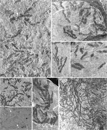
Specimens on GTM-19-5-1806, illustrated without description by Yang (Reference Yang2006, pl. 12, fig. 8) as Yuknessia sp., and those on GTB-12-3120 and GTB 19-2-201, illustrated as Yuknessia guizhouensis in Zhao et al. (Reference Zhao, Zhu, Babcock and Peng2011, figs. 241 and 242, respectively), are similar and consist of narrow elongate elements haphazardly arranged on the respective bedding surfaces and preserved as dark brown to black films without relief. Length is variable, the longest reaching 8 mm. All specimens are characterized by undulating sides, the undulations on one side of a specimen typically being slightly out of phase with those on the other (Fig. 1.2, 1.5). Specimens on a given slab have similar widths, but width varies slightly between slabs, those on GTB-12-3120 being narrower than those on GTB 19-2-201 (0.3 mm vs. 0.4 mm). None of the specimens show evidence of tapering. Element endings are abrupt, these being straight to slightly jagged and oriented at roughly 90 degrees to the long axis; no specimens show endings with definitively rounded or tapered forms. Definitive branching or connection between elements is not evident, but overlaps are present. Overall form is, in some cases, rectilinear; other specimens show gently curvature but no specimens show folding. Transverse banding or other microstructure was not observed with reflected light microscopy. Previously unpublished specimens examined during this study include those on GTMB-20-5-1119, GTBM-20-1-407a, and GTBM-16-100 (Fig. 1.1, 1.3, 1.4). These have an overall form comparable to GTB-12-3120, including distinctly undulating sides, and also lack definitive evidence of branching. Size is similar as well, but some specimens are somewhat wider, with widths reaching 1 mm (Fig. 3, Table 1). No transverse banding or other microstructure was observed in reflected light. BSE images of GTBM-20-5-1119 also lack any indication of small-scale structure (Fig. 1.6).
Specimens from the Cambrian of China assigned herein to Fuxianospira gyrata Chen and Zhou, Reference Chen and Zhou1997. (1–6) specimens from the Kaili Formation, Guizhou Province; (1) GTBM-20-5-1119; (2) GTB-19-2-201, illustrated in Zhao et al. (Reference Zhao, Zhu, Babcock and Peng2011) as Yuknessia guizhouensis; (3) GTBM-20-1-407a; (4) GTBM-16-100; (5) GTB-12-3120, illustrated in Zhao et al. (Reference Zhao, Zhu, Babcock and Peng2011) as Yuknessia guizhouensis; (6) BSE image of the area marked by the arrow in (1), sides of Fuxianospira specimen, nearly indistinguishable from the matrix, are marked by opposing arrows. (7) cluster of specimens from the Balang Formation, Guizhou Province, GY-374-1; (8) cluster of specimens from the Niutitang Formation, Guizhou Province, ZY-1668. All specimens photographed dry in direct light. Scale bar (1–5, 7, 8), 5 mm; (6), 1 mm.

Summary of morphometric data for previously unpublished material from the Kaili, Marjum, and Burgess Shale formations assigned herein to Fuxianospira gyrata.

Data sources: Kaili Formation, GTBM-16-100, GTB-19-2-201, GTBM-20-1-407a, GTBM-20-5-1119; Marjum Formation, KUMIP 373591, KUMIP 373592; Burgess Shale Formation, ROM 57145, ROM 63103. Abbreviations as follows: min.=minimum, max.=maximum, avg.=average, SD=standard deviation.
All measurements in millimiters.
Balang Formation material
Specimens on three small slabs were assigned with uncertainty to Yuknessia simplex in Fu et al. (Reference Fu, Wu, Liu, Peng and Zhao2010). Each slab bears numerous elongate elements arrayed in haphazard, often overlapping fashion, and preserved as dark films. Size and form are comparable to the Kaili Formation study material, including undulating sides (Figs. 1.7, 3). Transverse banding or other microstructure was not observed with reflected light microscopy.
Niutitang Formation material
Specimens on two small slabs (ZY-4-15, ZY-4-30) from the lower part of the Niutitang Formation comprise the type material of Yuknessia guizhouensis Yang and Zhao, Reference Yang, Zhao and Guo1999 (in Yang et al., Reference Yang, Zhao and Guo1999). Each slab bears numerous elongate elements arrayed in haphazard, often overlapping fashion, and preserved as dark films. Size and form are comparable to the Kaili Formation study material, including undulating sides. Yang and Zhao (Reference Yang, Zhao and Guo1999) regarded this material to differ from the type material of Yuknessia simplex by having fewer but longer stipes, and placed it in a separate species on that basis. The central holdfast mentioned in the original description is not evident, and any appearance of the elements being united at the base is regarded herein as a consequence of coincidental overlap. Material on a single slab (ZY-18-50) from the middle part of the formation illustrated as Yuknessia sp. in Yang et al. (Reference Yang, Zhang, Jiang and Gao2003) is comparable in every respect with material assigned to Y. guizhouensis in Yang et al. (Reference Yang, Zhao and Guo1999). Many similar specimens, previously unpublished, are present on additional slabs examined during this study. No evidence of branching or unification by a common holdfast is evident among this material, and size and shape agree with the type material of Y. guizhouensis. For SH-7882 and ZY-1668 (Fig. 1.8), width for the various specimens ranges from 0.3 to 0.45 mm and most are roughly 8 mm in length; for ZY-2660, width for the various specimens ranges from 0.2 to 0.35 mm and some appear to reach 12 mm in length, although overlapping of multiple specimens makes this measurement uncertain. Transverse banding or other microstructure was not observed with reflected light microscopy. A lack of microstructure was also noted in the original description of Y. guizhouensis.
Huaqiao Formation material
Numerous specimens distributed across three slabs were assigned to Yuknessia simplex by Babcock et al. (Reference Babcock, Peng, Wasserman and Robison2011). Each specimen is preserved as a dark film and comprises an elongate element 0.9 to 1.3 wide with subtly undulating sides; most are 10 to 16 mm in length. None of the specimens show evidence of tapering along their length or folding. Element endings are straight to slightly jagged and are oriented at roughly 90 degrees to the long axis; no specimens show endings with a definitively rounded or tapered form. Definitive evidence of branching is lacking and no microstructure was observed.
Yanjiahe Formation material
A single specimen (Yjh-HS81) preserved as a rust-colored compression was assigned to Yuknessia in open nomenclature by Guo et al. (Reference Guo, Li and Shu2010). The specimen comprises several elongate elements up to 16 mm in length with smooth sides and clearly arising from an ovoid basal structure with a width of 6 mm. Each element tapers from approximately 1.5 mm at the base to less than 0.5 mm at the distal termination. Branching of the elongate elements may be present, but is not definitive. BSE images of the specimen show no microstructure.
Marjum Formation material
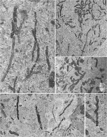
Conway Morris and Robison (Reference Conway Morris and Robison1988, fig. 11.1) illustrated specimens on KUMIP 204378 from the House Range of Utah as Yuknessia simplex and, in the associated caption, described this material as “fragments of stipes.” More than 100 of these “fragments” are distributed across the slab surface, each comprising a dark gray to black film in the form of an isolated elongate element. All lack evidence of folding, and branching or connection between elements is not apparent. Most specimens are 0.5 mm wide. Width is constant along specimen length and subtle undulation is evident along the sides of all specimens. The longest specimens reach 6 mm, but many are only half as long. Element endings are abrupt, straight to slightly jagged, and oriented at roughly 90 degrees to the long axis; no specimens show endings with definitively rounded or tapered forms. Specimens of similar size, form, and color, previously unpublished, are present on additional slabs recently collected from the House Range (Figs. 2.1, 2.4, 2.5, 4.1, 4.6). Width for this material ranges from 0.3 to 1.2 mm (Fig. 3, Table 1). All specimens show undulation along the sides. For some, this is subtle (Fig. 2.4), whereas for others it is pronounced, resulting in a sinuous form (Fig. 2.5). Specimens with various degrees of undulation intermediate between the aforementioned forms are also known, and in some cases variation in the amount of undulation is present along the length of individual specimens (e.g., left specimen in Fig. 2.1). BSE images of the study specimens show no transverse banding or other microstructure (Fig. 4.2, 4.7).
Fuxianospira gyrata Chen and Zhou, Reference Chen and Zhou1997 from the Cambrian of North America. (1) KUMIP 373591, Marjum Formation, House Range, Utah; (2) ROM 57145, Greater Phyllopod Bed, Burgess Shale Formation, British Columbia; (3) ROM 63103, Greater Phyllopod Bed, Burgess Shale Formation, British Columbia; (4) KUMIP 373592, Marjum Formation, House Range, Utah; (5) specimen from a different area of the same slab shown in (4) with pronounced axial twist. All specimens photographed dry in direct light except (2, 3) (cross-polarized light). Scale bar (1–4) 5 mm; (5) 3 mm.

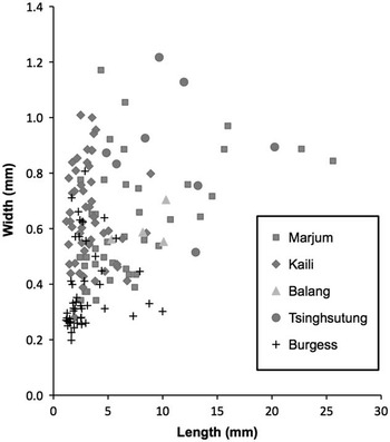
Length vs. width scatter plot for material assigned to Fuxianospira gyrata Chen and Zhou, Reference Chen and Zhou1997 herein and for material assigned to this taxon by Fu et al. (Reference Fu, Wu, Zhao, Zhu and Yang2012) from the Tsinghsutung Formation (circles). Data sources: Kaili Formation, GTBM-16-100, GTB-19-2-201, GTBM-20-1-407a, GTBM-20-5-1119; Balang Formation, GY-374-1b; Marjum Formation, KUMIP 373591, KUMIP 373592; Burgess Shale Formation, ROM 57145, ROM 63103; Tsinghsutung Formation, Q34-6-144, Q34-6-201, Q34-6-136, Q51-5-20.

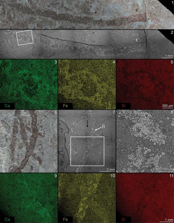
Reflected light and SEM images of Fuxianospira gyrata Chen and Zhou, Reference Chen and Zhou1997 from the Marjum Formation, House Range, Utah. (1) KUMIP 373593, reflected light; (2) BSE mosaic of specimen in (1); (3–5) EDS elemental maps for calcium, iron, and silicon, respectively, for the white rectangle in (2); (6) KUMIP 373594, reflected light; (7) BSE mosaic of specimen in (6), arrows A and B indicate specimen areas analyzed to obtain point data shown in Table 2; (8) higher magnification of area marked by white arrow in (7), showing fossil surface with iron oxide framboids (pseudomorphs after pyrite weathering); (9–11) EDS elemental maps for calcium, iron, and silicon, respectively, for the white rectangle in (7).

Wheeler Formation material
Handle and Powell (Reference Handle and Powell2012) described elongate elements from the House Range of Utah with a width of 0.5 mm and a length up to 10 mm under the name Linear Morphotype 1 (hereafter LM1). LM1 specimens comprise dark gray to black films and show no evidence of branching or connection between elements. All specimens illustrated as LM1 material show subtle undulation (Handle and Powell, Reference Handle and Powell2012, fig. 2E, 7A–D). Handle and Powell (Reference Handle and Powell2012, figs. 2G, 3, 9) described similar associated material but with pronounced undulation, resulting in a sinuous form, under the name Coiled Morphotype 2 (hereafter CM2). Length and width for this material is comparable to that for LM1 material. Notably, forms with undulation intermediate between that characteristic of LM1 and CM2 also occur on slabs illustrated by Handle and Powell (Reference Handle and Powell2012), indicating that these designations pertain to endmembers along a continuum of form. Handle and Powell (Reference Handle and Powell2012) reported a lack of transverse banding or other microstructure in BSE images for both LM1 and CM2 material (see Handle and Powell, Reference Handle and Powell2012, figs. 7 and 9).
Burgess Shale Formation material
Two slabs with specimens preserved as black films and comprising elongate elements with undulating sides previously referred to as “new Algae D” (Caron, Reference Caron2005; Caron and Jackson, Reference Caron and Jackson2008) were selected for this study and come from the Greater Phyllopod Bed (Fig. 2.2, 2.3). Width for this material ranges from 0.2 to 0.8 mm, with specimens on ROM 57145 being narrower than specimens on ROM 63103. The longest specimens reach 10 mm (Fig. 3, Table 1). No transverse banding or other microstructure is evident in reflected light.
Elemental composition
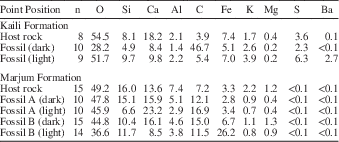
For Kaili Formation material, results of EDS point analyses applied to GTBM-20-5-1119, summarized in Table 2, show elevated levels of carbon throughout the analyzed specimen relative to the host rock. Most areas of the specimen show only minor carbon enrichment, but small, widely scattered areas that appear as thin dark patches in BSE images (Fig 1.6) are nearly 50% carbon on average. No enrichment in calcium, iron, magnesium, or aluminum was detected for the specimen, but elevated barite levels were observed in some areas.
Summary of EDS point spectral data for one study specimen and the surrounding host rock from the Kaili Formation, Guizhou Province, China, GTBM-20-5-1119, and for two study specimens and the surrounding host rock from the Marjum Formation, House Range, Utah, KUMIP 373594.
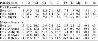
Data for Fossil A and Fossil B obtained from areas indicated by black and white arrows, respectively, in Figure 4.7; “dark” and “light” correspond to analyzed areas of specimens with relatively darker and lighter brightness levels in BSE images. All elemental point data quantified from K-shell electron emission X-rays and reported in normalized weight percentage. Elements are organized, from left to right, by abundance in Marjum Formation host rock.
For Marjum Formation study material, results of EDS point analyses show elevated carbon levels throughout all of the analyzed specimens relative to the host rock (Table 2). In addition, regions of calcium and iron enrichment are evident (Table 2; Fig. 4.3–4.4, 4.9–4.10), instances of the latter in some cases showing contiguous limonite/goethite framboidal clusters presumed to be oxidative pseudomorphs after original pyrite (Fig. 4.8). Regions of calcium and iron enrichment are somewhat irregular in distribution but show a tendency to be associated with the outer edges of the fossil along the bedding surface, and those for KUMIP 373593 appear to form irregular elongate patches that run diagonally across the specimen between corresponding undulations on either side (Fig. 4.3–4.4). Elemental maps for aluminum (not shown) are entirely coincident with silicon maps (Fig. 4.5, 4.11), and those for carbon (not shown) do not reveal elevated levels for any of the specimens.
On the basis of these findings, it can be concluded that the analyzed Kaili and Marjum formation specimens comprise thin films of carbon, the carbon film being so thin in most areas that it does not appear on BSE images of Kaili Formation material or on elemental maps of Marjum Formation material owing to the depth of electron beam penetration (Muscente and Xiao, Reference Muscente and Xiao2015) and the low backscatter coefficient for carbon. Regions of Marjum Formation specimens enriched in iron and calcium indicate localized diagenetic overprinting/replication. In some cases, this involved pyrite (later altered to iron oxides). In others, the composition points to calcium carbonate cements, and in still others, such as areas of “fossil B” that are dark in BSE images, iron-rich clay may be present, this having a composition distinct from the host rock clay by comparatively elevated iron and lower potassium and magnesium levels, as shown by point analysis data.
Discussion
From a morphological standpoint, all of the study specimens from the Kaili, Balang, Niutitang, and Huaqiao formations fall within a narrow range of size and can be characterized unequivocally as unbranched elongate elements lacking microstructure, such as transverse banding or longitudinal lineation, and displaying undulating sides, the undulations on one side being slightly out of phase with those on the other, a morphology consistent with the compressed remains of a simple twisted tube. Such an outcome supports assignment of this material to a single genus, as in the original reports. Study specimens from the Marjum Formation, including specimens on KUMIP 204378 attributed to Yuknessia simplex as fragmentary remains by Conway Morris and Robison (Reference Conway Morris and Robison1988), and the Burgess Shale Formation agree with the aforementioned Chinese material in terms of all key morphological features, including overall size and form. The same applies to specimens previously described as LM1 and CM2 by Handle and Powell (Reference Handle and Powell2012) from the Wheeler Formation. Specimens from the Marjum and Wheeler formations show in some cases a greater degree of undulation (axial twist) than noted for any of the Chinese specimens. For both of the Utah occurrences, however, specimens are known with clearly intermediate morphologies, thereby providing strong evidence against the possibility of two distinct morphotypes.
Previous work has indicated that original tissue histology may affect associated mineralogies within the Burgess Shale-type taphonomic mode (Gabbott et al., Reference Gabbott, Hou, Norry and Siveter2004; Page et al., Reference Page, Gabbott, Wilby and Zalasiewicz2008), and Handle and Powell (Reference Handle and Powell2012) extrapolated such a relationship to inform taxonomic interpretations for a variety of problematic taxa from several Cambrian units, including the Burgess Shale and Wheeler formations, and LM1 and CM2 material from the latter. Recent comparative analyses of Burgess Shale-type deposits, however, illustrate that late-stage diagenetic processes, including weathering and thermal history, play important roles in taphonomic mineralogical differences (e.g., Lin and Briggs, Reference Lin and Briggs2010; Forchielli et al., Reference Forchielli, Steiner, Kasbohm, Hu and Keupp2014). Moreover, even when mineralization processes occur early, they can differ due to local variations in the burial environment, including differing microbial metabolic zones related to differences in initial burial depth (Schiffbauer et al., Reference Schiffbauer, Xiao, Cai, Wallace, Hua, Hunter, Xu, Peng and J. Kaufman2014). Thus, although the analyzed Kaili Formation material is compositionally distinct from Marjum Formation material in that it lacks localized enrichment in iron or calcium, such a difference in this case cannot be regarded as having reliable taxonomic value. For LM1 and CM2 material from the Wheeler Formation, Handle and Powell (Reference Handle and Powell2012, table 1) summarized composition for the former as “major” for carbon and “trace” for illite, and for the latter as “major” for both chlorite and carbon. On this basis, they inferred an algal affinity for LM1 material and a coprolite affinity for CM2 material. It is not clear from that report if the analyzed specimens are from the same bedding plane, but they are from the same unit and general location. No point data were provided or discussed, but elemental maps (composite Fe, C, Ca, K, and Si) and Raman spectra were included. Elemental maps for LM1 and CM2 material are very similar (compare their fig. 7 and fig. 9), showing very little carbon signal and localized enrichment in iron and calcium in the fossils. Raman spectra for LM1 and CM2 material (Handle and Powell, Reference Handle and Powell2012, fig. 5) show only subtle differences. In general, the spectra for LM1 and CM2, with reversed peak intensities of the carbon D1 and O bands (labeled in fig. 5 of Handle and Powell, Reference Handle and Powell2012, as D and G, respectively), indicate a carbonaceous composition for both. CM2 also shows additional Raman shift peaks at ~275 and 700 cm-1 (as approximated from fig. 5 of Handle and Powell, Reference Handle and Powell2012) possibly, although not definitively, indicative of phyllosilicate compositions. These spectral bands may correspond to either di-octahedral or tri-octahedral phyllosilicates and therefore do not offer much resolution to clay mineralogy (Wang et al., Reference Wang, Freeman and Kuebler2002). Considered collectively, results reported in Handle and Powell (Reference Handle and Powell2012) do not identify conclusive compositional differences between LM1 and CM2 material, quantitative compositional data in each case indicating thin films of carbon with localized and irregularly distributed enrichments of iron and calcium and suggestions of clay minerals, the specific mineralogy of which cannot be determined from the available data. Overall, this composition is broadly consistent with that of the analyzed Marjum Formation study material reported here.
From the foregoing, it is concluded that all of the study material from the Kaili, Balang, Niutitang, Huaqiao, Marjum, Burgess Shale, and Wheeler formations is congeneric, including LM1 and CM2 material from the latter, and for the remainder of this section it will be referred to collectively as the twisted tube morphotype (TTM). The specimen from the Yanjiahe Formation assigned to Yuknessia sp. in Guo et al. (Reference Guo, Li and Shu2010), on the other hand, has a morphology that deviates from all of the other study material in several important regards, including a distinctly tapered form, lack of undulation, and the possible presence of branching. On the basis of these differences, this specimen is regarded as being taxonomically distinct from TTM material.
Comparison with Yuknessia
Morphological comparison between TTM material and Yuknessia, as recently redescribed by LoDuca et al. (Reference LoDuca, Caron, Schiffbauer, Xiao and Kramer2015) based on restudy of the Burgess Shale type material, associated complete specimens assigned to the genus in that report, and complete specimens from the Cambrian of Utah assigned to the genus by Conway Morris and Robison (Reference Conway Morris and Robison1988), reveals several key differences. First, the undulating nature of TTM specimens is not known for Yuknessia, the tubes of which show no tendency toward such a form. Second, transverse banding ubiquitous on the tubes of Yuknessia specimens examined with BSE imaging (e.g., LoDuca et al., Reference LoDuca, Caron, Schiffbauer, Xiao and Kramer2015, fig 2.6), and in some cases visible with reflected light microscopy, has not been observed among TTM material. The lack of such banding in Wheeler Formation TTM material (e.g., Handle and Powell, Reference Handle and Powell2012, fig. 7) despite its presence in intact specimens of Yuknessia from the same unit (e.g., LoDuca et al., Reference LoDuca, Caron, Schiffbauer, Xiao and Kramer2015, fig. 3) indicates that this difference is unlikely to be taphonomic in nature. Third, the tubes of TTM material do not taper along their length, whereas the erect tubes of Yuknessia consistently show a distinct taper (e.g., LoDuca et al., Reference LoDuca, Caron, Schiffbauer, Xiao and Kramer2015, fig. 1.3). Finally, new specimens formally assigned to Yuknessia from the Trilobite Beds type locality by LoDuca et al. (Reference LoDuca, Caron, Schiffbauer, Xiao and Kramer2015), as well as complete specimens assigned to the genus from the Cambrian of Utah by Conway Morris and Robison (Reference Conway Morris and Robison1988), clearly comprise clusters of branching tubes (e.g., LoDuca et al., Reference LoDuca, Caron, Schiffbauer, Xiao and Kramer2015, figs. 2.7–2.9, 3), whereas no definitive evidence of branching is present among TTM material, numbering well over 200 specimens. From a compositional standpoint, taphonomically described material of Yuknessia from the Wheeler Formation (LoDuca et al., Reference LoDuca, Caron, Schiffbauer, Xiao and Kramer2015) differs from TTM material from the same unit in that it commonly shows robust, electron microscopically visible remnants of carbon, this having a non-continuous, patchy distribution against a background of fossil-associated clays characterized by higher levels of iron and magnesium relative to the host rock. Collectively, these morphological and compositional discrepancies argue against a Yuknessia affinity for TTM material, a conclusion reached by Handle and Powell (Reference Handle and Powell2012) for the LM1 subset of the TTM material on the basis of broadly similar lines of evidence. The Yanjiahe Formation specimen described by Guo et al. (Reference Guo, Li and Shu2010) also differs markedly from Yuknessia. This specimen comprises a radiating cluster of elongate elements with smooth sides, like Yuknessia, but the elongate elements narrow distally rather than basally. In addition, like the TTM material, evidence of transverse banding is lacking, including in BSE images. Thus, this specimen, too, differs sufficiently from Yuknessia to preclude assignment to this taxon.
Recently, Maletz and Steiner (Reference Maletz and Steiner2015) interpreted much of the material regarded as Yuknessia by LoDuca et al. (Reference LoDuca, Caron, Schiffbauer, Xiao and Kramer2015), including material previously assigned to Yuknessia from the Cambrian of Utah by Conway Morris and Robison (Reference Conway Morris and Robison1988), as belonging to Sphenoecium Chapman and Thomas, 1936. It is important to note that this taxonomic matter, which we view as worthy of further investigation, is a separate issue from the foregoing. The same arguments presented above to exclude TTM material from Yuknessia apply equally to Sphenoecium.
Comparison with Fuxianospira
Chen and Zhou (Reference Chen and Zhou1997) erected the monotypic genus Fuxianospira on the basis of specimens from the Maotianshan Shale of Yunnan Province, China, and interpreted the assigned material as the compressed remains of a narrow unbranched organic tube with an axial twist. For this material, twisting of the tubes is indicated by undulation along the tube sides, this varying between specimens from subtle to pronounced. Recently, a second occurrence of the genus was described by Fu et al. (Reference Fu, Wu, Zhao, Zhu and Yang2012) from the Tsinghsutung (also published as Qingxudong) Formation (provisional Stage 4), which directly underlies the Kaili Formation in Guizhou Province. The undulating form characteristic of TTM material matches that known for Fuxianospira from both the Chengjiang biota and Tsinghsutung Formation occurrences, including between-specimen variation in the degree of undulation. Tube width for TTM material also compares well with that for specimens of Fuxianospira. In particular, the range between specimens (0.2–1.3 mm) overlaps with that for published specimens of Fuxianospira from both the type locality (0.65–1.1 mm) and the Tsinghsutung Formation (0.55–1.2 mm), with new material from the latter indicating a lower end closer to 0.3 mm (unpublished data). In addition, for TTM material, as for Fuxianospira, tube width for a given specimen remains constant along length. For tube length, specimens of Fuxianospira from the type locality are known to reach 200 mm. This surpasses the longest of the TTM specimens by a considerable margin. Other specimens from the type locality, however, are not nearly as long, and specimens of Fuxianospira from the Tsinghsutung Formation have a length comparable to that of many of the study specimens (Fig. 3). Additionally in this regard, it is likely that many of the study specimens are fragments of originally longer tubes, an inference supported by the jagged nature of the endings in many of the specimens. Finally, all TTM specimens, like specimens of Fuxianospira, lack branching, folding, and microstructure, including transverse banding. Thus, in all key respects, TTM material shows a morphology consistent with Fuxianospira. Accordingly, this material is here assigned to this genus. Such an assignment for the North American material expands both the paleogeographic and stratigraphic range of Fuxianospira, which was not known previously outside of China or in strata younger than Stage 4.
Systematic paleontology
?Division Chlorophyta
Genus Fuxianospira Chen and Zhou, Reference Chen and Zhou1997
Type species
Fuxianospira gyrata Chen and Zhou, Reference Chen and Zhou1997, by monotypy.
Fuxianospira gyrata Chen and Zhou, Reference Chen and Zhou1997
Figures 1.1-1.8, 2.1-2.5, 4.1-4.11
1988Yuknessia simplex Walcott; Conway Morris and Robinson, fig. 11.1.
1991Yuknessia sp.; Chen and Erdtmann, pl. 2, fig. 1.
1994Bosworthia simulans Walcott; Mao et al., p. 346, pl. 2, fig. 5.
1996Yuknessia sp.; Chen et al., p. 74, figs. 46, 48.
1996Algae sp.; Chen et al., p. 76, figs. 53, 54.
1997Fuxianospira gyrata Chen and Zhou, p. 88, figs. 143–145.
1999Fuxianospira gyrata; Luo et al., p. 105, pl. 28, fig. 9.
1999Yuknessia guizhouensis Yang and Zhao in Yang et al., p. 152, pl. 1, figs. 11, 13.
1999Yuknessia sp.; Hou et al., p. 14, fig. 15.
2001Yuknessia cf. Y. simplex; Babcock and Zhang, fig. 7.4.
2003Yuknessia sp.; Yang et al., pl. 1, fig. 4.
2004Fuxianospira gyrata; Hou et al., p. 30, fig. 7.1.
2005Alga/Algae D; Caron, Reference Caron2005, p. 270, 279, 295, 297, 303, 307, figs. 1.13, 2.7, 2.8, 3.1.
2006Yuknessia sp.; Yang, Reference Yang2006, pl. XII, fig. 8.
2008Alga/Algae D; Caron and Jackson, Reference Caron and Jackson2008, p. 226, 248, 249.
2010?Yuknessia sp.; Fu et al., p. 234, pl. I. figs. 5-8.
2011Yuknessia simplex; Babcock et al., p. 144, fig. 1D, 1E.
2011Yuknessia guizhouensis; Zhao et al., p. 189, figs. 241–242.
2012Fuxianospira gyrata; Fu et al., p. 59, pl. I, figs. 2, 3, 6, 7.
2012Linear Morphotype 1; Handle and Powell, Reference Handle and Powell2012, figs. 2E, 7.
2012Coiled Morphotype 2; Handle and Powell, Reference Handle and Powell2012, figs. 2G, 3, 9.
Holotype
ELRC80001 from the Yuanshan Formation, Chengjiang, Yunnan, China (Chen and Zhou, Reference Chen and Zhou1997, p. 88, fig. 143).
Material
GTB-19-2-201, GTB-15-19-1, GTB-12-3120, GTBM-9-2-4055, GTBM-9-5-00000, GTBM-9-5-0000, GTBM-9-5-2170, GTBM-16-100, GTBM-16-1056, GTBM-17-0000, GTBM-17-427, GTBM-17-554, GTBM-17-554b, GTBM-20-1-407a, GTBM-20-5-1119, GTM-19-5-1806, GY-34-1, GY-374-1a, GY-374-1b, GY-378-19, GY-378-21, ZY-4-15, ZY-4-30, ZY-18-50, ZY-641, ZY-1668, ZY-2660, ZY-2663, ZY-2665, SH-7882, NIGP 136371, NIGP 136374, NIGP 136376, KUMIP 204378, KUMIP 373591–373596, ROM 63103, ROM 57145.
Occurrence
Niutitang Formation, Guizhou Province, China (Cambrian Stages 2-3); Maotianshan Shale, Yunnan Province, China (Cambrian Stage 3); Qingxudong (Tsinghsutung) Formation, Guizhou Province (Cambrian Stage 4); Balang Formation, Guizhou Province, China (Cambrian Stage 4); Kaili Formation, Guizhou Province, China (Cambrian Stages 4-5); Greater Phyllopod Bed (Walcott Quarry), Burgess Shale Formation, Fossil Ridge, British Columbia (Cambrian Stage 5); Huaqiao Formation, Hunan Province, China (Cambrian Stage 5-Drumian Stage); Wheeler Formation, Marjum Pass and Lower Swasey Springs, Utah (Cambrian Stage 5-Drumian Stage); lower part of the Marjum Formation, House Range, Utah (Drumian Stage).
Remarks
Given that width varies widely between study specimens assigned herein to Fuxianospira (0.2–1.3 mm), it is reasonable to query whether distinct morphotypes might exist on the basis of size. In this regard, material from the Kaili and Marjum formations shows, in each case, most of the range of width known for the study material as a whole, with a fairly even and continuous distribution within this range (Fig. 3). Based on this distribution within individual assemblages, it is regarded best now to treat differences in width, as well as differences in the degree of axial twist, as variation within a single morphotype, pending further evidence to the contrary.
Other fossil taxa from Ediacaran–Cambrian strata preserved as flat carbonaceous films with a form and size roughly similar to F. gyrata include Sinocylindra yunnanensis Chen and Erdtmann, Reference Chen and Erdtman1991; Jiuqunaoella simplicis Chen in Chen and Xiao, Reference Chen and Xiao1991; Vendotaenia antiqua Gnilovskaya, Reference Gnilovskaya1971; Liulingjitaenia alloplecta Chen and Xiao, Reference Chen and Xiao1992; and Megaspirellus houi Chen and Erdtmann, Reference Chen and Erdtman1991. Each of these differs from F. gyrata, including material referred to this taxon herein, in one or more important respects. Sinocylindra yunnanensis is thinner and lacks twisting or pronounced undulation (see Chen and Erdtmann, Reference Chen and Erdtman1991; Chen et al., Reference Chen, Zhou, Zhu and Yeh1996; Xiao et al., Reference Xiao, Yuan, Steiner and Knoll2002). Jiuqunaoella simplicis is more irregular in shape, although it can be twisted (see Xiao et al., Reference Xiao, Yuan, Steiner and Knoll2002). Vendotaenia antiqua also shows occasional axial twisting but specimens typically lack the undulating nature characteristic of Fuxianospira and occasionally have longitudinal lineations (see Cohen et al., Reference Cohen, Bradley, Knoll, Grotzinger, Jensen, Abelson, Hand, Love, Metz, McLoughlin, Meister, Shepard, Tice and Wilson2009). Liulingjitaenia alloplecta was described as a twisted tubular form by Xiao et al. (Reference Xiao, Yuan, Steiner and Knoll2002), but a recent closer inspection revealed it to comprise twisted bundles of filaments (Xiao et al., Reference Xiao, Droser, Gehling, Hughes, Wan, Chen and Yuan2013). Megaspirellus houi, from the Chengjiang biota, is similar in terms of tube width and length, but has a more regular and open spiral form and the holotype shows apparent inclusions. Mention should also be made of a form described as “Problematicum type B” by Hofmann and Mountjoy (Reference Hofmann and Mountjoy2010) from the Ediacaran Miette Group of British Columbia. This form has distinctly undulating sides and was interpreted by Hofmann and Mountjoy (Reference Hofmann and Mountjoy2010) as the compressed remains of a three dimensional spiral, but it is substantially wider than Fuxianospira, with a width of approximately 7 mm.
In addition to the large subset of the study material assigned herein to Fuxianospira, specimens illustrated as Yuknessia simplex in Babcock and Zhang (Reference Babcock and Zhang2001) from the Chengjiang biota have a size and shape consistent with Fuxianospira. On this basis, this material is also transferred to Fuxianospira. The specimen illustrated as a fecal string in Steiner et al. (Reference Steiner, Zhu, Zhao and Erdtmann2005, fig. 7K) may also belong to Fuxianospira, but we did not have the opportunity to examine it directly.
Phylogenetic affinity of Fuxianospira
Fuxianospira was originally described as a noncalcified macroalga (Chen and Zhou, Reference Chen and Zhou1997). Steiner et al. (Reference Steiner, Zhu, Zhao and Erdtmann2005, p. 149), however, listed Fuxianospira, as well as the similar form Megaspirellus Chen and Erdtmann, Reference Chen and Erdtman1991, as a coprolite. This attribution was based on specimens with a three-dimensional form, indistinct borders, and incorporated sediment pellets (see figure 4 in Steiner et al., Reference Steiner, Zhu, Zhao and Erdtmann2005). Material assigned to Fuxianospira herein, as well as the F. gyrata type material, lacks all of these key features. All of this material is flattened, lacks evident inclusions, as determined from both light microscopy and SEM, and shows well-defined borders. The study material does bear some similarity in terms of overall form to carbonaceous material described as fecal strings recovered from the Cambrian Mount Cap Formation of Canada using acid maceration techniques (Butterfield and Nicholas, Reference Butterfield and Nicholas1996, fig. 3.8; Harvey and Butterfield, Reference Harvey and Butterfield2011, fig. 5). The Mount Cap material differs in that some specimens contain clear inclusions, including pellets and spheroidal acritarchs, and overall form in some cases grades into morphologies not observed for Fuxianospira, including blob-like and looped entities. The Mount Cap material is also significantly narrower (~0.1 mm) than Fuxianospira, but this may reflect the upper limit of what is recoverable by the maceration process. The study material also bears some similarity to material described as coprolites from the Cambrian (Stage 5) Gaotai Formation in Guizhou Province, particularly the “muddy string” and “muddy rod” morphotypes (Shen et al., Reference Shen, Pratt and Zhang2014). Like Fuxianospira, this material shows an elongate twisted form and lacks inclusions, and width for this material in this case overlaps with the lower end of the range shown by the study material. It differs, however, in its three-dimensional form, phosphatic composition, and tapered to rounded terminations, the latter emphasized by Shen et al. (Reference Shen, Pratt and Zhang2014) as pointing to a coprolite affinity. Specimens described as “corrugated/spiral bromalites” by Aldridge et al. (Reference Aldridge, Gabbott, Siveter and Theron2006) from the Ordovician Soom Shale of South Africa also have an elongate form with evident axial twisting. In this case, compositional analyses indicate a predominantly clay mineralogy. Important differences from the study material, like the material described by Shen et al. (Reference Shen, Pratt and Zhang2014), include three-dimensional form and distinctly tapered and rounded terminations. In addition, a notable size difference is evident, the Soom Shale material being wider than the widest Fuxianospira specimens by a factor of three. Handle and Powell (Reference Handle and Powell2012) proposed a coprolite affinity for CM2 material from the Wheeler Formation based on an inferred chloritic composition. As noted above, however, the data presented in that report leave some uncertainty with regard to the specific mineralogy of clay minerals present within both CM2 and LM1 specimens. In summary, a coprolite interpretation for Fuxianospira is difficult to establish because of the lack of inclusions and because of differences in preserved gross morphology and/or composition relative to other described examples of Cambrian and Ordovician coprolites.
Simple unbranched horizontal burrows have been described from many of the units that have produced Fuxianospira material, and these can have a diameter similar to Fuxianospira specimens. The burrows differ from Fuxianospira, however, by their three-dimensional form, lack of undulating sides, and lack of abrupt, jagged endings. In addition, apparent instances of current-alignment for some clusters of Fuxianospira specimens (e.g., Figs. 1.1, 2.1, 2.2) argue against a burrow affinity.
Living and fossil bacteria are known with a twisted tubular form similar to Fuxianospira. For living taxa, these include Titanospirillum and the cyanobacterium Arthrospira (Spirulina). Fossil forms include the Neoproterozoic–Cambrian taxon Obruchevella and the long-ranging calcareous taxon Girvanella. All of these, however, are at least an order of magnitude smaller.
From a size perspective, the tubes of Fuxianospira do overlap with organic tubes produced by some metazoans, including members of the Hemichordata, Cnidaria, and Annelida. In this case, however, a lack of distinctive constructional features, such as multi-layered structure or discrete growth increments, argues against a metazoan affinity.
In contrast to the previous comparisons, Fuxianospira can be readily understood as an alga. All aspects of form and size, as well as the lack of microstructural patterning, are consistent with the remains of a simple cylindrical thallus. From an ecological perspective, the lack of a holdfast among the material is compatible with a drifting habit, as known for a number of living macroalgal taxa, the available evidence in this regard allowing for either a near-bottom or surface-floating lifestyle. In addition, occurrences comprising dense accumulations of specimens with few and in some cases no associated taxa are in keeping with modern macroalgae characterized by opportunistic and/or stress-tolerant ecologies. In this regard, material from the type locality, in some cases with Sinocylindra, commonly forms an “organic hash” within otherwise largely barren intervals (Hu, Reference Hu2005; Steiner et al., Reference Steiner, Zhu, Zhao and Erdtmann2005), and similar occurrences are known for the Tsinghsutung, Balang, Huaqiao, Wheeler, and Marjum formations, these often directly associated with abundant specimens of the cyanobacterium Morania. Elemental composition for the study specimens, as described above, is consistent with that of originally organic material in general in Burgess Shale-type deposits, including macroalgae and cyanobacteria (e.g., Marpolia), the material being preserved as carbonaceous compressions but with indications in some instances of localized diagenetic overprinting/replication involving pyrite and possibly clay minerals (e.g., Butterfield, Reference Butterfield1995; Orr et al., Reference Orr, Briggs and Kearns1998, Reference Orr, Kearns and Briggs2009; Petrovich, Reference Petrovich2001; Gabbott et al., Reference Gabbott, Hou, Norry and Siveter2004; Hu, Reference Hu2005; Zhu et al., Reference Zhu, Babcock and Steiner2005; Butterfield et al., Reference Butterfield, Balthasar and Wilson2007; Gaines et al., Reference Gaines, Briggs and Zhao2008; Page et al., Reference Page, Gabbott, Wilby and Zalasiewicz2008; Lin and Briggs, Reference Lin and Briggs2010; Anderson et al., Reference Anderson, Schiffbauer and Xiao2011; Handle and Powell, Reference Handle and Powell2012; Cai et al., Reference Cai, Schiffbauer, Hua and Xiao2012; Meyer et al., Reference Meyer, Schiffbauer, Xiao, Cai and Hua2012; Forchielli et al., Reference Forchielli, Steiner, Kasbohm, Hu and Keupp2014; LoDuca et al., Reference LoDuca, Caron, Schiffbauer, Xiao and Kramer2015). Notably, Fuxianospira-bearing units commonly yield abundant undoubted benthic macroscopic phototrophs, indicating deposition within or close to the euphotic zone (e.g., Conway Morris and Robison, Reference Conway Morris and Robison1988; Yang et al., Reference Yang, Zhao and Guo1999; Fu et al., Reference Fu, Wu, Liu, Peng and Zhao2010; Babcock et al., Reference Babcock, Peng, Wasserman and Robison2011; Wu et al., Reference Wu, Zhao, Tong and Yang2011; Zhao et al., Reference Zhao, Zhu, Babcock and Peng2011).
A modern algal analogue for Fuxianospira is provided by the cladophoralean green alga Chaetomorpha, the wiry thallus of which has the form of an elongate unbranched tube often exceeding 100 mm in length. Thallus diameter is comparable to that known for Fuxianospira. Specimens of C. crassa, for example, are between 0.3 and 0.7 mm wide, and flattening would extend the upper end of this range. In addition, like Fuxianospira, Chaetomorpha thalli typically have a twisted character, a consequence of helically arranged microfibrils within the cellulose cell wall (Frei and Preston, Reference Frei and Preston1961; Lloyd and Chan, Reference Lloyd and Chan2002). Chaetomorpha reproduces vegetatively by breaking into fragments, each fragment comprising a segment of the elongate mother thallus. Fragment size for Chaetomorpha, as reported by Odom and Walters (Reference Odom and Waters2014), broadly coincides with specimen lengths recorded herein for Fuxianospira (Fig. 3). Some species of Chaetomorpha attach to the seafloor with a simple holdfast, but others are drifters and lack such a structure. In addition, Chaetomorpha is both opportunistic and stress-tolerant, forming sizable blooms that blanket nearshore marine settings to the exclusion of most other taxa (Hurd et al., Reference Hurd, Harrison, Bischof and Lobban2014), ecological characteristics consistent with occurrences of Fuxianospira. Finally, macroalgae with a wall composition and structure similar to Chaetomorpha have been shown to have remarkably high degradation resistance (Graham et al., Reference Graham, Cook, Wilcox, Graham, Taylor, Wellman and Lewis2013).
Considered collectively, the available evidence indicates that Fuxianospira, as defined by the type material and including specimens assigned to this taxon herein, is best regarded as a noncalcified macroalga, although a coprolite affinity cannot be definitively excluded. Despite the many points of similarity with the living alga Chaetomorpha, however, Fuxianospira is retained as a separate taxon and assigned to the Chlorophyta only with uncertainty. Convergent evolution of thalli with simple cylindrical gross morphologies is a common theme in algal evolutionary history, and such forms are found among the red, brown, and green algal clades. Molecular and fossil data indicate that divergence of the major algal clades had occurred before the end of the Neoproterozoic (Knoll, Reference Knoll2014), and material of Fuxianospira does not provide information that might allow confident assignment to a particular division, such as pigment composition, reproductive structures, and cellular organization. With regard to the latter, the thallus of Chaetomorpha comprises a uniserial chain of relatively giant cells. No evidence for such an organization has been detected in Fuxianospira, but it is possible that fine details of this nature could have escaped preservation.
Conclusions
Reexamination of material from the Cambrian of China previously attributed to Yuknessia indicates that all of this material differs sufficiently from the type species, Y. simplex, recently redescribed as a hemichordate by LoDuca et al. (Reference LoDuca, Caron, Schiffbauer, Xiao and Kramer2015), to preclude assignment to this genus and to hemichordates more generally. On the basis of comparison with type and related material from the Chengjiang biota, most of this material is reassigned to Fuxianospira. Material examined from the Cambrian of Utah and British Columbia is similarly assigned to Fuxianospira, thereby expanding both the paleogeographic and stratigraphic range of this taxon.
Morphological and taphonomic characteristics of Fuxianospira are consistent with a macroalgal affinity, as proposed in Chen and Zhou (Reference Chen and Zhou1997) for the genus and in original reports of the other Chinese material examined herein. Interpreted as an alga, Fuxianospira was a widespread and abundant primary producer in Cambrian seas, roughly comparable in overall form, size, and ecology to drifting forms of the living green alga Chaetomorpha.
Acknowledgments
We thank A. Kramer for donating KUMIP 373591-373596. Assistance with electron microscopy was provided by G. Moore (University of Michigan), J. Sheets, and S. Welch (both Ohio State University). E. O’Neil (EMU) assisted with the collection of morphometric data. Study of material in China was assisted by Jin Peng, Xinglian Yang, Haijing Sun, and Yuning Yang (Guizhou University), by Zhuo Feng, Peiyun Cong, and Xiguang Zhang (Yunnan University), and by Junfeng Guo (Chang’an University). Work on this project by S.L. was funded in part by the National Science Foundation (EAR 1250756 and EAR 1332320), D. Cooper, and the College of Arts and Sciences and Graduate School at Eastern Michigan University. M. W. was supported by a grant from the Science and Technology Foundation of Guizhou Province (QKHJZ [2012]2018). Y. Z. acknowledges support from the National Basic Research Program of China (Grant No. 20BCB835002), J.-B. C. acknowledges support from an NSERC Discovery Grant (#341944), and J. D. S. acknowledges support from the Donors of the American Chemical Society Petroleum Research Fund (PRF# 53833-DNI8), the Virginia Tech Nanoscale Characterization and Fabrication Laboratory, and the Mizzou Electron Microscopy Core. Work by S. X. was funded in part by NSF grant EAR 1250800. The initial manuscript was improved by insightful comments and suggestions provided by the reviewers, R. Gaines and T. Harvey, associate editor N. Butterfield, and editor B. Pratt. This is Royal Ontario Museum Burgess Shale project number 66.