Case report
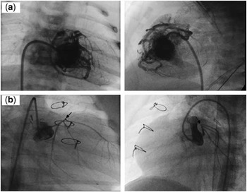
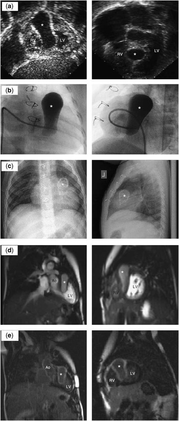
A male infant with pulmonary atresia with muscular infundibular atresia and numerous right ventricle-to-coronary artery fistulae underwent a modified Blalock–Taussig shunt in the newborn period. The right ventricle was not decompressed at the time of surgery because of the extensive nature of the right ventricle–to-coronary artery connections, as demonstrated by the angiography (Fig 1a). He had an uneventful post-operative course and was discharged home on post-operative day 8. He presented at 2 months of age for a routine outpatient visit. An echocardiogram was obtained, which revealed a large septal cavitary lesion that was not seen at birth or in the foetal period (Fig 2a); however, thinning of the superior aspect of the ventricular septum was noted at these times. The left ventricular function was normal and the patient was asymptomatic.
(a) Right ventriculogram of the patient in the newborn period before the modified Blalock–Taussig shunt surgery. Anteroposterior and lateral projections show numerous coronary cameral fistulae but no obvious coronary interruptions and no evidence for a ventricular septal aneurysm. (b) Selective left coronary angiography at 2 months of age shows a diminutive left coronary artery with the anterior interventricular coronary artery blindly ending at the apex of the septal aneurysm (marked by black arrow). The vessels off the left main coronary artery all course posteriorly.

(a) The transthoracic echocardiogram of the patient's 2-month status after modified Blalock–Taussig shunt placement. The subcostal long-axial oblique view and apical four-chamber view show a large ventricular septal aneurysm (marked by an asterisk). (b) The angiogram of the ventricular septal aneurysm taken at 2 months of age. The catheter is positioned across the tricuspid valve with the tip positioned within the mouth of the aneurysm. (c) The anteroposterior and lateral chest radiograph reveals a large, “light bulb”-shaped septal aneurysm, which is heavily calcified. (d) Magnetic resonance bright-blood imaging shows the large ventricular septal aneurysm, which runs from the base of the heart to the origin of the pulmonary artery. (e) Magnetic resonance imaging shows the borders of the septal aneurysm light up with delayed enhancement, consistent with myocardial scarring (RV = right ventricle; LV = left ventricle; Ao = aorta).

Following the echocardiogram, a cardiac catheterisation was repeated. The angiography confirmed the presence of a massive aneurysm that was in communication with the right ventricular cavity (Fig 2b). Selective coronary angiography was performed, which showed that the anterior interventricular branch of the left coronary artery ended blindly at the apex of the aneurysm (Fig 1b), suggesting that the aneurysm could have developed from stenosis and eventual occlusion of the proximal anterior interventricular coronary artery with resultant septal infarct and aneurysm formation. Alternatively, the aneurysm could represent progressive dilation of the distal anterior interventricular branch of the left coronary artery, which remained in continuity with the hypertensive right ventricle via a right ventricle-to-coronary fistula. This connection was visible on the newborn catheterisation (Fig 1a).
The patient remained asymptomatic and subsequently underwent staged single-ventricle palliation with right-sided cavopulmonary anastomosis at 4 months of age and Fontan completion at 27 months of age. Before the Fontan procedure, a catheterisation was repeated, which showed excellent left ventricular function. The aneurysm remained stable but had become heavily calcified as evident by the “light bulb” shape visible on a plain chest radiograph (Fig 2c). Cardiac magnetic resonance imaging was performed to further define the anatomy of the aneurysm and to elucidate the possible causes for its formation. On coronal and sagittal bright-blood imaging (Fig 2d), a large 3.4 × 3.4 × 1.4-centimetre ventricular septal aneurysm was seen originating from the right ventricle and extending the full superoinferior extent of the ventricular septum towards the level of the atretic pulmonary valve. The aneurysm was confined to the proximal one-third of the length of the septum, with the os of the aneurysm opening into the right ventricle. On viability sequences, the entire aneurysmal area is determined to be scar tissue consistent with remote infarction (Fig 2e). Despite these findings, the child has remained asymptomatic from a cardiovascular standpoint, with normal left ventricular function at the most recent follow-up, that is, at age 3.5 years.
Discussion
Pulmonary atresia with intact ventricular septum, which was first described by HunterReference Hunter1 in 1783, is a rare congenital cardiac anomaly representing less than 1% of all congenital cardiac disease. This lesion has significant anatomic variability with a high rate of coronary artery abnormalities – ranging from 31% to 68% – including coronary-cameral fistulae, coronary stenoses, and atresia.Reference Guleserian, Armsby, Thiagarajan, del Nido and Mayer2, Reference Freedom, Anderson and Perrin3 In some patients, the distal coronary perfusion is dependent on the right ventricle. This so-called “right ventricular-dependent coronary circulation” can lead to acute myocardial ischaemia and infarct of the left ventricle if the right ventricle is decompressed as part of surgical palliation.Reference Freedom, Anderson and Perrin3 Myocardial ischaemia and infarct may also develop over time because of the progression of coronary arterial stenoses.Reference Freedom, Anderson and Perrin3 To the best of our knowledge, acquired large aneurysms of the ventricular septum have not been previously reported in patients with pulmonary atresia and intact ventricular septum.
Congenital ventricular septal aneurysms are rare and have been reported in three patients with pulmonary atresia and intact ventricular septum with thinning of the septal myocardium noted in utero or at birth.Reference Nguyen, Srivastava, Ko and Lai4 The exact cause of aneurysm formation in this patient is unclear. Several mechanisms have been postulated, including prenatal or postnatal myocardial infarction of the septum due to irregularities of coronary flow; progressive dilation of the distal anterior interventricular branch of the left coronary artery due to exposure to suprasystemic right ventricular pressure; and/or myocardial weakening or structural abnormality with thinning of the ventricular septum. The prognostic significance of this finding is unknown. However, if we consider this an issue of coronary perfusion comparable to patients with pulmonary atresia and intact ventricular septum with right ventricular-dependent coronary circulation, based on actuarial survival analysis, the post-Fontan outcome should be comparable to other cardiac lesions requiring single ventricle palliation – 81% survival at 15 years.Reference Guleserian, Armsby, Thiagarajan, del Nido and Mayer2, Reference Powel, Mayer, Lang and Lock5