

Introduction
TiB2/Al composites are widely used in aerospace and automobile industries due to their high modulus, high strength, light weight, and excellent processability [Reference Han, Liu and Bian1, Reference Wang, Ma, Li, Li and Wang2]. However, the properties of the composites are significantly influenced by the morphology and distribution of the reinforced TiB2 particles, because of different stress states and interface bonding that exist around particles with different morphologies. The sharp corners in the particles may scratch the matrix in the service process and ultimately degrade the material performance dramatically. Therefore, it is necessary to understand the growth mechanism of TiB2 and the influence of different elements and then to realize the shape control during the formation of in situ TiB2 particles.
Generally, TiB2 exhibits hexagonal prism in aluminum matrix and the diameter ranges from few tens of nanometers to a few micrometers [Reference Guo, Sun, Jiang, Wu and Chen3, Reference Li, Wu and Liu4, Reference Ding, Liu and Nie5, Reference Lü, Lai, Su, Teo and Feng6]. Due to their excellent properties, TiB2 attracts increasing attention as a reinforcement of composite materials [Reference Wang, Xie, Zhou, Shi and Wu7]. The size and morphology of particles will significantly influence the mechanical property of the composites due to different stress states existing around the particles with different shapes [Reference Uddin, Mahmud, Wolf, Glanz, Kolaric, Volkmer, Höller, Wienecke, Roth and Fecht8, Reference El-Kady and Fathy9, Reference Deng, Wang, Wu, Hu, Wu and Gan10]. Therefore, the shape control of the TiB2 particles in in situ process is very important. However, the morphology of TiB2 is rather difficult to control due to the complexity of in situ reaction. Recently, it has been found that the TiB2 morphology is significantly influenced by the matrix composition. Li [Reference Li, Chen, Wang, Lavernia and Shan11] fabricated the in situ nanoscale TiB2 particles-reinforced Al composites, and the tensile strength was approximately 9 and 5 times higher than those of the initial coarse-grained Al. Cha [Reference Cha, Lartigue-Korinek, Walls and Mazerolles12] suggested that in Fe matrix, TiB2 exhibited screw-shaped, hexagonal plates or shapes like T with a straight edge in two-dimensional (2D) view. It was examined that in Ti alloy matrix the morphology of TiB2 was generally flaky or rod-like [Reference Wang, Lin, He, Wang and Chen13, Reference Wang, Lin, He, Wang and Chen14]. In Guo’s work [Reference Guo, Shen and Wang15], the TiB2 particles exhibited rectangular or irregular hexagonal shape in the cross section in Cu matrix. Sun [Reference Sun, Xiang, Dai, Liu and Zhou16] provided a theoretical explanation for the easy grain coarsening of the TiB2 crystals by considering the anisotropic surface stability. According to the analysis, the author suggested that different bonding conditions of Ti and B in different crystal surfaces contribute to the different surface energies which inhibits or facilitates adsorption of atoms. Based on Sun’s [Reference Sun, Xiang, Dai, Liu and Zhou16] research, the hexagonal plate-like grain morphology was predicted. In our previous work [Reference Sun, Zhang, Zhang, Ma and Wang17], it was found that TiB2 morphology was influenced significantly by different alloy elements in Al matrix.
Polyhedral or spherical structures, such as octahedral, cuboctahedral, truncated cubic, and truncated hexagon, can effectively decrease the stress concentration trend and therefore have attracted much attention. The polyhedron or spherical particles possess interesting properties, such as high symmetry, nanosized dimension, and the existence of critical surfaces [Reference Cao, Fan, Bai, Hu, Yang, Yuan and Chen18, Reference Zasada, Piskorz, Cristol, Paul, Kotarba and Sojka19]. However, very little research has been conducted in the control of the spherical particles growth in the chemistry field. Firstly, some noble metal nanopolyhedrons were obtained by a special preparation and they exhibited excellent properties [Reference Zeng, Zheng, Rycenga, Tao, Li, Zhang, Zhu and Xia20, Reference Ma, Zeng, Li, McKiernan, Xie and Xia21]. Then, the polyhedrons of some metallic oxides or ceramics, such as α-Fe2O3 [Reference Wang, Song, Dang, Chen, Yang and Zeng22, Reference Yang, Xu, Sun, Zhang and Gao23], ZnO [Reference Gao, Li, Gao, Qiu, Gan, Wang and Leng24], LaB6 [Reference Pengting, Chong, Jinfeng, Jun and Xiangfa25], TiC [Reference Yang, Jin and Jiang26], NbB2 [Reference Jin, Shen, Li, Zhou and Jiang27], were synthesized. However, the preparation of polyhedron or spherical morphology of TiB2 particle has not been found.
Huang [Reference Huang, Yan, Ma and Wang28] studied the influence of different Sc additions on the formation of TiB2 particles and suggested Sc promoted the TiB2 particle growth and coarsened the particles. However, in the present experiment, TiB2 crystals with polyhedral or near-spherical morphologies were prepared in Al–Sc systems. Under the influence of Sc element, the TiB2 morphology transformed from the equilibrium hexagonal plates into the nearly spherical morphology, and moreover, the different growth mechanism of TiB2 particles in Al and Al–Sc systems were demonstrated. Additionally, with the different contents of Sc addition, the in situ TiB2 exhibited various morphologies and diameter distributions.
Results and discussion
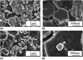
The in situ TiB2 particles morphology in the CPAl matrix and the Al–0.2Sc matrix is illustrated in Figs. 1(a)–1(d), respectively. It can be seen that in TiB2/CPAl composite, the majority of the reinforced particles exhibited hexagonal platelets surrounded by heaps of aggregated nanoscale reinforcements. Closer examination indicates that the isolated hexagonal particles primarily had sharp corners and edges, as shown in Fig. 1(b). Conversely, when Sc element was added, the TiB2 morphology changed markedly. As illustrated in Fig. 1(c), the TiB2 particles in Al–0.2Sc matrix appeared as polyhedron or nearly spherical shape. Meanwhile, in the Al–Sc matrix, the aggregation of the reinforced particles was improved substantially. The reinforcement distribution in the discontinuously reinforced composites significantly affected the comprehensive properties of the materials. The ideal situation was the reinforced particulates were distributed uniformly while the micro–nano reinforcements, especially the nanoscale particulates, tended to exist as clusters to maintain a minimum energy system. Consistently, Gao [Reference Gao, Wu, Lü, Xiong, Du and An29] concluded that the dispersively distributed TiB2 particles in the Aluminum matrix by ultrasonic vibration treatment can increase the tensile strength by 84%. Compared with the Fig. 1(a), the particles in Fig. 1(c) were individually scattered rather than gathered into a mass. Further inspection under higher magnification in Fig. 1(d) indicated the modified TiB2 particles by Sc packaged with multiple planes.
In situ TiB2 morphology (a, b) in the CPAl matrix and (c, d) in the Al–0.2Sc matrix.

According to the Bravais–Friedel law [Reference Liu, Chen, Zhou, Tian, Pereira and Ferreira30] and Wuff’s theorem [Reference Benson and Patterson31], the equilibrium crystal morphology of TiB2 was hexagonal enclosed by the faces (0001) and  $\left( {10\bar{1}0} \right)$. Typically in the ceramics, based on the faceted growth mode (because of the larger Jackson factor α > 2), the TiB2 crystals displayed anisotropic growth characteristics, exhibiting hexagonal morphology, close to the CPAl matrix. Similarly, in Abdel-Hamid’s [Reference Abdel-Hamid, Hamar-Thibault and Hamar32] work, the TiB2 compounds were fabricated by the liquid AlTi–B using the electromagnetic phase separation technique, and the scanning electron microscopy results showed that the obtained crystals presented a plate-like growth limited by large (0001) faces and smaller
 $\left( {01\bar{1}0} \right)$ facets. In Hamid’s experiment, the TiB2 generated in the liquid and the growth conditions approached the equilibrium conditions. According to the crystal growth theory, the growth rate of the TiB2 close-packed lattice planes conformed to the following law: (0001) <
 $\left( {10\bar{1}0} \right)$, which contributed to the hexagonal platelet formation of the TiB2 crystals. Nonetheless, the moderate rare element addition may alter the growth mode of the crystals by adjusting the surface energy of the different lattice planes, simultaneously, deviating from the equilibrium morphology.
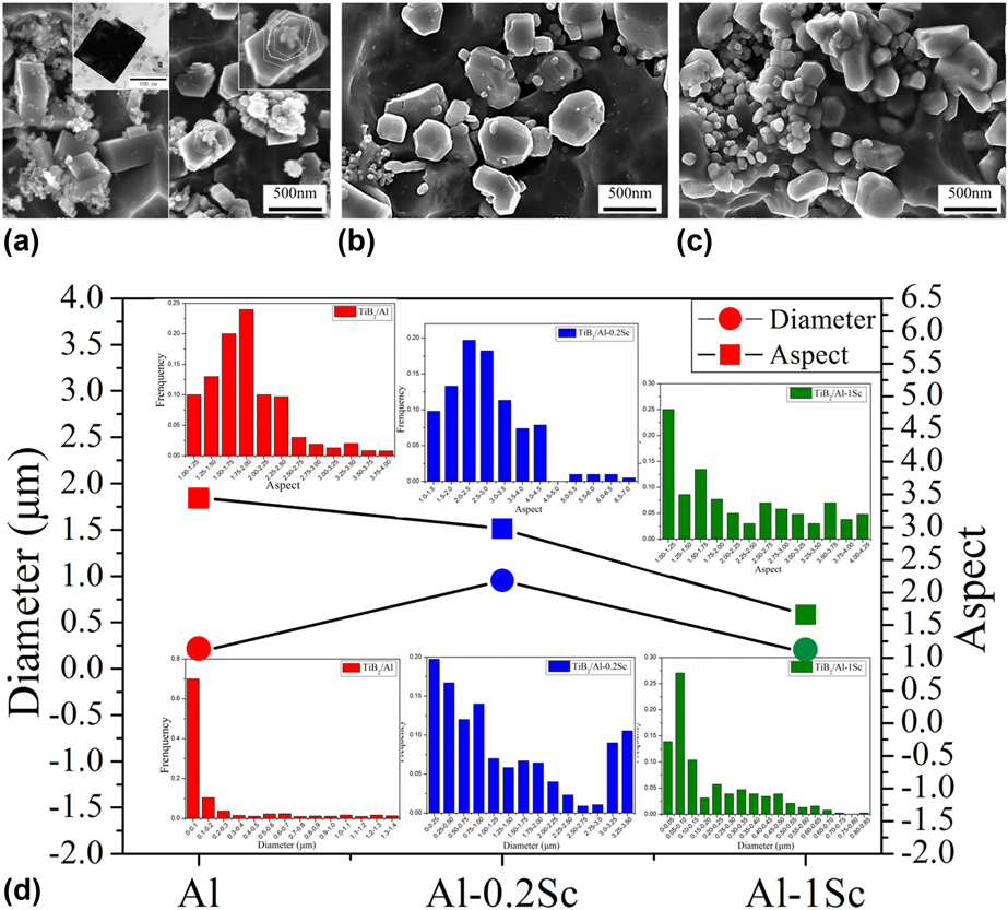
The morphology evolution of TiB2 particles with the increase in Sc is shown in Figs. 2(a)–2(c). The particle size distribution (PSD) and the particle aspect distribution (PAD) in different matrices are shown in Fig. 2(d). Here, the aspect referred to the ratio of the particle diameter and thickness, and it can be easily concluded that the smaller the aspect value, the closer to sphere the particle was. The TiB2 particles in CPAl exhibited hexagonal plate structure accompanied with abundant powdery fine particles [Fig. 2(a)]. Further examination indicated that the hexagonal plates had smooth surface with sharp corners and the powdery fine particles with the diameter of about dozens of nanometers generally existing as clusters. Moreover, the magnified observation of TEM showed that the fine particles in clusters exhibited hexagon or quadrangle with clear corners in 2D view [Fig. 2(a)]. In Fig. 2(a) closer observation showed that parallel growth stripes existed along the C-axis on some hexagonal particles. However, the trace of the spiral growth, such as the continuous winding line or the pointed tail at the end top, was not found. It can be seen from Fig. 2(d) that the most PSD in CPAl fell in the scope of 0–0.2 μm and the average diameter was about 0.19 μm. In Al–0.2Sc, the majority of the particles presented as polyhedrons with the degeneration of the sharp corners and edges. The sharp corners were replaced by small triangular planes and the edges were replaced by chamfered planes resulting in the polyhedral morphology of TiB2 particles in Al–0.2Sc matrix [Fig. 2(b)].
SEM images of microcrystals in (a) CPAl, (b) Al–0.2Sc matrix, (c) Al–1Sc matrix, and (d) the size and aspect distribution in different matrices.
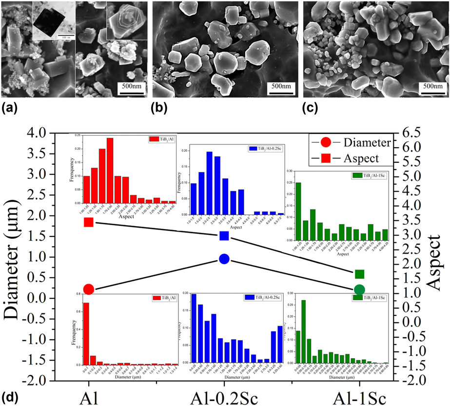
When the Sc content was increased to 0.2%, the PSD ranged in the scope of 0–3.5 μm while the majority of them were distributed in the range of 0–1 μm; thus, the average diameter of TiB2 particles in Al–0.2Sc was 0.95 μm. Moreover, the big particles with micrometer dimension mostly exhibited hexagonal morphology. As the Sc content increased to 1%, the majority of TiB2 particles exhibited nearly spherical morphology with the disappearance of corners and edges. The average diameter of TiB2 particles in Al–1Sc was the smallest (0.17 μm), which was close to the value in CPAl, but the PSD was relatively centralized with small standard deviation under the influence of Sc. The PAD in CPAl and Al–0.2Sc was probably subjected to normal distribution, while the PAD in Al–1Sc was relatively discrete with larger standard deviation probably resulting from the uneven distribution of Sc. Furthermore, the average value of the aspect was greatest in Al (3.46) and minimum in Al–1Sc (1.66) indicating that the TiB2 particles were obviously spheroidized with the existence of Sc. Therefore, with the increase in Sc, the morphology of TiB2 particles transformed from hexagonal to polyhedron and finally spherical shape with the particle size decreasing. Further, compared with CPAl, in Al–Sc systems, the amount of powdery particles decreased significantly, indicating the improvement of the particle aggregation.
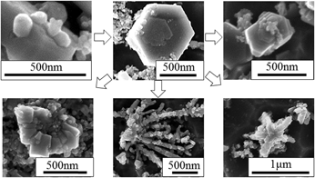
Comparing the particle morphology in CPAl and Al–Sc systems showed that the Sc solvent as a kind of impurity has a great impact on the TiB2 growth mechanism. The images in Figs. 3 and 4 provide clear information about the growth process of TiB2 particles in Al and Al–Sc systems by extracting typical morphology at different growth stages, respectively. Figure 3 showed the growth of TiB2 particles in CPAl. It showed that TiB2 grain exhibited a quasispherical morphology at the initial stage, and then a preplatelet was generated as the spherical grain grew. Subsequently, 2D nucleus developed on the preplatelet and finally, a complete TiB2 particle with the morphology of hexagonal prism with growth stripes, pentagram, dendrite-structure particles or clusters stacked by flaky particles (Fig. 3) would be fabricated by repeating the two steps above [Reference Li, Wu and Liu4].
Typical morphology evolution of TiB2 crystals at different stages in CPAl matrix.

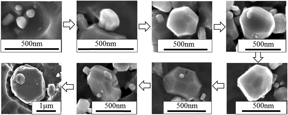
Typical morphology transformation of TiB2 particles at different stages in Al–0.2Sc matrix.

The typical TiB2 morphology at different stages in Al–0.2Sc was extracted as shown in Fig. 4. Similarly, the TiB2 microcrystals presented nearly spherical shape with a diameter of less than 100 nm at the earlier stage. As the growth proceeded, small flat facets appeared on the surface of the sphere to retain low interfacial energy and polyhedral TiB2 particles with different sizes were fabricated at different stages. As the polyhedral TiB2 particles continued to grow, the higher-index planes degraded and were replaced by low-index ones progressively. Therefore, as some low-index planes grew bigger, sharp corners and edges emerged on the particle surface. Finally, the morphology of TiB2 particle will appear as complete hexagonal plate with a diameter of about 1–2 μm. However, further observation demonstrated that there were no growth stripes like that in CPAl appearing on the surface of hexagonal particles in Al–Sc systems. Based on the experimental observation, it was speculated that the TiB2 particles conformed to different growth mechanisms in CPAl and Al–Sc systems.
According to the crystal growth theory, calculations of the attachment energy of the TiB2 crystal suggested the following hierarchy of the growth rates: (0001) <  $\left( {10\bar{1}0} \right)$ <
 $\left( {10\bar{1}1} \right)$ <
 $\left( {1\bar{2}10} \right)$ <
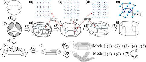
 $\left( {1\bar{2}11} \right)$ [Reference Hyman, McCullough, Levi and Mehrabian33]; thus, the equilibrium crystal shape of TiB2 was hexagonal platelets. Figure 3 shows the schematic model of morphology evolution in Al and Al–Sc systems. In the initial stage, TiB2 nucleus undergoes an isotropic growth both in CPAl and Al–Sc, consequently producing a spherical TiB2 grain with nanoscale diameter as shown in Fig. 5(a). With further growth, the spherical grain became unbalanced as the result of decrease in surface energy and some small facets emerged on the surface of the sphere [Fig. 5(f)]. Then, the growth mechanism of TiB2 in Al and Al–Sc split and TiB2 grew according to mode (II) dominantly in CPAl matrix. Mode (II) conformed to the 2D nucleation mechanism as illustrated in Fig. 5. Firstly, the thin hexagonal preplatelets were formed enclosed by {0001} and
 $\left\{ {10\bar{1}0} \right\}$ in the melt through the dissolved and adsorption of the Ti and B atoms [Fig. 5(k)]. As the growth rate along
 $\left\langle {10\bar{1}0} \right\rangle$ was faster than 〈0001〉, the Ti and B atoms concentrated on the top of {0001} which would promote the formation of the 2D nucleus. The repetition of the 2D nucleation and the growth of the platelets will finally form the hexagonal plates with parallel growth steps along the 〈0001〉 direction as schematically shown in Fig. 5(m). Sometimes, several 2D-nucleis simultaneously appeared on one preplatelet and subsequently the pentagram or dendrite-like structure particles composed by small plates with a displacement between the two adjacent plates were fabricated as shown in Fig. 5(n).
Schematic illustration of the morphology evolution model of TiB2 in CPAl (II) and Al–0.2Sc, Al–1Sc (I) matrixes. (a) Initial spherical crystal nuclei; (b) atomic arrangement in  ${\left( {1\bar{2}12} \right)_{{\rm{Ti}}{{\rm{B}}_{\rm{2}}}}}$; (c) atomic arrangement in
 ${\left( {11\bar{2}0} \right)_{{\rm{Ti}}{{\rm{B}}_{\rm{2}}}}}$; (d) atomic arrangement in
 ${\left( {1\bar{1}01} \right)_{{\rm{Ti}}{{\rm{B}}_{\rm{2}}}}}$; (e) unit cell of TiB2 crystal; (f) facets in the spherical nuclei; (g, h, i) polyhedron morphology of TiB2 crystals; (j) complete hexagonal morphology of TiB2 crystal; (k, l) formation of the 2D nucleus in the hexagonal preplatelet; (m) hexagonal prism of TiB2 with parallel growth steps along the 〈0001〉 direction; and (n) pentagram or dendrite-like structure TiB2 particles.
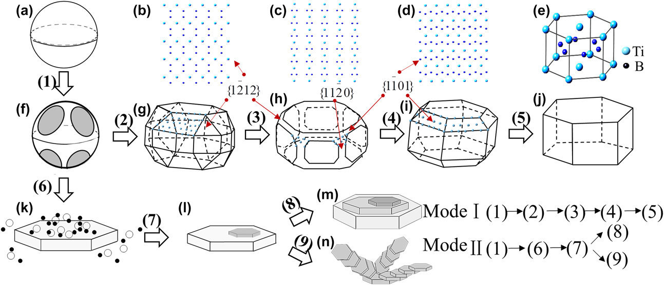
It was supposed that the growth of TiB2 in Al–Sc acted as mode (I) as illustrated in Fig. 5. Firstly the spherical nucleus generated in the melt and then the isotropic spherical nucleus will lose their stability with further growth, resulting in the facets with curved surfaces gradually covering the spherical surface. As the faceting process proceeded, the facets grew bigger and flatter with the higher-index planes gradually replaced by close-packed or nearly close-packed planes. Based on the Bravais–Friedel law [Reference Liu, Chen, Zhou, Tian, Pereira and Ferreira30], the particle morphology tended to be polyhedron enclosed by {0001},  $\left\{ {10\bar{1}0} \right\}$, the prismatic plane
 $\left\{ {10\bar{1}1} \right\}$,
 $\left\{ {11\bar{2}0} \right\}$ and the pyramidal plane
 $\left\{ {1\bar{2}12} \right\}$ as shown in Fig. 5(g). The atomic arrangement of the prismatic
 $\left\{ {1\bar{2}12} \right\}$ and pyramidal planes
 $\left\{ {10\bar{1}0} \right\}$ and
 $\left\{ {11\bar{2}0} \right\}$ is shown in Figs. 5(b)–5(d). With the further growth of the microcrystal, the non-close-packed planes degraded gradually. The triangular plane on the chamfered corners
 $\left\{ {1\bar{2}12} \right\}$ degenerated firstly as the sparse atomic arrangement [Fig. 5(b)], followed by the chamfered side prismatic plane
 $\left\{ {11\bar{2}0} \right\}$. Finally, the
 $\left\{ {10\bar{1}1} \right\}$ disappeared as shown in Fig. 5(i) and a complete hexagonal prism enclosed by {0001} and
 $\left\{ {10\bar{1}0} \right\}$ formed with clear corners and edges as illustrated in Fig. 5(j).
In Al–Sc systems, the morphology of TiB2 particles exhibited polyhedrons or nearly spherical shapes with curved surfaces and vague edges. Based on the experimental results and the growth process of the particles, it was assumed that Sc was preferentially adsorbed on some high-index crystal surfaces to inhibit the prior growth of the advancing surfaces, which will lead to the morphology transformation. Different levels of Sc will make the particles stay in different stages of mode (II) as shown in Fig. 5. In Al–0.2Sc, the TiB2 particles mainly presented as polyhedrons with the average diameter of 0.95 μm, while in Al–1Sc the particles exhibited as nearly spherical morphology with a smaller diameter of about 0.17 μm, indicating that abundant Sc maintained particle morphology in the earlier period.
In the research by Chen [Reference Chen, Wang, Wang, Lin and Wang34], the interfacial kinetics had an important effect on the morphological stability of the spherical particles. Furthermore, the growth velocity of the particles decreased with the increase in interfacial kinetics parameters.
According to the analysis above, the adsorption energy (ΔE ads/surf) of Sc on the planes at the chamfered corners or edges of TiB2 was calculated. For the adsorption of Sc, the ΔE ads/surf was expressed as Eq. (1) [Reference Chen, Wang, Li, Zha and Jiang35]
 $$\eqalign{\Delta {E_{{{{\rm{ads}}} / {{\rm{surf}}}}}} = - {{\left( {{E_{{{{\rm{Sc}}} / {{\rm{Ti}}{{\rm{B}}_{\rm{2}}}}}}} - {E_{{\rm{isolated}}\;{\rm{Sc}}}} - {E_{{\rm{isolated}}\;{\rm{Ti}}{{\rm{B}}_{\rm{2}}}}}} \right)} / \cr{{n_{{\rm{surf}}}}\quad ,}}$$
where  ${E_{{{{\rm{Sc}}} / {{\rm{Ti}}{{\rm{B}}_{\rm{2}}}}}}}$ is the energy of the interface system and E isolated Sc and
 ${E_{{\rm{isolated}}\;{\rm{Ti}}{{\rm{B}}_{\rm{2}}}}}$ are the energies of the Sc layer and the TiB2 substrate, respectively. n surf is the number of adsorption sites in the calculated cell, and Sc atom was added in the Bridge (B) position [Reference Li, Sakidja and Ching36]. The results are listed in Table I.
The ΔE ads/surf of Sc on different surfaces of TiB2.

The calculation results confirmed that Sc was strongly adsorbed on the surface of TiB2 particles and Sc was preferentially adsorbed on  $\left\{ {1\bar{2}12} \right\}$,
 $\left\{ {11\bar{2}0} \right\}$, and
 $\left\{ {10\bar{1}1} \right\}$ which would inhabit the growth of these faces effectively and retain a lower-energy state of the polyhedral or quasispherical TiB2 particles in Al–Sc systems.
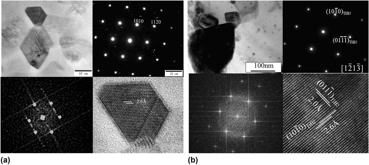
The chamfered surface specification under the influence of Sc can be confirmed from the results of TEM and HR-TEM images as shown in Fig. 6. Figure 6(a) shows a typical distorted basal facet (0001) enclosing the TiB2 particles of hexagonal morphology. Figure 6(b) shows that the polyhedron TiB2 particles exposed a nearly close-packed side plane  $\left( {1\bar{2}1\bar{3}} \right)$ on the influence of Sc. The HRTEM results were consistent with the analysis as shown in Fig. 5, showing that the TiB2 particles exposed the nearly close packed crystal planes in Al–Sc matrix, consequently leading to the polyhedron or nearly spherical morphology. Based on the adsorption energy calculated in Table I, precisely because of the Sc preferential adsorption in the nearly close packed planes of TiB2, the nearly spherical transformation of the particles occurred.
TEM and HR-TEM images of (a) TiB2 particles in CPAl matrix; (b) TiB2 particles in Al–0.2Sc matrix with the chamfered face.

The morphology control of the in situ reinforcements TiB2 was a complex process, involving the addition method, the holding time, and distribution of the addition element and so on. Meaningfully, the present paper provided a method to fabricate spherical in situ TiB2-reinforced Al composites. By finely adjusting the Sc addition content, the spherical reinforced TiB2 particles can be obtained. Further examination was conducted continuously.
Conclusions
TiB2 particles with polyhedron or near-spherical morphology were prepared in CPAl matrix under the influence of Sc element. The morphology of TiB2 particles in three-dimensional space was examined using SEM. With the modifier content of Sc, increasing TiB2 particles transformed from hexagonal plates to polyhedron or near-spherical morphology with more high-index crystal planes exposed. The different growth mechanism of TiB2 particles in Al and Al–Sc matrix was discussed by the observation of extracted typical particle morphology in different growth stages. It was the main reason for the morphology transformation that the selective adsorption of Sc element in the crystal surfaces decreased the total surface energy, and therefore, the morphology of TiB2 can be restrained at an intermediate stage. Sc was preferentially adsorbed on  $\left\{ {1\bar{2}12} \right\}$,
 $\left\{ {11\bar{2}0} \right\}$, and
 $\left\{ {10\bar{1}1} \right\}$, which would inhabit the growth of these faces effectively and retain a lower-energy state of the polyhedral or quasispherical TiB2 particles in Al–Sc systems. The present study demonstrates a simple method to prepare near-spherical TiB2 particles in Al matrix by in situ method, and it provides instructions for the morphological control of other ceramic particles. It was supposed that TiB2 particles with different morphologies (quasispherical shape or polyhedron) and size will be fabricated by adjusting the Sc levels. The influence of alloy element on TiB2 morphology is very complicated and the detailed mechanism of the particle morphology transformation should be investigated further.
Experiments
The material used in the experiment was prepared from a commercial pure aluminum (CPAl) with different contents of Sc (0, 0.2 wt%, and 1 wt%, respectively), and TiB2 particles were formed by reactions of mixed salts (K2TiF6 and KBF4). The metal ingot was melted in steel mold which was placed in the resistance furnace and heated to about 750 °C. Then, the mixed salts were added in a proper proportion and the melt was preserved at 850 °C for 1 h. The content of the generated TiB2 both in the commercial Al matrix and Al–Sc matrices was 10 wt%. The temperature of the melt containing the in situ TiB2 particles was reduced to 750 °C and held for about half an hour before pouring into a preheated steel mold.
Metallographic samples were prepared from a piece of the solidified material, about 30 mm from the center of the cast ingot by mechanical grinding and polishing, followed by etching in a 0.5% HF aqueous solution for about 3–4 min. The microstructure of both the TiB2/Al composite and the TiB2/Al–Sc composite was examined using a scanning electric microscope (Nova Nano SEM, FEI, Brno, Czech Republic) and transmission electron microscope (TEM; JEM2010, Japan Electronics Co. Ltd., Tokyo, Japan). Statistical analysis on the size and shape factor of the primary silicon was carried out using computer image processing facility.