I. INTRODUCTION
New types of nanostructured materials, such as nanoflowers, urchin-like structures, dendritic structures, and multipods, have recently attracted considerable attention as microwave-absorbing and shielding materials.Reference Tong, Wu, Guan, Qian and Yuan1–Reference Tong, Guan, Xiao, Mou, Wang and Yan6 This is because of the ability of these complex nanostructures to maintain the basic features of the original material and confer novel absorbing properties and mechanisms. More importantly, their high Brunauer–Emmett–Teller (BET) specific surface area (S BET) and adjustable shape parameters allow control of subsequent microwave electromagnetic (EM) and absorbing characteristics. In particular, Fe3O4 nanomaterials have aroused much interest because of their potential applications as catalysts, gas sensors, magnetic filters, electrode materials, magnetic storage media, and as materials for spin electronics and biomedical research.Reference Yu, Chen, Rice, Wang, White and Sun7–Reference Peng and Sun10 Fe3O4 complex nanostructures could also be utilized as microwave absorbing materials.Reference Chen, Gao, Wang, Zhu, Wang, Cao and Jin11–Reference Zhao, Jia, Wei, Pu and Liu13 However, studies on microwave absorption properties of Fe3O4 nanomaterials are few. Chen et al.Reference Chen, Gao, Wang, Zhu, Wang, Cao and Jin11 found that porous Fe3O4/SnO2 core/shell nanorods exhibited dual-frequency absorption characteristics, with a maximum reflection loss of −27.38 dB at 16.72 GHz on a thickness of 4 mm. Fu and coworkersReference Cao, Fu, Yang, Yu, Zhang, Wang, Zhao, Sui, Zhou, Zhao, Leng, Zhao, Chen and Qi12 reported that flower-like ZnO/Fe3O4 nanocomposites showed markedly high EM wave absorption compared with the individual nanostructures (bare flower-like tubular ZnO or Fe3O4 nanoparticles). This difference was ascribed to the three-dimensional conductive networks of the nanocomposites and the diffuse reflections produced by them. In addition, Zhao et al.Reference Zhao, Jia, Wei, Pu and Liu13 studied the microwave absorption characteristics of hierarchically nanostructured Fe3O4 microspheres. They attributed two microwave magnetic loss peaks at 4.0−5.0 GHz and 16.0−17.0 GHz to the effect of the morphology of the hierarchical structure. Although the aforementioned complex nanostructures display some increased EM characteristics and new absorption mechanism, the corresponding absorbing properties are unsatisfactory for practical application. Developing novel nanostructured absorbers with high microwave absorbing properties remains a great challenge.
In this article, morphological dependence of static magnetic and microwave absorption properties of polymorphic Fe3O4 nanomaterials, namely, nanospheres, urchins, and sponges, are reported. Compared with Fe3O4 nanospheres, Fe3O4 urchins and sponges exhibited significantly enhanced microwave absorption properties. The possible mechanisms for the observed properties are discussed in detail.
II. EXPERIMENTAL
A. Preparation
All chemical reagents were from Shanghai Chemical Reagents Company. All were of analytical grade and were used as received without any further purification.
1. Synthesis of Fe3O4 nanospheres
Fe3O4 nanospheres were prepared through a previous oxidation–precipitation method.Reference Yu, Zhang, Zhang, Qiao, Yang and Liu14 In a typical synthesis, 100 mL of 0.15 M NaOH solution was placed in a round-bottom flask and mechanically stirred at 50 °C. Afterward, 100 mL of 0.06 M FeSO4 solution was slowly added to the solution under N2 protection, and the resulting mixture was stirred vigorously for 10 min. A constant stream of air was then introduced into the reaction mixture at 40 L·h−1 for 3 h. The resulting precipitates were magnetically collected and sequentially washed with distilled water and ethanol. Spherical Fe3O4 nanoparticles were obtained upon drying the precipitates under vacuum at 60 °C for 12 h.
2. Synthesis of Fe3O4 sponges
In a typical procedure, 17.80 g of D-(+)-glucose (C6H12O6·H2O) and 20.20 g of iron (III) nitrate [Fe (NO3)3·9H2O] were dissolved in 100 mL of deionized water. The obtained solution was vigorously stirred at 70 °C until the water evaporated and a yellow-green precipitate remained. This precipitate was filtered, washed several times with deionized water, and then dried in vacuum at 60 °C for 12 h. Finally, Fe3O4 sponges were obtained after carbothermic reduction of the products at 500 °C for 2 h under nitrogen. This procedure is similar to a protocol reported in the literature.Reference Yan, Guan and Wang15
3. Synthesis of Fe3O4 urchins
First, the urchin-like α-FeOOH precursors were prepared by a glucose-guided hydrolysis approach according to the procedure in a previous work.Reference Tong, Guan and Zhang16–Reference Tong18 The α-FeOOH precursors were annealed at 300 °C for 1 h and then reduced in a mixed gas of H2/Ar (1:4 volume ratio) at 350 °C for 2 h to obtain Fe3O4 urchins. The flow rate of the mixed gas and the constant heating rate were 1.0 L·min−1 and 5 °C·min−1, respectively. Finally, the system was allowed to cool to room temperature under N2, and the product was collected for characterization. A similar H2 reduction route for Fe3O4 has been reported earlier.Reference Osterhout and Craik19
B. Characterization
The phase analysis of the samples was performed by an x-ray diffraction (XRD) (D/MAX-RB; Rigaku, Tokyo, Japan), using CuKα radiation with 2θ from 10–80°. The S BET was measured using a surface area analyzer (Autosorb-1; Quantachrome, Boynton Beach, FL). The surface morphologies of the as-prepared samples were observed under a field-emission scanning electron microscope (FE-SEM) (S-4800; Hitachi, Tokyo, Japan), operated at 10.0 kV.
The static magnetic properties for the samples were carried out at room temperature using a vibrating sample magnetometer (4HF; ADE, Chicago, IL) with a maximum magnetic field of 17.5 kOe. The complex permittivity and permeability of the samples were measured using a vector network analyzer (N5230; Agilent, Santa Clara, CA) at 2–18 GHz. The samples used for EM measurements were prepared by homogeneously mixing the Fe3O4 samples with the wax melted at 60 °C in a mass ratio (Fe3O4/wax) of 1:1. The mixture was subsequently made into toroidal-shaped samples with an outer diameter of 7.0 mm, inner diameter of 3.0 mm, and thickness of ∼3.0 mm while hot. In general, microwave absorption property is denoted by the reflection loss (R L). In the case of a metal-backed single layer, R L was calculated by the following equationsReference Tong, Wu, Guan, Qian and Yuan1:


where c is the light velocity, Z 0 is the characteristic impedance of free space, and Z in is the input impedance between free space and the materials.
III. RESULTS AND DISCUSSION
A. Structural and morphological characterization
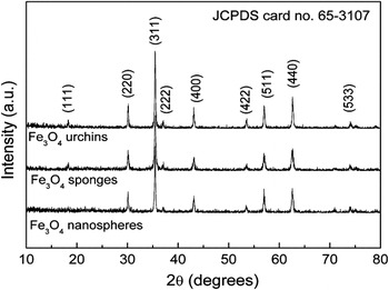
The phase structures of the three synthesized samples were characterized by their XRD patterns. All the diffraction peaks in Fig. 1 can be indexed to face-centered cubic Fe3O4 (JCPDS Card No. 65-3107). Also, the Mössbauer spectra of three samples (see Fig. S1 in the supporting information) can further confirm that all the samples were Fe3O4. The average crystal size and the internal strain of the products were calculated by the Hall–Williamson equation.Reference Wang, Gong, Li, Liu and Cheng20, Reference Kim, Chung, Kim and Jeon21 The S BET was determined through a multipoint BET method. These results are listed in Table I. The Fe3O4 nanospheres obtained at relatively low temperature had a high S BET, small crystal size, and high internal strain (Table I). However, the Fe3O4 urchins possessed a slightly higher S BET than the Fe3O4 sponges due to the morphological variation.
X-ray diffraction patterns of Fe3O4 nanospheres, sponges, and urchins.

Average crystal size (δ), internal strain (ε), Brunauer–Emmett–Teller specific surface area (S BET), and static magnetic and electromagnetic (EM) wave absorption properties of Fe3O4 with diverse morphologies.

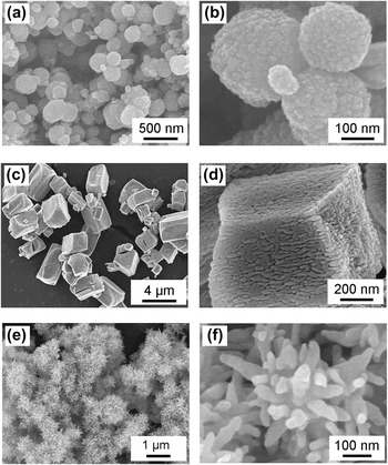
The surface morphologies of the samples were further observed by FE-SEM. The FE-SEM images of the Fe3O4 nanospheres [Figs. 2(a) and 2(b)] show 10-nm nanocrystals that formed 100- to 450-nm-diameter spherical structures. In the experiment, ferrous ion in alkaline solution formed a suspension of ferrous hydroxide, which was partially oxidized to ferric hydroxide in the presence of moderate levels of oxidant. Subsequently, magnetite particles began to nucleate, grow, and settle. The morphology and structure of Fe3O4 nanoparticles could be controlled by varying air flow rate, reaction temperature, reaction time, and alkali content.
Scanning electron microscope images of Fe3O4 nanospheres, sponges, and urchins.

In comparison, images of the Fe3O4 sponges produced [Fig. 2(c)] show large-scale submicropolyhedra with lengths of 0.5−6.0 μm and aspect ratios of 1−2. A higher magnification view [Fig. 2(d)] clearly reveals that the submicropolyhedra comprise uniform sponge-like porous structures. The porous structures were formed by ordered 10- to 20-nm-diameter nanofibers. In the current experiment, Fe(NO3)3·9H2O reacted with D-(+)-glucose and show 10-nm nanocrystals that formed 100- to 450-nm-diameter spherical structures and formed polyhedral iron gluconate, which occurred as crystals upon evaporation of water. The Fe3O4 submicropolyhedra were then obtained through a carbothermic reduction process. Kinetic factors, such as the feeding ratio of Fe(NO3)3·9H2O to D-(+)-glucose and carbothermic reduction temperature and time, were varied to modulate the morphology, structure, and aspect ratio of the products. The morphological evolution, formation mechanism, and properties of these products will be studied in subsequent work.
The Fe3O4 urchins [Figs. 2(e) and 2(f)] consisted of rod-like nanoparticles with 20- to 50-nm diameter and 50- to 200-nm length that radially spread and formed spherical aggregates with 0.5- to 1.0-μm diameter. A detailed account of the morphological and structural modulation of the products was presented in a previous work.Reference Tong, Wu, Guan, Qian and Yuan1
B. Static magnetic properties
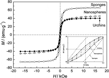
Figure 3 presents the hysteresis loops of polymorphic Fe3O4. The magnetic properties are listed in Table I. All samples were ferromagnetic and exhibited saturation below a magnetic field of 5 kOe. From Table I, the Fe3O4 nanospheres had low saturation magnetization (M s = 45.49 emu·g−1) and coercivity (H c = 28.4 Oe). The former is ascribed to the small crystal size that causes surface spin canting and thermal fluctuation,Reference Wang, Sun, Sun and Chen22 and the latter is attributed to its isotropic geometry. In contrast, Fe3O4 sponges exhibited markedly enhanced Ms and H c. In particular, the M s value of 65.55 emu·g−1 is about 1.5 times higher than those of Fe3O4 nanospheres (45.49 emu·g−1) and Fe3O4 urchins (40.17 emu·g−1). In general, a change in the value of M s is associated with grain size,Reference Li, Chen, Ong and Yang23 crystallinity, superexchange interactions,Reference Mørup, Madsen, Franck, Villandsen and Koch24 and noncollinear magnetic structure.Reference Li, Ong, Yang, Wei, Zhou, Zhao and Morrish25, Reference de Bakker, De Grave, Vandenberghe and Bowen26 The H c variation is related to grain size and stress and to shape and surface anisotropy.Reference Li, Chen, Ong and Yang23 Except for the increased grain size and crystallinity, the specific morphology may be substantially responsible for the higher M s of the Fe3O4 sponges. The magnetic easy axis of sponge-like Fe3O4 submicropolyhedra is aligned along the long axis of the submicropolyhedra, whose orientation is greatly affected by the magnetic field of surrounding Fe3O4 sponges. Eventually, M s increases.Reference Wang, Han, Xu, Wang, Du, Wang, Qin and Zhang27, Reference Niu, Chen, Ning, Jia and Wang28 On the other hand, the ordered array of nanofibers/nanorods along the long axis of the submicropolyhedra can also increase magnetic interactions and result in enhanced M s. Furthermore, the decreased M s of Fe3O4 urchins may not be dependent on factors such as grain size and crystallinity but rather on radial arrangement of the easy axis, which bates the magnetic interaction.
Magnetic hysteresis loops of Fe3O4 nanospheres, sponges, and urchins.

However, the pronounced increase of H c of the Fe3O4 sponges may be mainly associated with the higher shape anisotropy originating from the overall structure and subparticles. The Fe3O4 urchins were also found to display higher H c compared with Fe3O4 nanospheres; this implies that the urchin-like nanostructure could increase the H c. Similar coercivity behavior was also observed in Fe2O3 complex nanoarchitectures.Reference Tong, Guan, Xiao, Huang and Guan29 In this study, the increased H c may be relevant to the novel hierarchical structure constructed from a radial array of nanorods, which results in a combination of the coupling effect and the spatial confinement effect.Reference Tong, Guan, Xiao, Huang and Guan29
C. Dynamic magnetic properties
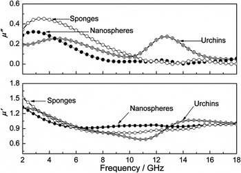
Figure 4 shows the frequency dependence of the complex permeability of the wax composites with 50 wt%Fe3O4 powders. For all samples, the values of the real part (μ′) and imaginary part (μ″) of the complex permeability slowly varied within 0.7−1.55 and 0−0.45, respectively, as the frequency increased from 2 to 18 GHz. This is due to the eddy current loss induced by EM waves.Reference Tong, Hua, Wu, Qin, Li and Gong30 The μ″ value of Fe3O4 nanospheres is nearly constant at high frequency, with wide resonance peak at 2−7 GHz. In comparison, Fe3O4 sponges exhibited a stronger and wider resonance peak, whereas Fe3O4 urchins produced two strong resonance peaks and higher μ″ value at high frequency. This suggests that Fe3O4 sponges and urchins caused larger magnetic loss.
Frequency dependence of the real part μ′ and imaginary part μ″ of the complex permeability of wax composites containing 50 wt%Fe3O4 nanospheres, sponges, and urchins.

Generally, the microwave magnetic loss of magnetic materials mostly originates from magnetic hysteresis, domain wall resonance, eddy current loss, natural resonance, and exchange resonance. However, magnetic hysteresis from irreversible magnetization only takes place in a strong applied field, whereas domain wall resonance from multidomain materials only occurs in the low frequency range (<2 GHz). Thus, the permeability of the obtained Fe3O4 samples is mainly due to eddy current loss, natural resonance, and exchange resonance rather than magnetic hysteresis or domain wall resonance.
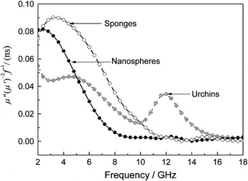
To investigate resonance behavior, the equation,  ,Reference Yang, Xu, Xia, Wang and Li31 where μ 0 is vacuum permeability and f is frequency, was used to explain the eddy current loss contribution to μ″ in relation to the thickness (d) and the electric conductivity (σ) of the composites. According to the equation, if the magnetic loss only stems from the eddy current loss, then the values of μ″(μ′)−2 f −1 should be constant when the frequency is changed. The values of μ″(μ′)−2 f −1 of the Fe3O4 nanospheres and sponges were similar at 9−18 and 12−18 GHz, respectively (Fig. 5). This means that the eddy current loss greatly contributed to μ″ and magnetic loss over this frequency range. A sharp resonance peak at 10−16 GHz was observed in the μ″(μ′)−2 f −1 plot of Fe3O4 urchins. This suggests that the magnetic loss of Fe3O4 urchins at high frequency is chiefly caused not only by eddy current loss but also by natural resonance and exchange resonance.Reference Tong18 This demonstrates that the multiresonance behavior of the ferromagnetic material is sensitive to its morphology.
Plots of μ″(μ′)-2f-1 versus frequency for Fe3O4 with diverse morphologies.

D. Dielectric properties
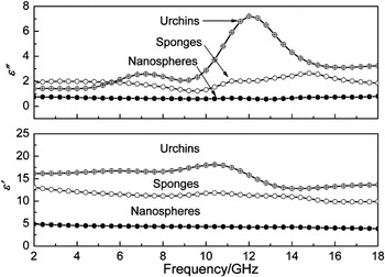
Figure 6 shows the frequency dependence of the real (ε′) and imaginary (ε″) parts of the relative complex permittivity of the wax composites containing 50 wt%Fe3O4 powders. The ε′ and ε″ values of Fe3O4 nanospheres present a flat trend and are ∼5.0 and 0.8, respectively. In comparison, Fe3O4 urchins and sponges produced noticeably increased ε′ and ε″, with two broadened resonance peaks, which indicate higher storage and loss capability for electric energy.
Frequency dependence of the real part ε′ and imaginary part ε″ of the complex permittivity of wax composites with 50 wt%Fe3O4 nanospheres, sponges, and urchins.

Dielectric loss mainly originates from conductance loss, dielectric relaxation loss, and resonance loss, among others.Reference Tong, Wu, Hua, Miao, Guan and Qian32 These are described by the composition, dimension, morphology, conductivity, and polarization model.Reference Tong, Guan, Fan, Wang and Li33 The complex nanostructures have better dielectric characteristics than those of traditional bulk materials because of their morphologies, larger surface volume, and quantum size effect. Nanomaterials with high defects and dangling bonds are apt to create space-charge polarization in an alternating EM field. On the other hand, complex nanostructures usually have high surface-to-volume ratio and shape anisotropy, which can generate strong interfacial and orientation polarizations. Thus, from Table I, the marked improvement of dielectric characteristics of Fe3O4 sponges and urchins are chiefly due to enhanced orientation polarization. Meanwhile, Fe3O4 urchins produced higher ε′ and ε″ values than Fe3O4 sponges did. This indicates that the urchin-like nanostructure could enhance dielectric loss due to its radial nanostructure, and thus generate orientation and interfacial polarizations.
Two strong peaks at 5−6 GHz and 10−15 GHz can be observed in the ε″ plots of Fe3O4 urchins and sponges. Similar multiresonance behaviors were reported for Fe3O4 microspheres,Reference Ni, Sun, Wang, Zhou, Yang, Wang and He34 Ni–B/Fe3O4 core/shell particles,Reference Li, Han, Tan and Xu35 and Ni-Fe3O4 hollow spheres.Reference Li, Beng, Shen and Hu36 These are attributed to the presence of polarizable Fe2+ ions in the heterogeneous spinel structure, the heterogeneous structure of the composite, and the vacancy and space-charge polarization, respectively. Hence, permittivity resonance behavior is intrinsic to the urchin-like and sponge-like nanomaterials.
E. Microwave absorption properties
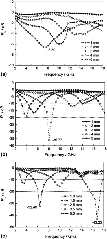
Figure 7 shows the calculated reflection loss curves for the paraffin-based composites with 50 wt%Fe3O4 powders. The composites containing isotropic Fe3O4 nanospheres showed poor absorption at 2–18 GHz, with minimal R L of –6.59 dB at 7.6 GHz. The absorption bandwidth (R L ≤ –10 dB) is zero. However, the composites containing anisotropic Fe3O4 sponges and urchins possessed conspicuously increased absorption properties, with a minimum R L of –35.77 dB at 8.0 GHz and –43.23 dB at 16.8 GHz, respectively. This corresponds to a matching thickness of 3.0 and 1.5 mm, respectively. Moreover, the absorption bandwidth (R L ≤ –10 dB) is extended from 0 GHz to 14.93 and 14.78 GHz, respectively; these correspond to a coating thickness of 1–5 mm. The above absorption property is better than those of previously reported Fe3O4 nanocrystalsReference Ni, Sun, Wang, Zhou, Yang, Wang and He34 and flower-like ZnO/Fe3O4.Reference Cao, Fu, Yang, Yu, Zhang, Wang, Zhao, Sui, Zhou, Zhao, Leng, Zhao, Chen and Qi12
Reflection loss curves of the paraffin-based composites containing 50 wt% (a) Fe3O4 nanospheres, (b) Fe3O4 sponges, and (c) Fe3O4 urchins.

Urchin-like and sponge-like nanomaterials demonstrated EM characteristics different from the spherical samples in terms of (i) enhanced value of all parameters and (ii) extra multiresonance behaviors. The microwave attenuation of the nanomaterials in this study has certain notable characteristics. First, the complex nanostructures are prone to form a microcurrent network, which can enhance conductivity loss.Reference Zhou, Qiao, Feng, Chen, Yan, Wu and Yan4, Reference Yan, Cheng, Zhou, Chen, Feng, Feng, Li, Wu, Wang and Yan5 For sponge-like submicropolyhedra formed by interconnected nanorods/nanofibers, the alternating electric field can excite the vibrating microcurrent inside the submicropolyhedron. The linking of urchin-like particles, which forms continuous micronetworks, may generate the microcurrent on a large scale.Reference Zhou, Qiao, Feng, Chen, Yan, Wu and Yan4, Reference Cao, Fu, Yang, Yu, Zhang, Wang, Zhao, Sui, Zhou, Zhao, Leng, Zhao, Chen and Qi12 Second, the complex nanostructures randomly distributed in the matrix can produce multiple scattering and further attenuation of EM waves. Third, urchin-like nanomaterials can act as isotropic quasi-antennas, which favor penetration of EM waves in the composites containing urchin-like nanostructures. The above EM energy is further converted, induced into a dissipative current, and then consumed in the discontinuous networks.Reference Zhou, Qiao, Feng, Chen, Yan, Wu and Yan4 Further experimental and theoretical work is in progress to elucidate the mechanisms of these phenomena.
IV. CONCLUSIONS
The properties of Fe3O4 complex nanostructures strongly depend on the aggregation morphology of the original materials. Compared with Fe3O4 nanospheres and urchins, Fe3O4 sponges present significantly enhanced M s and H c due to increased magnetic interactions and shape anisotropy, respectively. The microwave attenuation in composites with 50 wt%Fe3O4 sponges and urchins are as high as –35.77 dB at 8.0 GHz and –43.23 dB at 16.8 GHz, respectively. The excellent microwave absorption characteristics are ascribed to their unique morphologies, which result in larger EM parameters, multiresonant behavior, and dissipative current. The results give insight into the absorption mechanism of these nanostructures. These Fe3O4 nanostructures may have application in many fields, such as catalysis, energy conversion, EM devices, electrode materials, sensors, biomedicine, and microwave-absorbing and shielding materials.
ACKNOWLEDGMENTS
This work is financially supported by Natural Science Foundation of Zhejiang Province (Project Nos. Y4100022, Y4110025, and Y4090636), Special Grand Science-Technology Project in Zhejiang Province (No. 2010C11053), the Key Innovative Team of Magnetic Materials in Zhejiang Province (2011R09006-06), and Science and Technology Projects from Jinhua City (Grant Nos. 2010A12066 and 2011A13173).