Introduction
Mythimna separata (Walker) is a polyphagous pest damaging corn and other cereal crops, with the characteristics of wide host range, strong reproduction capacity, and long-distance migration ability (Jiang et al., Reference Jiang, Luo, Zhang, Sappington and Hu2011, Reference Jiang, Zhang, Cheng and Luo2014; Liu et al., Reference Liu, Qi, Chi and Wuriyanghan2016). Chemical pesticides are mainly used to control M. separata to prevent serious crop losses caused by larval feeding (Liu et al., Reference Liu, Qi, Chi and Wuriyanghan2016, Reference Liu, Wang, Lei and Zhu2017; Duan et al., Reference Duan, Gong, Wu, Miao, Jiang, Li, Wu and Wu2017). However, the use of chemical pesticides in a large area can easily lead to serious consequences such as insecticide resistance and residues, and damaging beneficial insects and natural enemies (Lv et al., Reference Lv, Wu and Liu2014; Younas et al., Reference Younas, Khan, Wakil, Shaaban and Prager2016; Duan et al., Reference Duan, Gong, Wu, Miao, Jiang, Li, Wu and Wu2017). Therefore, it is of great practical significance to explore new green and efficient pest control measures.
Chemosensory proteins (CSPs) are important for insect sensory system to sense external stimuli to obtain information, and complete important processes such as feeding, mating, spawning, and avoiding natural enemy (Kaupp, Reference Kaupp2010; Leal, Reference Leal2013; Wang et al., Reference Wang, Yang, Chen, Jiang, Li, Wang and Kang2015; Antony et al., Reference Antony, Soffan, Jakše, Abdelazim, Aldosari, Aldawood and Pain2016; Pelosi et al., Reference Pelosi, Iovinella, Zhu, Wang and Dani2018). CSPs mainly bind and dissolve small hydrophobic compounds, and are widely distributed in various chemical receptors of insects (Gong et al., Reference Gong, Luo, Rizwan-ul-Haq and Hu2012; Pelosi et al., Reference Pelosi, Iovinella, Felicioli and Dani2014). In addition to olfactory perception, they also play the functions of pheromone carrier, nutrient absorption, visual pigment carrier, insecticide resistance, regeneration, and development in insects (Pelosi et al., Reference Pelosi, Iovinella, Zhu, Wang and Dani2018). Although a great number of researches on CSPs have been reported in insects these years, most of them focused on the roles of CSPs in insect olfactory system. Other functions exercised in non-olfactory organs have been poorly studied.
In this study, two highly expressed CSPs were identified from the larval integument transcriptome of M. separata. The spatio-temporal expression patterns and functions of these two CSPs during larval development were analyzed, expecting to provide new insights of CSP function in insects. The results will lay a foundation for deeply analyzing the molecular regulation mechanism of M. separata larva growth, which is helpful to explore novel molecular targets for pest control.
Materials and methods
Insects
Mythimna separata was collected in the field of Yu'an District, Lu'an City, Anhui Province, and a stable population was established in the laboratory in Anhui Agricultural University. The larvae were reared with fresh corn leaves after hatching, and the adults were fed with 10% honey water after eclosion. Insects were maintained under the conditions of temperature 25 ± 1℃, relative humidity 70%, and photoperiod 12 L:12 D.
Cloning of MsCSP17 and MsCSP18
MsCSP17 and MsCSP18 were identified from the larval transcriptome dataset of M. separata (Huang et al., Reference Huang, Yin, Zhu, Jiang, Li, Dong, Sheng, Liao and Cao2021), with their open reading frame (ORF) sequences predicted using the ORF Finder (http://www.ncbi.nlm.nih.gov/orffinder/). The online tool Primer3web 4.1.0 (https://primer3.ut.ee/, Untergasser et al., Reference Untergasser, Cutcutache, Koressaar, Ye, Faircloth, Remm and Rozen2012) was used to design specific primers MsCSP17-(S/A) and MsCSP18-(S/A) for the ORF cloning (Table S1). Genes were amplified by PCR with the cDNA template synthesized from the RNA of 4th instar larval integuments using PrimeScript™ RT reagent Kit with gDNA Eraser (Perfect Real Time, Takara, Dalian, China) according to the manufacturer's protocol. The 25 μl PCR reaction contained 12.5 μl Prime STAR max Premix 2 × , 1 μl each primer, 1 μl cDNA template, and 9.5 μl RNase-free H2O. The reaction procedure was: pre-denaturation at 98°C for 3 min; 35 cycles of denaturation at 98°C for 10 s, annealing at 53°C for 15 s, and extension at 72°C for 2 s; and extension at 72°C for 5 min. The PCR products were detected by 1% agarose gel electrophoresis, and the target bands were purified. After T-vector ligation and verification, monoclone was sent to Tsingke Biotechnology Co., Ltd (Nanjing, Jiangsu) for sequencing. The plasmids with right ORF sequences were extracted and stored at −20°C until use.
Sequences analysis and phylogenetic tree construction
The amino acid sequences of MsCSP17 and MsCSP18 were obtained by A plasmid Editor (ApE, Davis and Jorgensen, Reference Davis and Jorgensen2022), with their physicochemical properties such as relative molecular mass and isoelectric point predicted using ExPASy (https://web.expasy.org/protparam/, Gasteiger et al., Reference Gasteiger, Hoogland, Gattiker, Duvaud, Wilkins, Appel, Bairoch and Walker2005). The protein tertiary structures of CSPs were predicted by SWISS-MODEL (https://swissmodel.expasy.org/interactive, Waterhouse et al., Reference Waterhouse, Bertoni, Bienert, Studer, Tauriello, Gumienny, Heer, de Beer, Rempfer, Bordoli, Lepore and Schwede2018). The signal peptides were predicted by SignalP4.1 (http://www.cbs.dtu.dk/services/SignalP/, Almagro Armenteros et al., Reference Almagro Armenteros, Tsirigos, Sønderby, Petersen, Winther, Brunak, von Heijne and Nielsen2019). The similarity of MsCSP17 and MsCSP18 was compared using the Blastp from the National Center for Biotechnology Information (NCBI), and multiple sequence comparisons of amino acids were performed using DNAMAN 9.0 (Lynnon Biosoft, San Ramon, CA). The phylogenetic tree was constructed using the Maximum-likelihood method in MEGA7 with Bootstrap of 1000 replicates as previous report (Kumar et al., Reference Kumar, Stecher and Tamura2016; Huang et al., Reference Huang, Liao, Yang, Xiao, Hu, Zhou and Cao2018), with orthologs from other insect species collected from the NCBI database.
Spatio-temporal expression analysis of MsCSP17 and MsCSP18
The cDNAs of different developmental stages of M. separata (eggs, 1st–6th instar larvae, pupae, male adults, and female adults), different tissues of 4th instar larvae (head, thorax, abdomen, midgut, Malpighian tubule, fat body, integument, ventral nerve cord, brain, and mandible), integuments of 1st–6th instar larvae, mandibles of 3rd–6th instar larvae, and integuments of 3rd–4th instar larvae during molting were synthesized as templates for RT-qPCR analysis of two CSP genes. Ribosomal protein 49 (rp49) was used as the internal reference gene (Table S1, Huang et al., Reference Huang, Yin, Zhu, Jiang, Li, Dong, Sheng, Liao and Cao2021; Zhao et al., Reference Zhao, Zhu, Xing, Li, Zhou, Wang, Li, Cao and Huang2022). RT-qPCR reactions contained 5 μl of TB Green Premix Ex Taq II (Tli RNaseH Plus) (2 × ), 0.5 μl cDNA sample, 0.5 μl each primer (10 μM), and 3.5 μl RNase-free water. The reaction procedure was: pre-denaturation at 95℃ for 30 s; 40 cycles of denaturation at 95℃ for 5 s, annealing and extension at 60℃ for 30 s; and a simultaneous solubility curve from 60 to 95℃ to verify the specificity of each primer pair. Three biological replicates were set for each genes and samples. The results were analyzed by the comparative cycle threshold (CT) method referring to the reference gene (Huang et al., Reference Huang, Liao, Yang, Shi, Xiao and Cao2020).
Microinjection of dsRNA
Primers with a T7 promoter sequence at the 5' end were designed to amplify PCR products to synthesize dsRNA for MsCSP17 and MsCSP18 using the online website https://primer3.ut.ee/, with green fluorescent protein (GFP) gene as a negative control (Table S1). PCR amplification was performed with MsCSP17-, MsCSP18-, and GFP-containing plasmids. The correctly sequenced products were used as templates for dsRNA synthesis in vitro according to the instructions of the T7 RiboMAX™ Express Large Scale RNA Production System kit (Takara, Dalian). DsMsCSP17, dsMsCSP18 and dsGFP were injected into healthy third instar larvae by Nanoliter2010 microinjection system (World Precision Instruments, Sarasota, FL, USA). The injection site was at the front of the abdomen (1st–2nd abdominal segments). Each larva was injected with 2 μl of dsRNA at a concentration of 1000 ng/μl, and 24 larvae were injected as a group, with three replicates for each gene.
Phenotypes of larvae after RNAi
After injection, the larvae were fed with fresh corn leaves, weighing daily. The abnormal phenotypes were counted and recorded by a QImaging Micropublisher 3.3 digital camera mounted on an Olympus BS41 stereo microscope (Tokyo, Japan). Larvae injected with dsMsCSP17, dsMsCSP18 and dsGFP for 24 hours were randomly selected for RNA extraction and cDNA synthesis, with three biological replicates and two larvae in each sample. The silencing efficiency was detected by RT-qPCR technique.
Data statistics and analysis
Data were processed using Microsoft Excel 2019, with data plots produced using GraphPad Prism 8 (GraphPad Software, Boston, MA). One-way analysis of variance (ANOVA) was performed on gene spatiotemporal expression using SPSS 16.0 (IBM, New York, NY), followed by Duncan's test (p < 0.05). The significance of RNAi silencing efficiency was analyzed by independent sample t-test with p < 0.05.
Results
Sequences characterizations of MsCSP17 and MsCSP18
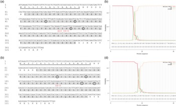
Two CSP genes of M. separata were cloned based on the sequences from the transcriptome dataset, which were named as MsCSP17 and MsCSP18. The ORF of MsCSP17 was 378 bp which encoded 125 amino acids (aa), containing a 15 aa of signal peptide at the N-terminal (fig. 1a, c). The relative molecular weight of MsCSP17 was 14.00 kD, and the isoelectric point was 6.42. Similarly, the ORF of MsCSP18 was 387 bp which encoded 128 aa, containing an 18 aa of signal peptide at the N-terminal (fig. 1b, d). The relative molecular weight of MsCSP18 was 14.84 kD, and the isoelectric point was 6.83. Both of MsCSP17 and MsCSP18 contained four conserved cysteine sites (C × (6)-C × (18)-C × (2)-C), which were involved in the formation of two pairs of disulfide bonds (fig. 1a, b).
Sequence analysis of MsCSP17 and MsCSP18. The nucleotide sequence and putative amino acid sequence of MsCSP17 (a) and MsCSP18 (b), where the circles indicate the conserved cysteine sites. Signal peptide prediction of MsCSP17 (c) and MsCSP18 (d).

Alignment and phylogenetic analysis of MsCSP17 and MsCSP18
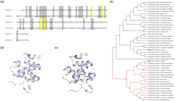
Alignment analysis showed that sequences of MsCSP17 and MsCSP18 shared 32% similarity with each other (fig. 2a). Using SWISS-MODEL, the structures of MsCSP17 and MsCSP18 were constructed based on CSPsg4 from Schistocerca gregaria, which showed that MsCSP17 and MsCSP18 were both α-helix-rich globular proteins consisting of six α-helices and containing four typical cysteine sites (fig. 2b, c). Phylogenetic analysis showed that MsCSP17 had high homology with HaCSP23 and SeCSP12, while MsCSP18 was clustered into the different clade which was close to AiCSP3 and MsCSP2 (fig. 2D).
Characteristics of MsCSP17 and MsCSP18. (a) Sequence similarity alignment of MsCSP17 and MsCSP18. Predicted protein structures of MsCSP17 (b) and MsCSP18 (c). The purple and yellow regions indicate the α-helix and cysteine in the structure, respectively. (d) The phylogenetic tree of CSPs. Proteins marked with red dots are MsCSP17 and MsCSP18.

Expressions of MsCSP17 and MsCSP18 in different developmental stages and tissues
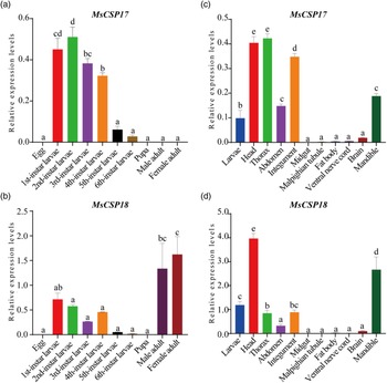
The expression of MsCSP17 and MsCSP18 at different developmental stages of M. separata was detected by RT-qPCR. The results showed that MsCSP17 was expressed in all 1st–6th instar larvae, with the highest expression in 2nd and 1st instar larvae but low in non-larva stages (fig. 3a). Differently, MsCSP18 was significantly highly expressed in female and male adults, followed by the expression in 1st instar larvae (fig. 3b). Among different tissues of 4th instar larvae, MsCSP17 was highly expressed in the head, thorax and integument, followed by the mandible (fig. 3c), while MsCSP18 was significantly highly expressed in the head and mandible, followed by the thorax and integument (fig. 3d).
Expression profiles of MsCSP17 and MsCSP18 among different developmental stages and tissues. The relative expression levels of MsCSP17 (a) and MsCSP18 (b) at different developmental stages. Relative expression levels of MsCSP17 (c) and MsCSP18 (d) in different tissues of 4th instar larvae. Data represent means of three replicates. One-way ANOVA was performed followed by Duncan's test (p < 0.05).

Expressions of MsCSP17 and MsCSP18 in larval integuments and mandibles
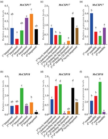
Since the high expression of MsCSP17 and MsCSP18 in larval integuments and mandibles, the expression of two genes in integuments of 1st–6th instar larvae, larval integuments during molting, and mandibles of 3rd–6th instar larvae were detected by RT-qPCR. The results showed that the expression of MsCSP17 was the highest in the integument of 5th instar larvae and the lowest in the integument of 2nd instar larvae (fig. 4A), while MsCSP18 was significantly highly expressed in the integument of 3rd instar larvae but lowly in the integument of 6th instar larvae (fig. 4B). Interestingly, both of MsCSP17 and MsCSP18 lowly expressed in larval integuments when larvae molted for 6 h and 9 h from 3rd to 4th instar, but highly at the beginning and end phase during molting (fig. 4C, D). Moreover, the expression of MsCSP17 was high in the mandible of 3rd instar larvae, while the expression of MsCSP18 was high in the mandible of 5th instar larvae and low in 6th instar larval mandibles (fig. 4E, F).
Expressions of MsCSP17 and MsCSP18 in larval integuments and mandibles. The relative expression of MsCSP17 (a) and MsCSP18 (b) in larval integuments at different developmental stages. The relative expression of MsCSP17 (c) and MsCSP18 (d) in larval integuments during molting from 3rd to 4th instar. The relative expression of MsCSP17 (e) and MsCSP18 (f) in mandibles of 3rd–6th instar larvae. Data represent means of three replicates. One-way ANOVA was performed followed by Duncan's test (p < 0.05).

Silencing efficiency of MsCSP17 and MsCSP18
A total of 2 μg of dsMsCSP17, dsMsCSP18, and dsGFP were injected into a 3rd instar larva, respectively. Compared with control groups injected with dsGFP, the mRNA expressions of MsCSP17 and MsCSP18 were significantly down-regulated in larvae injected with dsMsCSP17 and dsMsCSP18, with a decrease of 66 and 56%, respectively (fig. 5A, B), indicating the high silencing efficiency of dsRNA microinjection on CSPs in M. separata.
Knockdown efficiency of MsCSP17 and MsCSP18 in M. separata larvae. Relative mRNA expression levels of MsCSP17 (a) and MsCSP18 (b) after microinjection of dsRNA for 24 h. Data represent means of three replicates. Black bar and the asterisk represent the significant difference analyzed by Student's t-test (p < 0.05).

Effects of RNAi on the development of M. separata
The dsRNA-injected larvae were fed with fresh maize leaves to analyze their phenotype and growth. The larvae injected with dsGFP showed normal growth and development, while 15.0 and 17.5% of the larvae injected with dsMsCSP17 and dsMsCSP18 showed retarded growth and smaller size compared to the control, respectively, with old integument stuck between the head and thorax (fig. 6a). Most of the abnormal larvae died due to not feeding (fig. 6b), and a few ones gradually returned to normal at the end of 5th instar or the beginning of 6th instar, but failed to implement eclosion after pupation (fig. 6c). According to the results, the mortalities were 40.5 and 36.0% for dsMsCSP17- and dsMsCSP18- injected larvae after 12 days, respectively, while the mortality of the control was 4.2%. There was no significant difference in the pupation rate among different groups, while the emergence rates were 87.5, 75.0 and 95.7% for dsMsCSP17-, dsMsCSP18- and dsGFP-injected groups, respectively. The RNAi larvae without obvious phenotype were weighed every 24 h after injection. Compared with the control group, the body weight of the larvae injected with dsMsCSP17 and dsMsCSP18 was significantly reduced before pupation, respectively (fig. 6d).
Effects of RNAi on the development of M. separata larvae. (a) Changes of larval size after injection of dsRNA. (b) Larvae with abnormal phenotypes died after injection of dsMsCSP17 and dsMsCSP18. (c) M. separata failed eclosion after injection of dsMsCSP17 and dsMsCSP18. (d) Changes in larval body weight after injection of dsRNA.

Discussion
CSPs play a crucial role in insect survival and reproduction (Pelosi et al., Reference Pelosi, Iovinella, Felicioli and Dani2014), which have a stable and compact structure that binds and solubilizes hydrophobic small molecules, such as sex pheromones, nutrients, visual pigments, growth and development-related hormones. In this study, two CSP genes, MsCSP17 and MsCSP18 were cloned from M. separata, both of which have four conserved cysteine sites that are typical characteristics of CSPs in insects (Cao et al., Reference Cao, Liu, Wei, Liao, Walker, Li and Wang2014; Wang et al., Reference Wang, Zhang, Li, Guo and Luo2016; Xue et al., Reference Xue, Fan, Zhang, Xu, Han, Sun and Chen2016). MsCSP17 and MsCSP18 shared 32% sequence similarity with each other, while they have 70–88% sequence similarity with their homologs of other insects such as Agrotis ipsilon and Mamestra brassicae, which was consistent with the high conservation of CSPs among different species (Wanner et al., Reference Wanner, Willis, Theilmann, Isman, Feng and Plettner2004). The functional importance of conserved amino acid residues can infer that CSPs probably play an important role in insect physiology (Gu et al., Reference Gu, Wang, Zhang, Ji, Liu, Wang, Wu, Gou, Zhou and Zhang2012).
Gene spatio-temporal expression pattern is helpful for grasping the dynamic changes of gene expression levels and predicting their functions in insects. In this study, MsCSP17 was significantly highly expressed in the larval stage, especially in head, thorax, integument, and mandible, while MsCSP18 was expressed highly in adults and larvae, and the head and mandible of larvae. This was consistent with reports that CSPs were not only expressed in the antennae, but also in non-olfactory organs such as the head, thorax, feet, wings, and gonads of insects (Wanner et al., Reference Wanner, Willis, Theilmann, Isman, Feng and Plettner2004; Zhang and Lei, Reference Zhang and Lei2015). The high levels of MsCSP18 in adults indicated that it may have the potential role of finding host or mating location. The broad-spectrum expression of MsCSP17 and MsCSP18 in M. separata larvae suggested that CSP genes may have important functions other than chemosensory (Guo et al., Reference Guo, Wang, Ma, Xue, Han, Yu and Kang2011, Gu et al., Reference Gu, Wang, Zhang, Ji, Liu, Wang, Wu, Gou, Zhou and Zhang2012; Zhang et al., Reference Zhang, Jin, Jin, Xia, Zhou, Deng and Dong2013).
With the high expression levels in larval integuments, the expressions of two genes in larval integuments at different stages were compared. MsCSP17 had higher expression in 5th instar larval integuments, while MsCSP18 expressed highly in 3rd instar larval integuments. Combined with the variations of mRNA levels during molting, MsCSP17 and MsCSP18 might be involved in the formation of new integument in M. separata. Moreover, with the high expression levels in larval mandibles, the relative expression levels of two genes were compared among mandibles of 3rd–6th instar larvae. The results showed that MsCSP18 highly expressed in the mandibles of 3rd–5th instar but lowly in 6th instar, which indicated its potential role in the development of mandibles (Zhao et al., Reference Zhao, Zhu, Xing, Li, Zhou, Wang, Li, Cao and Huang2022).
RNAi is widely used in the field of insect functional genomics, especially in screening lethal target genes, which are involved in important physiological and biochemical processes such as reproductive development, energy metabolism, immunity, and neuromodulation in insects. In this experiment, RNAi was effectively applied to explore the functions of two CSPs in M. separata larvae, with the expression of MsCSP17 and MsCSP18 significantly silenced after injection of dsRNA. Compared with the control, CSP-injected larvae showed stunted growth, smaller size, increased mortality, and reduced eclosion rate. The abundant expression of CSPs in metabolically active tissues and organs was closely related to the development of insects, hence the reduced expressions of CSPs can affect growth and reproduction negatively (Gong et al., Reference Gong, Luo, Rizwan-ul-Haq and Hu2012; Ma et al., Reference Ma, Cui, Tian, Zhang, Chen, Gao, Tian, Chen, Gou and Zhou2019). For instance, CSPs were involved in the foot regeneration in Periplaneta americana (Kitabayashi et al., Reference Kitabayashi, Arai, Kubo and Natori1998), molting process in Pheidole megacephala larvae (Pelosi et al., Reference Pelosi, Iovinella, Zhu, Wang and Dani2018), and reproduction in Spodoptera exigua (Gong et al., Reference Gong, Luo, Rizwan-ul-Haq and Hu2012). Moreover, inhibition of SiCSP9 expression has been reported to affect fatty acid biosynthesis and inhibit cuticle development and molting in Solenopsis invicta (Cheng et al., Reference Cheng, Lu, Zeng, Liang and He2015). Thus, MsCSP18 and MsCSP19 were functional in the development of M. separata.
In conclusion, MsCSP17 and MsCSP18 were important for the development of M. separata larvae, with high expressions in larvae and their heads, integuments, and mandibles. Our findings expanded our current understanding of the expression patterns of CSPs in non-olfactory tissues and their functions in M. separata larvae, providing potential targets to develop effective pest control strategies.
Supplementary material
The supplementary material for this article can be found at https://doi.org/10.1017/S0007485323000354.
Acknowledgements
This work was supported by the National Natural Science Foundation of China (31901877), the Young Talent Promotion Project of Anhui Association for Science and Technology (RCTJ202003), and the College Student Innovation Project of Anhui Agricultural University (X202210364233).
Competing interests
None.