

Humans are continually exposed to dangerous pollutants in the environment. The kidney is the main organ for xenobiotics excretion and renal cells are thus exposed to high doses of metabolites, causing nephrotoxicity. Chemical- and drug-induced nephrotoxicity is a principal cause of acute kidney injury which may be due to altered intraglomerular haemodynamics, inflammation, rhabdomyolysis, microangiopathy, and tubular cell toxicity. It is reported as a significant cause of mortality and morbidity (Dashti-Khavidaki et al. Reference Dashti-Khavidaki, Shahbazi, Khalili and Lessan-Pezeshki2012). CCl4, an environmental contaminant, induced biochemical, haematologic and oxidative stress parameter alterations in experimental animals (Bellassoued et al. Reference Bellassoued, Ben Hsouna, Athmouni, Pelt, Makni Ayadi, Rebai and Elfeki2018). Its greatest concern is as a toxic agent in liver tissue, attributed to the trichloromethyl radical produced during oxidative stress (Stoyanovsky & Cederbaum, Reference Stoyanovsky and Cederbaum1999). In addition to causing classic hepatic injury CCl4 can also cause damage in many other tissues such as kidney (Mbarki et al. Reference Mbarki, Alimi, Bouzenn, Elfeki and Hfaiedh2017), heart (Hamed et al. Reference Hamed, Chaari, Ghannoudi, ElFeki, Chaabouni Ellouz and Gargouri2017) and brain (Ma et al. Reference Ma, Luo, Jiang and Liu2016). Among these tissues, the adverse effects of CCl4 on the kidney have gained interest. Although the precise mechanism of kidney toxicity caused by metabolism of CCl4 through the cytochrome P450 CYP2E1 to produce highly toxic trichloromethyl (–CCl3) and peroxytrichloromethyl (–OOCCl3) is similar to the liver, kidney is at additional risk due to the predominant presence of P450 cytochrome in the cortical tubule cells. Several studies demonstrated that CCl4 induced lipid peroxidation, protein oxidation and cell membrane damage in the kidney (Safhi, Reference Safhi2017). In addition, the damage in kidney can include glomerular necrosis and alteration in proximal and distal tubules progressing ultimately to detachment of the epithelial cells and tubular necrosis (Ogeturk et al. Reference Ogeturk, Kus, Colakoglu, Zararsiz, Ilhan and Sarsilmaz2005).
The human body has integrated an antioxidant defence system to oppose oxidative stress, including both non-enzymatic as well as enzymatic mechanisms such as catalase (CAT), superoxide dismutase (SOD) and glutathione peroxidase (GPx).
Fermented milk has been used successfully to manage various inflammatory diseases (Desrouillères et al. Reference Desrouillères, Millette, DangVu, RTouja and Lacroix2017). Several studies have confirmed that bioactive peptides derived from fermented milk proteins have biological benefits such as antimicrobial, antioxidant, anticytotoxic, antihypertensive and immunomodulatory action (Gobbetti et al. Reference Gobbetti, Minervini and Rizzello2004). Few studies have examined the nephroprotective activities of fermented camel milk against CCl4 toxicity. Thus, the current study designed to investigate the possible preventive effects of fermented camel milk against CCl4-induced kidney damage.
Materials and methods
Chemicals
All reagents used in the present study were of analytical grade. Carbon tetrachloride was obtained from sd Fine Chemicals, Bhoisar, Mumbai, India. 5,5’-Dithiobis (2-nitrobenzoic acid) (DTNB), L-Glutathione (reduced form), and all current chemicals were bought from Sigma Chemical Co., (St. Louis, MO, USA).
Bacterial strains and growth conditions
Lactococcus lactis subsp.cremoris was isolated in a previous study. Standard cultures were prepared by inoculation of 10 mL M17 broth (Biokar Diagnostics) or MRS (Accumix) with 100 µl of frozen strains stock and incubated for 24 h at 37 °C. Then, 100 µl of the 24 h grown strain was reinoculated into 10 ml MRS broth for 24 h at 37 °C. To adapt bacteria to milk fermentation conditions, strains working cultures were prepared by inoculating (1%) fresh precultures in sterile (100 °C, 20 min) camel milk and incubated (12 h, 37 °C).
Sample preparation
The fresh camel milk sample was collected from the local farm on May 2016 (Sfax, Tunisia). The sample was cooled immediately at 4 °C and then blended to obtain a homogenous sample followed by the pasteurisation process. Fresh milk was heated at 80 °C for 20 min and cooled to 43–45 °C (Moslehishad et al. Reference Moslehishad, Mirdamad, Ehsani, Ezzatpanah and Moosavi-Movahed2013).
Batch culture experiments were performed in the pasteurised fresh milk. Pure and mixed cultures were carried out without any mechanical agitation. After 24 h of fermentation, the samples were centrifuged for 15 min at 15 000 × g and supernatant used in the assays.
Animals and experimental design
This study was carried out on 28 female mice weighing 29 ± 2 g, purchased from the Central Pharmacy of Tunis (SIPHAT). After 15 d adaptation period, the mice were randomly and equally divided into four groups of seven animals each: group1 (control group [C]); Group 2 (CCl4 group [CCl4]); Group 3 (Fermented Camel Milk + CCl4 group [FCM + CCl4]); Group 4 (Fermented Camel Milk group [FCM]).
Groups 2 and 3 received a single dose of CCl4 (10 ml/kg in 0·3% olive oil. ip) at 14 d. Mice of the control group (group 1) received an equal volume of olive oil. Groups 3 and 4 were administrated camel milk fermented using lactococcus lactis subsp cremoris by oral gavage (0·4 ml/kg b.w/day) during 15 d before CCl4 injection. Twenty-four hours after the CCl4 challenge, all animals were weighed and then sacrificed by cervical decapitation to avoid stress.
The plasma was obtained by centrifugation (4000 rpm g, 15 min, 4 °C) and stored at −20 °C for biochemical assays. The kidneys of each animal were removed, cleaned, weighed and rinsed with a physiological saline solution. Samples were rinsed, homogenised in tris buffer solution (50 mm; pH 7·4) using an Ultra-Turax Homogeniser and centrifuged (5000 × g for 25 min at 4 °C). The supernatant was kept at − 20 °C until analyses. In parallel, portions of kidney were immediately fixed into in 10% buffered formalin solution for histological studies.
Biochemical parameters
The levels of urea, uric acid, and creatinine in plasma were assayed by autoanalyser (Erba xl 200, Mannheim, Germany) using Erba diagnostic kit.
The levels of sodium, potassium, calcium, chloride, and phosphorus in plasma were assayed by autoanalyser (Erba Lyte pro, Mannheim, Germany).
Lactate dehydrogenase (LDH) activities in the kidney and plasma were measured using a commercially available diagnostic kit (Biomaghreb, Tunisia, Ref. 200126).
Oxidative damage assays
The level of lipid peroxidation in kidney was estimated according to the method of (Yagi, Reference Yagi1976). Briefly, an aliquot of kidney extracts supernatant was mixed with 1 ml of 5% trichloroacetic acid and centrifuged at 2500 × g for 10 min. An amount of 1 ml of thiobarbituric acid reagent (0·67%) was added to 500 µl of supernatant and heated at 90 °C for 15 min. The mixture was cooled and the absorbance was recorded at 765 nm. Likewise, protein carbonyl (PCO) content in kidney tissue was measured using the DNPH method of Reznick & Packer (Reference Reznick and Packer1994). Briefly, after precipitation of protein with an equal volume of 1% trichloroacetic acid, the pellet was resuspended in 10 mm/l DNPH plus 2N HCl, or in 2N HCl as a control blank. Next, after the washing procedure with 1 : 1 ethanol-ethylacetate, the final palette was dissolved in 6 m/l guanidine. The carbonyl group was determined from the absorbance at 370 nm. Results were expressed in nm/mg protein. Protein content was evaluated as described by Lowry et al. (Reference Lowry, Rosebrugh and Farr1951) using bovine serum albumin as standard.
Assessment of antioxidant status
Superoxide dismutase (SOD) activity was estimated according to Beyer & Fridovich (Reference Beyer and Fridovich1987).
Catalase activity was assayed by H2O2 consumption method (Aebi, Reference Aebi1984). GPx activity was measured according to the method of Flohe & Gunzler (Reference Flohe and Gunzler1984). GSH in plasma was measured by the method of Ellman (Reference Ellman1959) modified by Jollow et al. (Reference Jollow, Mitchell, Zampaglione and Gillette1974).
The determination of vitamin C content in the kidney tissue was performed according to Jacques-Silva et al. (Reference Jacques-Silva, Nogueira, Broch, Flores and Rocha2001).
Histopathological examination
Kidneys removed from the control and tested mice were cleaned and fixed in Bouin solution. Then they were embedded in paraffin and stained with haematoxylin–eosin for histopathological studies. All sections were evaluated for the degree of tubular and glomerular injury and necrosis. Six slices from each kidney were examined and assigned for severity of changes using scores on a scale of none (−), mild (+), moderate (++), and severe (+++).
Statistical analysis
Statistical analyses were performed using one-way ANOVA followed by Fisher's post-hoc test (PLSD) for comparison between groups. Data are expressed as mean ± se. Differences were considered significant if P < 0·05.
Results
Effects of FCM on serum biochemical parameters
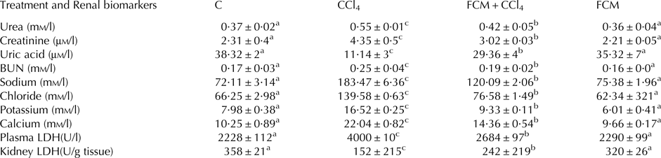
Levels of biomarkers of kidney function in all mice group are given in Table 1. Results show that CCl4 significantly raised serum urea, creatinine and BUN levels in mice kidney as compared to control animals (+48·6%, +68·9%, and +47%, respectively, P < 0·05). Besides, we have found a significantly decreased level of uric acid following CCl4 administration (−71%, P < 0·05). Pretreatment of CCl4-intoxicated mice with FCM significantly attenuated all of these effects (P < 0·05) without totally restoring control values. Treatment with FCM alone had no effect on the levels of any of the biomarkers.
Effect of CCl4 or FCM and their combination on kidney and plasma biomarkers

BUN, blood urea nitrogen: calculated following: BUN (mm/l) = [urea (mm/l)]/2·14; LDH, Lactate dehydrogenase
Values are mean ± sd (n = 7 per group). Values having different letters on the same row showed significant difference (P < 0·05)
Effect of FCM on LDH activities
LDH activity in the kidney was significantly lower in the CCl4 group (−57%) but increased in plasma (by + 79%, P ≤ 0·001) compared to the normal group (Table 1). Pretreatment with FCM attenuated both effects, partially restoring kidney LDH activity whilst reducing plasma LDH (Table 1). Pretreatment mice with FCM alone showed no significant change in LDH activities when compared to control group.
Effects of FCM on serum electrolytes
Table 1 presents the change in the levels of electrolytes in serum. In this study, sodium (Na+), chloride (Cl+), potassium (K+) and calcium (Ca) in the CCl4 group were markedly increased (P ≤ 0·001) compared to control group. However, FCM prevents increases of these electrolytes in the serum of mice intoxicated by CCl4. Treatment with FCM alone didn't change the electrolyte levels in serum of normal animals.
Effects of FCM on kidney oxidative damage
Data concerning kidney MDA and PCO are given in Table 2. Both measures were significantly increased (+89·23% P ≤ 0·001) in kidney tissue of CCl4 treated animals when compared to control untreated mice. In the group that received FCM plus CCl4, levels were significantly decreased compared with the CCl4 group. Treatment with FCM alone did not change either measure when compared to control animals.
Effect of CCl4 or FCM and their combination on biomarkers of oxidative stress

Values are mean ± sd (n = 7 per group). Values having different letters on the same column showed significant difference (P < 0·05)
Similarly, a significant increase in PCO content was observed in kidney tissues of CCl4 intoxicated mice. Pretreatment with FCM reduced the level of PCO as compared to CCl4 group values. The administration of FCM alone to normal mice did not change PCO level.
Effect of FCM on non-enzymatic antioxidant
Acute intoxication of animals by CCl4 caused significant decreases in GSH content compared with the negative control group (P < 0·001). Animals treated with CCl4 along with FCM showed a significant recovery of GSH towards control level. FCM alone had no effect on GSH (Table 2).
In the kidney of untreated mice, vitamin C level was 0·78 µm/mg protein. However, this level decreased by 70% (P ≤ 0·01) in CCl4 group. While the vitamin C level in animals pretreated with FCM was significantly higher (P < 0·001) than the CCl4-treated group.
Effect of FCM on antioxidant enzymes activities
The concentrations of enzymatic antioxidants (SOD, CAT, and GPx) in the kidney tissues of normal and experimental animals group after 15 d are shown in Table 2.
In CCl4 treated group, a significant decrease in enzymatic antioxidants viz. SOD, CAT, and GPx were identified in kidney homogenates (−41·7%, −47·8% and −50·5%; P < 0·001), respectively, as compared to those of normal control group of mice. As with other measures, pretreatment with FCM partially reversed these effects and FCM alone did not change any antioxidant enzyme activities compared to control group.
Histopathological findings
The histology of the kidney sections of the control group showed a normal kidney histological structure with normal tubules and intact glomeruli (online Supplementary Fig. S1: [A,A’]). The administration of CCl4 caused severe glomerular and tubulointerstitial necrosis indicated by the detachment of tubular epithelial cells and vacuolisation of tubules in the kidney. CCl4 produced a marked injury to Bowman's capsule, destruction of glomeruli, in addition to the infiltration of inflammatory leucocytes (online Supplementary Fig. S1: [B,B’]), changes that were largely reversed by pretreatment with FCM (online Supplementary Fig. S1: [D,D’]). In addition, the prevention with FCM against CCl4 reduced markedly the glomeruli fragmentation and the Bowman's space enlargement. FCM alone did not alter kidney histology (online Supplementary Fig. S1: [C,C’]). The histological changes in treated mice were graded and are summarised in Table 3.
Grading of the histopathological changes in the renal tissues in mice treated with CCl4 or FCM and their combination

Scoring was done as follows: none (−), mild (+), moderate (++) and severe (+++)
Discussion
In recent years, there has been increasing interest in the use of probiotics and related fermented products in the diet. It has been reported that they may have benefit for the prevention and treatment of certain diseases (Shiby & Mishra, Reference Shiby and Mishra2013). Fermented camel milk inoculated with lactococcus lactis subsp.cremoris showed a strong biological activity compared to samples fermented using other strains in all in vitro tests, as previously described (Hamed et al. Reference Hamed, Chaari, Ghannoudi, ElFeki, Chaabouni Ellouz and Gargouri2017). This property makes it a suitable product to be tested for prevention of kidney damage. In this study, we assessed the response of the kidney to acute exposure of CCl4 in mice and demonstrated that camel milk fermented using lactococus lactis subsp cremoris could play a potential role in preventing the renal toxicity induced by this toxic agent.
The kidney is the main organ of excretion, metabolism, and detoxification of xenobiotic and absorption of minerals but is susceptible to damage. Determination of plasma creatinine is a classic marker of renal dysfunction. In addition, urea is easily measured as another biomarker of renal impairment. The present study confirms that administration of CCl4 induced damage in the kidney as indicated by the high levels of urea and creatinine (El-kholy et al. Reference El-kholy, Hassanen and Abbas2013). Uric acid was another parameter used in the present work to assess kidney's function. Our results showed a decrease of uric acid level in plasma of mice treated by a single dose of CCl4. Furthermore, blood urea nitrogen (BUN) increased in CCl4 intoxicated mice as compared with controls, indicating kidney disease with a decrease in glomerular filtration rate. This observation is in agreement with that of Sajid et al. (Reference Sajid, Khan, Shah, Ullah, Younis, Majid, Ahmad and Nigussie2016) in mice exposed to CCl4.
Considering that electrolytes levels could change acutely with kidney damage, Na, Cl, K and Ca levels in serum were evaluated. This study showed that intoxication with CCl4 caused a significant increase of these parameters. This is consistent with recent studies with CCl4 (Karakus et al. Reference Karakus, Değer and Yıldırım2016), who suggested that CCl4 contributes to the alteration of the cell membrane or to cell death. FCM significantly (P < 0·001) attenuated the increase in the serum electrolyte levels. Pretreatment with FCM, attenuated the kidney impairment as suggested by a significant amelioration of the renal biomarkers indicated earlier.
In order to understand the mechanism by which the FCM prevented CCl4-induced nephrotoxicity, we evaluated markers of oxidative stress, including MDA and PCO, the stable marker of lipid and protein oxidation respectively and antioxidative enzyme activities. Our findings confirm that the increase in the level of renal thiobarbituric acid reactive substances (TBARS) is generally thought to be a consequence of damage and failure of antioxidant defence mechanism to prevent the increased production and liberation into the circulation of tissue lipid peroxides (Khan et al. Reference Khan, Rizvi, Khan, Khan and Sheen2009).
In addition, the large generation of ROS following CCl4 exposure was correlated with the high levels of protein oxidation reaction, leading to the formation of protein carbonyl (PCO). Pretreatment with FCM prevented oxidative damage induced by CCl4 in the renal tissue and accompanied by a decrease in the levels of MDA and PCO. This could be due to beneficial nutritional characteristics of fermented milk which provide a lower substrate for lipid peroxidation and consequently decreases the generation of free radicals (Moreno-Fernandez et al. Reference Moreno-Fernandez, Diaz-Castro, Alferez Maria, Boesch, Nestares and Lopez-Aliag2017). Fardet & Rock (Reference Fardet and Rock2017) reported that fermented milk may inhibit oxidative stress, especially due to the production of antioxidant peptides.
Antioxidant defence of renal cells against oxidative damage is preserved via several mechanisms involving enzymatic antioxidants. In the current study, dramatic decreases in these antioxidant enzymes activities were found in CCl4 intoxicated mice. Conversely, FCM could markedly impede the decline of CAT, SOD and GPx activities. This antioxidant effect of FCM on kidney tissue could be mainly attributed to milk bioactive peptide, vitamins, and oligosaccharides (Fernández et al. Reference Fernández, Hudson, Korpela and Reyes-Gavilán2015).
To investigate also the mechanisms underlying CCl4 nephrotoxicity, the non-enzymatic antioxidant status was estimated. The degradation of GSH, observed in this study, as a result of CCl4 intoxication, may be due to the involvement of antioxidants to prevent the development of free radicals (Güven et al. Reference Güven, Güven and Gülmez2003).
Our observations revealed that FCM supplementation ameliorated the impaired antioxidant defence system in mice kidney, as indicated by the restoration of GSH level.
In addition, our data revealed that administration of CCl4 induced a significant decrease of vitamin C levels in comparison with normal control. Interestingly, when the CCl4 treated mice were pretreated with FCM, the kidney Vitamin C was maintained at near-normal level. Osman et al. (Reference Osman, Abdel Rahman and Dirar2010) indicated that fermented milk is considered a source of vitamin C, and this could relate to antioxidant capacity and the free radical scavenging activity. Overall, the protein fraction contains antioxidant activity, especially casein. Other potential antioxidants in the fermented milk include casein-derived peptides, lactoferrin, conjugated linoleic acid, coenzyme Q10, vitamins E, A and D, equol, uric acid, carotenoids and mineral activators of antioxidant enzymes (Fardet & Rock, Reference Fardet and Rock2017). Thus, FCM could prevent oxidative damage in the organism through its ability to scavenge free radicals and to inhibit ROS accumulation.
Conclusion
Therapeutic interventions that inhibit the oxidative stress have the potential for attenuating renal dysfunction and accelerating recovery after acute renal failure. This study indicates that camel milk fermented using lactococcus lactis subsp cremoris has a preventive effect against tetrachloride carbon induced nephrotoxicity in mice. This renoprotective effect can be explained by its ability to inhibit lipid peroxidation and improve the antioxidant status in kidney tissues.
Supplementary material
The supplementary material for this article can be found at https://doi.org/10.1017/S0022029918000250
The authors acknowledge the ‘Ministry of Higher Education, Scientific Research and Technology, Tunisia’, for the support of this study.