Introduction
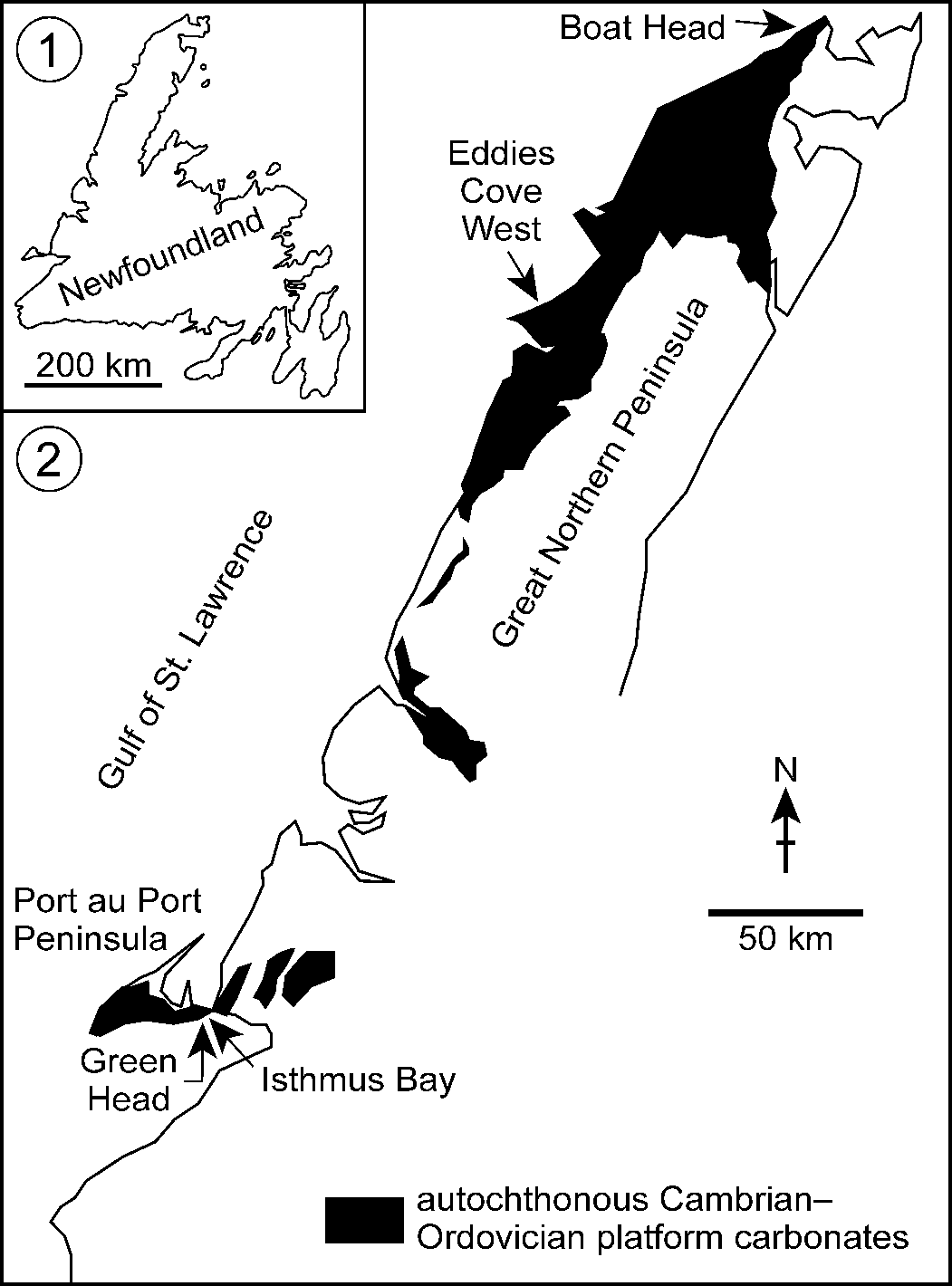
Modular coral-like fossils from Lower Ordovician (Tremadocian) strata within the St. George Group in western Newfoundland, Canada—initially identified as the tabulate coral Lichenaria Winchell and Schuchert, Reference Winchell, Schuchert, Lesquereux, Woodward, Thomas, Schuchert, Ulrich and Winchell1895 (Pratt and James, Reference Pratt and James1982, Reference Pratt, James, Geldsetzer, James and Tebbutt1989a, Reference Pratt, James, Geldsetzer, James and Tebbuttb)—recently have been assigned to Amsassia Sokolov and Mironova, Reference Sokolov and Mironova1959 (Elias et al., Reference Elias, Lee and Pratt2021; Lee et al., Reference Lee, Elias and Pratt2021). Amsassia terranovensis Lee, Elias, and Pratt, Reference Lee, Elias and Pratt2021, occurs in the Watts Bight Formation at Green Head, and A. diversa Lee, Elias, and Pratt, Reference Lee, Elias and Pratt2021, appears in the middle member of the overlying Boat Harbour Formation at Eddies Cove West and Boat Head (Figs. 1, 2). Here, a closely related genus, Reptamsassia n. gen., is erected for materials from two stratigraphic levels within the Lower Ordovician (Floian) Barbace Cove Member of the Boat Harbour Formation in Isthmus Bay (Figs. 1, 2). Reptamsassia divergens n. gen. n. sp. occurs at both levels, whereas a comparatively small-module species, R. minuta n. gen. n. sp., is confined to the upper level.
(1) Map of Newfoundland. (2) Map of western Newfoundland showing autochthonous lower Paleozoic platform carbonates (based on Knight et al., Reference Knight, Azmy, Boyce and Lavoie2008, fig. 1), which are present from Port au Port Peninsula to the northern tip of Great Northern Peninsula. Samples of Amsassia described by Lee et al. (Reference Lee, Elias and Pratt2021) were collected at Green Head, Eddies Cove West, and Boat Head; samples of Reptamsassia n. gen. for the present study were collected at Isthmus Bay (for detailed map see Knight and James, Reference Knight and James1987, fig. 3d).

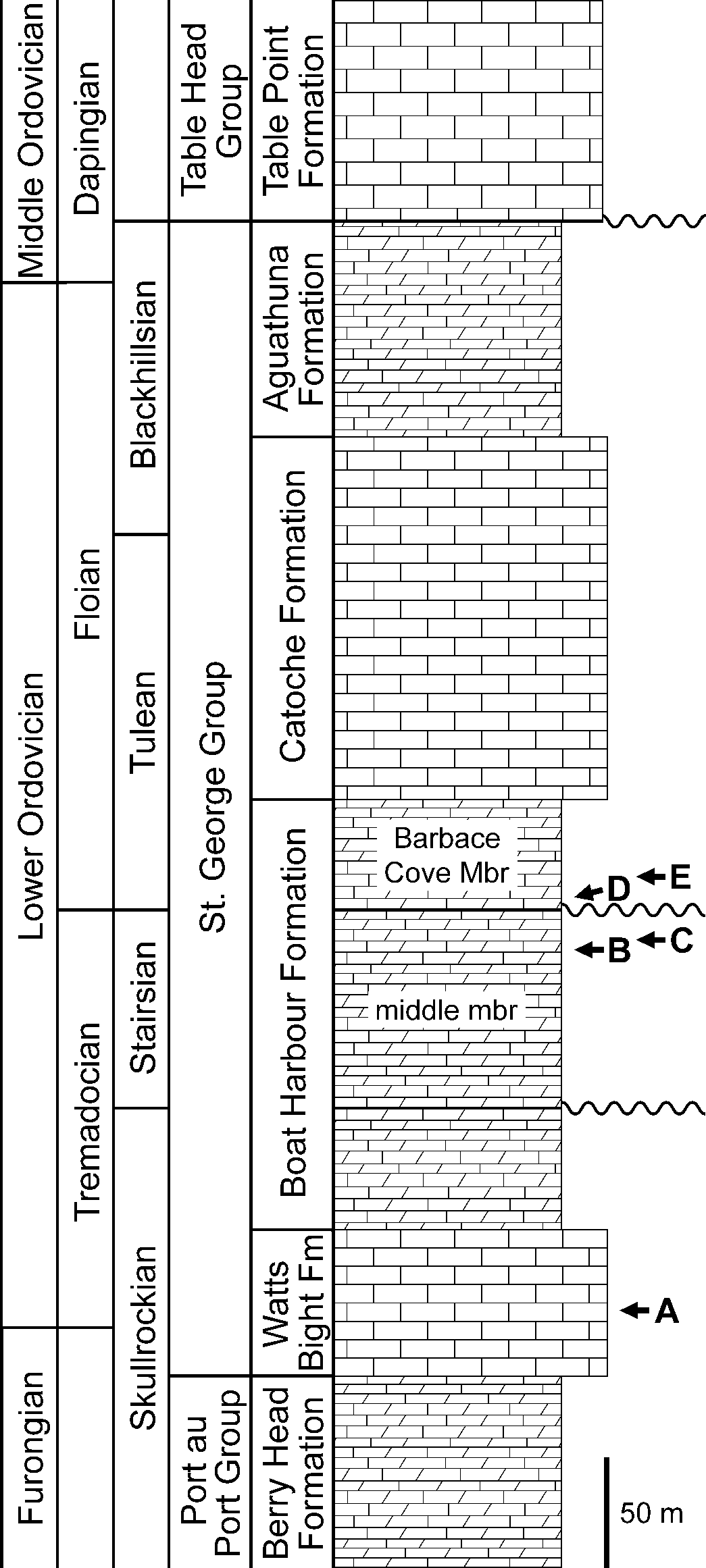
Generalized stratigraphic section of the St. George Group in western Newfoundland (based on Knight et al., Reference Knight, Azmy, Boyce and Lavoie2008, fig. 2; with updated chronostratigraphy in Boyce et al., Reference Boyce, McCobb and Knight2013, fig. 2). The North American Skullrockian through Blackhillsian stages comprise the Ibexian Series. Besides the contact between the St. George Group and the overlying Table Head Group, there are two significant regional disconformities within the Boat Harbour Formation (shown with wavy lines). The middle member and Barbace Cove Member of the Boat Harbour Formation are labeled, but other members are not. Lithologic symbols: rectangles = limestone; right trapezoids = interbedded limestone and dolostone. Stratigraphic positions of Amsassia and Reptamsassia n. gen. are marked with arrows (geographic locations are shown in Fig. 1.2): A, Amsassia terranovensis and Amsassia? sp. A, ~26–33 m above base of Watts Bight Formation at Green Head; B, Amsassia diversa and Amsassia? sp. B, ~18 m below top of middle member in Boat Harbour Formation at Boat Head; C, Amsassia diversa, ~13.5 m below top of middle member in Boat Harbour Formation at Eddies Cove West; D, Reptamsassia divergens n. gen. n. sp., ~4 m above base of Barbace Cove Member in Boat Harbour Formation at Isthmus Bay (locality B86); E, Reptamsassia divergens n. gen. n. sp. and R. minuta n. gen. n. sp., ~15 m above base of Barbace Cove Member in Boat Harbour Formation at Isthmus Bay (locality B97).

Regarding biologic affinity, the Ordovician genus Amsassia has been excluded from corals and calcareous sponges, as well as cyanobacteria, and referred to calcareous algae (Sun et al., Reference Sun, Elias and Lee2014; Lee et al., Reference Lee, Park, Tien, Choh, Elias and Lee2016, Reference Lee, Elias, Choh and Lee2018, Reference Lee, Elias and Pratt2021; Elias et al., Reference Elias, Lee and Pratt2021). Its characteristic morphologic features are different from those of tetradiids, and suggest that Amsassia may have been a green alga (Lee et al., Reference Lee, Elias and Pratt2021). The morphology of Reptamsassia n. gen. is consistent with that interpretation. The two genera are considered to represent a distinct group, the Amsassiaceae n. fam.
The Early Ordovician origination of Amsassia followed by Reptamsassia n. gen. contributed to the rise of calcareous macroalgae as part of the distinctive Ordovician marine flora (Chuvashov and Riding, Reference Chuvashov and Riding1984; LoDuca et al., Reference LoDuca, Bykova, Wu, Xiao and Zhao2017), and coincided with the beginning of the major increase in global diversity during the Great Ordovician Biodiversification Event (Fan et al., Reference Fan, Shen, Erwin, Sadler and MacLeod2020, fig. 1). Reptamsassia minuta n. gen. n. sp. was an obligate symbiont that colonized living areas on the surface of its host, R. divergens n. gen. n. sp., in a reefal environment. Isolated modules of R. divergens n. gen. n. sp. were able to persist as symbionts in the resulting intergrowths with R. minuta n. gen. n. sp. This is the earliest known symbiotic association involving the intergrowth of macroscopic modular species, thereby providing a new example of the development of ecologic specialization and ecosystem complexity in Early Ordovician reefs, as part of the Great Ordovician Biodiversification Event (Stigall et al., Reference Stigall, Edwards, Freeman and Rasmussen2019).
Geologic setting
The uppermost Cambrian (Furongian) to lowermost Middle Ordovician St. George Group of western Newfoundland is a carbonate platform succession deposited on the lower Paleozoic passive margin of Laurentia. It crops out discontinuously from the Port au Port Peninsula in the southwestern part of the island to the northern tip of the Great Northern Peninsula (Knight and James, Reference Knight and James1987, Reference Knight and James1988; Fig. 1.1, 1.2). The group consists of limestones and dolostones represented by a variety of shallow-marine and peritidal facies. These include bioturbated lime mudstone and wackestone, wave-ripple cross-laminated grainstone and lime mudstone, stromatolites, and microbial laminites (Pratt and James, Reference Pratt and James1986). Numerous beds contain thrombolite patch reefs up to ~2 m in thickness, and larger microbial reef complexes are also present (Pratt and James, Reference Pratt and James1982, Reference Pratt, James, Geldsetzer, James and Tebbutt1989a, Reference Pratt, James, Geldsetzer, James and Tebbuttb).
The St. George Group is subdivided into four formations: the Watts Bight, Boat Harbour, Catoche, and Aguathuna formations, in ascending stratigraphic order (Fig. 2). Two regionally extensive subaerial exposure surfaces are present in the lower and upper Boat Harbour Formation, termed the lower disconformity and Boat Harbour disconformity, respectively, which serve to subdivide the formation into the lower, middle, and Barbace Cove members (Knight et al., Reference Knight, Azmy, Boyce and Lavoie2008). Units in the St. George Group are well dated biostratigraphically using conodonts and trilobites (Fortey, Reference Fortey1979; Ji and Barnes, Reference Ji and Barnes1994; Boyce and Stouge, Reference Boyce and Stouge1997).
Coral-like fossils assigned to Amsassia occur in microbial patch reefs in the Watts Bight Formation and the middle member of the Boat Harbour Formation, in rocks of Early Ordovician (Tremadocian) Skullrockian and Stairsian ages, respectively (Elias et al., Reference Elias, Lee and Pratt2021; Lee et al., Reference Lee, Elias and Pratt2021; Figs. 1.2, 2). The coral-like fossils described here are assigned to a new genus, Reptamsassia. They are from two beds of Early Ordovician (Floian) Tulean age, in the Barbace Cove Member of the Boat Harbour Formation cropping out along the shore of Isthmus Bay at the eastern end of Port au Port Peninsula (Knight and James, Reference Knight and James1987, fig. 3d; Knight et al., Reference Knight, Azmy, Boyce and Lavoie2008, fig. 3; Figs. 1.2, 2). There, the member measures 52 m thick (Knight et al., Reference Knight, Azmy, Boyce and Lavoie2008). The two beds with Reptamsassia n. gen. are from separate localities described in ascending stratigraphic order.
Locality B86
Reptamsassia divergens n. gen. n. sp. is present in a 0.8-m-thick thrombolite bed capped by thin grainstone; the thrombolite is flanked by dolomitized burrowed limestone. The top is an undulating erosion surface overlain by dolomite intraclasts in the depressions, followed by a dolomitized microbial laminite that passes laterally to a breccia of angular laminite clasts. The Reptamsassia-bearing bed is ~4 m above the Boat Harbour disconformity marking the base of the Barbace Cove Member of the Boat Harbour Formation, and ~47 m below the Catoche Formation (unit 190 in Pratt, Reference Pratt1979, p. 217; just above sample site BH-42 at +84 m in Knight et al., Reference Knight, Azmy, Boyce and Lavoie2008, fig. 4, column E; Fig. 2).
Locality B97
Reptamsassia divergens n. gen. n. sp. and R. minuta n. gen. n. sp. are present in a 2.3-m-thick thrombolite bed recorded in the field as a burrowed wackestone. This occurrence is ~15 m above the Boat Harbour disconformity marking the base of the Barbace Cove Member of the Boat Harbour Formation, and ~34 m below the Catoche Formation (unit 207 in Pratt, Reference Pratt1979, p. 216; between sample sites BH-45 and BH-46 at +92 m in Knight et al., Reference Knight, Azmy, Boyce and Lavoie2008, fig. 4, column E; Fig. 2).
Lithology
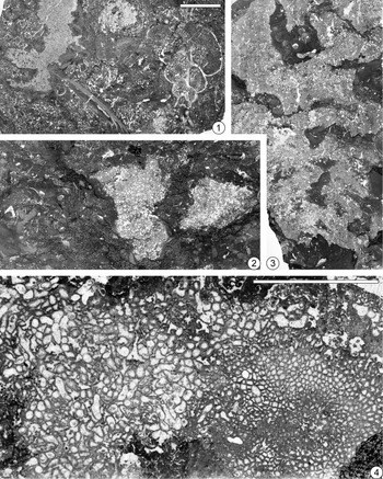
The thrombolite framework at both localities (B86 and B97) consists of irregular to crudely columnar masses of microbialite composed of clotted micrite and micrite with embedded skeletons of Reptamsassia n. gen., some preserved in growth position (Fig. 3.1–3.3). Microbialite locally exhibits centimeter-scale surfaces of abrasion. Flanking sediment is mainly a grainstone consisting of micrite, peloids, small intraclasts of clotted micrite, and bioclasts, all of which are sporadically incorporated in the microbialite. The similarity between the framework and matrix and places where matrix grades into microbialite make it somewhat difficult to distinguish between the two.
(1–3) Representative dolomitic limestone fabrics of thrombolite mounds containing Reptamsassia n. gen. in the Barbace Cove Member (Floian) of the Boat Harbour Formation, Isthmus Bay, Port au Port Peninsula, western Newfoundland; back-lit photographs of thin sections oriented horizontally (1; locality B86) and vertically (2, 3; locality B97) with respect to bedding; scale bar (shown in 1) = 10 mm. (1) Rudstone composed of peloids, rare bioclasts, a fragment of nautiloid (lower center), a turriform gastropod (right), and intraclasts of biolithite, including one containing Reptamsassia n. gen. (top center). (2) Boundstone composed of Reptamsassia n. gen. (center and right) encased in microbialite, flanked by wackestone composed of sparse biomicrite and locally pelsparite, with burrows conforming to Palaeophycus and Planolites where backfilled. (3) Boundstone composed of Reptamsassia n. gen. in upright growth position, mostly enveloped by a thin microbialite coating and flanked by lime mudstone consisting of burrowed micrite and locally pelsparite. (4) Direct association of Reptamsassia n. gen. species; back-lit photograph of thin section. Transverse view of large-module species R. divergens n. gen. n. sp. (left; GSC 142673) and the small-module species R. minuta n. gen. n. sp. (right; GSC 142674) from floatstone/rudstone at locality B97; scale bar = 5 mm.

Small intraclasts range from rounded to angular. Some peloids are hollow capsules. Bioclasts are sparse, broken, and commonly abraded, and include trilobite sclerites, brachiopod valves, gastropods, and nautiloids, along with rare Nuia Maslov, Reference Maslov1954, stylophoran echinoderm ossicles, rounded fragments of the radiating calcimicrobe Botomaella Korde, Reference Korde1958, and blunt-tipped calcisponge spicules. Gastropods have turbinate, turriform, and planispiral shapes; nautiloids are orthocones. Micrite envelopes on bioclasts are common. Small monaxon and hexactine siliceous sponge spicules are present in some micritic areas and intraclasts. Subrounded clasts of microbialite and Reptamsassia n. gen. are also present. Both the matrix and micritic areas of microbialite are penetrated by spar- and partly sediment-filled burrows conforming to Palaeophycus Hall, Reference Hall1847; local backfilled burrows at locality B97 conform to Planolites Nicholson, Reference Nicholson1873. Inclusion-rich, isopachous fibrous calcite cement is confined to portions of the Reptamsassia n. gen. skeletons.
Paleoenvironment
Reefal deposits at both localities (B86 and B97) record a relatively high-energy setting, where parts of the microbial framework, including some of the incorporated Reptamsassia n. gen. skeletons, were subjected to physical breakage and disintegration. Fragments were variably rounded by wave action, and small areas of the reef surface were abraded. Vigorous water movement through the elevated framework elements promoted submarine cementation in the Reptamsassia n. gen. skeletons. The reef surface was populated sparsely by invertebrates. Shells and sclerites also suffered breakage and rounding, commonly with bioerosion forming micrite envelopes. Quiescent periods allowed accumulation of lime mud on the accreting reef surface, locally accompanied by growth of small siliceous sponges. Both the microbial framework and matrix were affected by burrowing before sufficient cementation stiffened the sediment.
Symbiotic intergrowth of species
Reptamsassia divergens n. gen. n. sp. and R. minuta n. gen. n. sp. co-occur in the Floian-age Barbace Cove Member of the Boat Harbour Formation at locality B97 in Isthmus Bay, western Newfoundland. Although skeletons of the two species may seem intergradational where they are in contact with one another, these species can be differentiated on the basis of distinctive characteristics (Fig. 3.4). The larger, more common species, R. divergens n. gen. n. sp., attained a maximum skeletal size >80 mm across and 55 mm high. Skeletons of R. minuta n. gen. n. sp., which occur only in direct contact with R. divergens n. gen. n. sp., are up to 10 mm across and 4 mm high. Both species have phacelocerioid skeletal structure with predominantly densely packed and some loosely packed modules, but areas of loosely packed modules are less conspicuous in R. minuta n. gen. n. sp. Growth directions of modules are more variable in R. divergens n. gen. n. sp., resulting in greater variation of module shapes in transverse section. In R. minuta n. gen. n. sp., modules tended to grow upward, appearing predominantly polygonal to subpolygonal in transverse section. Module size is the most obvious difference between the two species. In R. minuta n. gen. n. sp., size ranges from tiny to 0.30 mm, with the largest 10% of modules having diameters of 0.19–0.30 mm. Reptamsassia divergens n. gen. n. sp. is the comparatively large-module species; size ranges from tiny to 0.53 mm, with the largest 10% of modules having diameters of 0.40–0.53 mm. Thus, although the overall size range in R. minuta n. gen. n. sp. falls within that in R. divergens n. gen. n. sp., the sizes of large modules in the two species are distinctly different. In each species, the characteristic range of module size occurs in all growth stages of the skeleton. Therefore, the difference in maximum size, here considered to indicate the presence of two species, cannot be attributed to different life-history stages within a single species. Furthermore, R. divergens n. gen. n. sp. occurs at locality B86, where skeletons having the characteristics of R. minuta n. gen. n. sp. are absent, ruling out an interpretation that such skeletons represent a life-history stage of R. divergens n. gen. n. sp. Regarding growth habit, both R. divergens n. gen. n. sp. and R. minuta n. gen. n. sp. were encrusters, with the base of the skeleton conforming to the underlying substrate. Skeletons of R. divergens n. gen. n. sp. were usually initiated on sedimentary surfaces, but in some cases encrusted one another. Reptamsassia minuta n. gen. n. sp. attached exclusively to skeletal surfaces of R. divergens n. gen. n. sp.
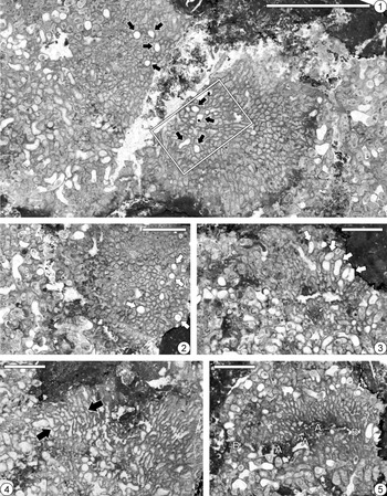
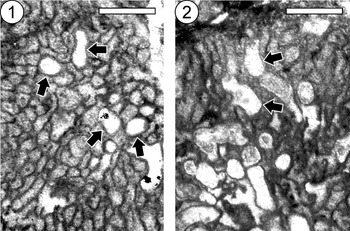
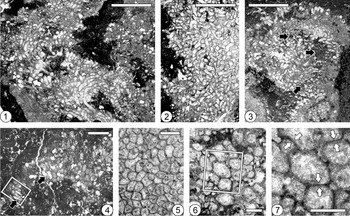
At locality B97, many skeletons of R. divergens n. gen. n. sp. occur in direct association with R. minuta n. gen. n. sp., which has been found only in such associations (Figs. 3.4, 4.1, 4.2). These associations were recognized in 16 specimens, occurring predominantly in boundstone and in floatstone/rudstone. Longitudinal sections reveal that surfaces of R. divergens n. gen. n. sp. were colonized and partially overgrown by R. minuta n. gen. n. sp. (Fig. 4.3, 4.4), with the two species becoming intergrown as some modules of R. divergens n. gen. n. sp. persisted by directing their growth upward (Figs. 4.3, 5.2). In a few cases, peripheral areas of R. divergens n. gen. n. sp. subsequently overgrew R. minuta n. gen. n. sp. (Fig. 4.5). Transverse and longitudinal sections show that isolated modules of R. divergens n. gen. n. sp. influenced the shape and growth of adjoining smaller modules of R. minuta n. gen. n. sp. (Fig. 5). Isolated modules of R. divergens n. gen. n. sp. have not been observed to extend beyond the terminal growth surface of the surrounding R. minuta n. gen. n. sp. skeleton (Fig. 5.2), suggesting that such modules were unable to survive on their own.
Thin-section photomicrographs showing transverse (1, 2), longitudinal (3, 4), and oblique longitudinal (5) views of intergrowths of large-module species Reptamsassia divergens n. gen. n. sp. and the small-module species R. minuta n. gen. n. sp. from boundstone at locality B97. (1) Reptamsassia minuta n. gen. n. sp. (middle of photograph) associated with R. divergens n. gen. n. sp. (left and right sides); R. minuta encloses isolated modules of R. divergens (arrows; outlined area enlarged in Fig. 5.1); GSC 142675a (holotype of R. minuta n. gen. n. sp.); scale bar = 5 mm. (2) Reptamsassia minuta n. gen. n. sp. (right side) associated with R. divergens n. gen. n. sp. (left side); isolated modules of R. divergens (arrows) are surrounded by R. minuta; GSC 142675b (holotype of R. minuta n. gen. n. sp.); scale bar = 2 mm. (3) Reptamsassia divergens n. gen. n. sp. overgrown by R. minuta n. gen. n. sp.; isolated modules of R. divergens (arrows) are intergrown with R. minuta; GSC 142676; scale bar = 2 mm. (4) Reptamsassia minuta n. gen. n. sp. overgrowing and intergrown with R. divergens n. gen. n. sp.; tabula-like structures, associated with constrictions of modules, are developed at same level in adjacent modules of R. minuta (between arrows); GSC 142677a (paratype of R. minuta n. gen. n. sp.); scale bar = 2 mm. (5) Reptamsassia divergens n. gen. n. sp. (bottom) partially overgrown by R. minuta n. gen. n. sp. (center), which it subsequently overgrew (top); patch of microbial clotted micrite (A) with continuous module growth on both sides (B); GSC 142677b (paratype of R. minuta n. gen. n. sp.); scale bar = 2 mm.

Thin-section photomicrographs showing transverse (1) and longitudinal (2) views of intergrowths of large-module species Reptamsassia divergens n. gen. n. sp. and the small-module species R. minuta n. gen. n. sp. from boundstone at locality B97; scale bars = 1 mm. (1) Enlargement of outlined area in Figure 4.1, showing isolated modules of R. divergens n. gen. n. sp. (arrows) enclosed in R. minuta n. gen. n. sp.; GSC 142675a (holotype of R. minuta n. gen. n. sp.). (2) Reptamsassia divergens n. gen. n. sp. (lower left) overgrown by R. minuta n. gen. n. sp., showing isolated modules of R. divergens (arrows) intergrown with R. minuta, and terminal growth surface (upper left); GSC 142678.

The association of R. minuta n. gen. n. sp. and R. divergens n. gen. n. sp. is considered to represent a symbiotic relationship of two modular species that interacted with one another in a reefal environment. Reptamsassia minuta n. gen. n. sp. was apparently an obligate symbiont, in that it has not been found to encrust other surfaces on the growing reef. It presumably benefited by colonizing the relatively stable growth surface of its larger host, R. divergens n. gen. n. sp., where it would have been slightly elevated above the substrate. Although the surface of R. divergens n. gen. n. sp. was negatively affected in areas overgrown by R. minuta n. gen. n. sp., isolated modules of R. divergens were nevertheless able to persist in intergrowths of the two species (Figs. 4.3, 5.2). The shape and growth direction of R. minuta n. gen. n. sp. modules were affected in the immediate vicinity of isolated R. divergens n. gen. n. sp. modules, but the overall effect on R. minuta was negligible (Fig. 5). Both species continued to grow upward and outward, with their modules generally parallel to and keeping pace with one another (Figs. 4.3, 5.2). The areas of R. divergens n. gen. n. sp. not overgrown by R. minuta n. gen. n. sp. were unaffected, and in some cases eventually expanded to partially or completely cover R. minuta (Fig. 4.5). The symbiotic relationship of these two species may be a form of commensalism, with R. minuta n. gen. n. sp. benefiting substantially while not significantly harming its host, R. divergens n. gen. n. sp. Modern encrusting calcareous algae are known to tolerate overgrowth with little negative effect (Airoldi, Reference Airoldi2000). The coexistence of algal crusts covered by filamentous algal turf has been related to their abilities to tolerate overgrowth and to overgrow, respectively (Airoldi, Reference Airoldi2000).
The appearance of Reptamsassia n. gen. in the Floian contributed to the rise in diversity on a global scale and in reefal settings early in the Great Ordovician Biodiversification Event (Fan et al., Reference Fan, Shen, Erwin, Sadler and MacLeod2020, fig. 1; Harper et al., Reference Harper, Cascales-Miñana and Servais2020, fig. 5f). The symbiotic relationship of R. divergens n. gen. n. sp. and R. minuta n. gen. n. sp. exemplifies the development of ecologic specialization and ecosystem complexity in Early Ordovician reefs (Stigall et al., Reference Stigall, Edwards, Freeman and Rasmussen2019). This is the earliest known symbiotic intergrowth of macroscopic modular species. Although various types of symbiotic interactions have been recognized in the Cambrian, most simply involved one organism attached to another; modular organisms were rarely hosts, and endobionts were microscopic and rare (Vinn, Reference Vinn2017). A number of taxa with modular coral-like skeletons are known from reefs dating to Cambrian Epoch 2, including Flindersipora Lafuste in Lafuste et al., Reference Lafuste, Debrenne, Gandin and Gravestock1991, Moorowipora Fuller and Jenkins, Reference Fuller and Jenkins1994, Arrowipora Fuller and Jenkins, Reference Fuller and Jenkins1995, Harklessia Hicks, Reference Hicks2006, and Blinmanipora Fuller and Jenkins, Reference Fuller and Jenkins2007. Symbiotic intergrowths involving these taxa have not been reported or observed in published photographs, although attached archaeocyaths and encrusting calcimicrobes have been documented in some cases (Lafuste et al., Reference Lafuste, Debrenne, Gandin and Gravestock1991; Savarese et al., Reference Savarese, Mount, Sorauf and Bucklin1993; Hicks, Reference Hicks2006; Fuller and Jenkins, Reference Fuller and Jenkins2007).
Prior to discovery of the R. minuta-R. divergens association, the earliest symbiotic intergrowth of macroscopic organisms was thought to date from the late Middle Ordovician (Darriwilian). It involved a worm-like animal, represented by the bioclaustration Anoigmaichnus odinsholmensis Vinn et al., Reference Vinn, Wilson, Mõtus and Toom2014, hosted by the bryozoan Mesotrypa bystrowi Modzalevskaya, Reference Modzalevskaya1953 (Vinn et al., Reference Vinn, Wilson, Mõtus and Toom2014). In the Late Ordovician, during the culmination of the Great Ordovician Biodiversification Event, symbiotic intergrowths became more common, and modular organisms became more common as hosts (Vinn and Wilson, Reference Vinn and Wilson2015; Vinn, Reference Vinn2017). Bioclaustrations of soft-bodied organisms and less commonly skeletonized organisms such as cornulitids, conulariids, and solitary rugosans occur in modular hosts including tabulate corals and more commonly bryozoans (Vinn et al., Reference Vinn, Ernst and Toom2018a, Reference Vinn, Ernst and Toomb, Reference Vinn, Ernst, Wilson and Toom2019). However, symbiotic intergrowths of modular organisms remained rare; the bioclaustration Catellocaula vallata Palmer and Wilson, Reference Palmer and Wilson1988, which may record a colonial hydroid or tunicate, occurs in the bryozoan Amplexopora persimilis Nickles, Reference Nickles1905 (Palmer and Wilson, Reference Palmer and Wilson1988).
Materials and methods
Materials referred to in the systematic paleontology section (see below) were obtained from two thrombolite beds in the Barbace Cove Member of the Boat Harbour Formation, each at a separate locality (B86 and B97) in Isthmus Bay, western Newfoundland (Figs. 1.2, 2). Rock samples were cut into slabs and many large-format thin sections (51 × 76 mm), including some serial sections spaced ~1.5 mm apart, were prepared: 14 from locality B86 and 47 from locality B97. The thin sections revealed numerous small skeletons of Reptamsassia n. gen., which were studied petrographically. For determination of module diameters, transverse thin-sections and a stage micrometer were photographed at the same magnification with a stereoscopic microscope. Dimensions were measured on enlarged photomicrographs, using a ruler calibrated with the micrometer.
Repository and institutional abbreviation
Types, figured specimens, and other materials examined in this study are deposited in the Geological Survey of Canada (GSC), Ottawa, Ontario, Canada.
Systematic paleontology
Family Amsassiaceae new family
Genera
Amsassia Sokolov and Mironova, Reference Sokolov and Mironova1959 (type genus), and Reptamsassia n. gen.
Diagnosis
Growth form massive, structure phacelocerioid; growth direction of modules upward, lateral, or reptant. Transverse shape of modules polygonal to subpolygonal in densely packed areas, subcircular to circular in loosely packed areas, or irregular. Adjoining modules have back-to-back walls. Species have maximum module diameters ranging from 0.28–1.66 mm. Module increase by longitudinal fission involving infolding(s) of wall: bipartite, tripartite, quadripartite, or hexapartite. Modules resulting from fission remain in contact, separate slightly, or diverge in opposite directions with an inter-module angle up to 90°. Tabulae absent. Tabula-like structures form incomplete to complete, concave-up bases of rejuvenated or regenerated modules. Calices shallow to deep with concave-up bottoms.
Remarks
Amsassia was originally considered to be a tabulate coral and assigned to Family Cryptolichenariidae Sokolov in Sokolov and Mironova, Reference Sokolov and Mironova1959. Sokolov and Mironova (Reference Sokolov and Mironova1959) noted the similarity of Amsassia to representatives of Lichenariidae Okulitch, Reference Okulitch1936, Tetradiidae Nicholson, Reference Nicholson1879, and Chaetetidae Milne-Edwards and Haime, Reference Milne-Edwards and Haime1850. Since then, Cryptolichenariidae (including Amsassia) has been placed in the orders Lichenariida Sokolov, Reference Sokolov1950 (e.g., Yu and Zhang, Reference Yu, Zhang, Yu, Wu, Zhao and Zhang1963), Tetradiida Okulitch, Reference Okulitch1936 (e.g., Sokolov, Reference Sokolov, Sokolov and Orlov1962; Lin et al., Reference Lin, Tchi, Jin, Li and Yan1988), and Chaetetida Okulitch, Reference Okulitch1936 (Hill, Reference Hill and Teichert1981, who noted that Amsassia is possibly a tetradiid). The nominative lichenariid genus, Lichenaria, is definitely a tabulate coral (Laub, Reference Laub, Oliver, Sando, Cairns, Coates, Macintyre, Bayer and Sorauf1984; Elias et al., Reference Elias, Lee and Woo2008, Reference Elias, Lee and Pratt2021). The nominative cryptolichenariid genus, Cryptolichenaria Sokolov, Reference Sokolov1955, was included by Scrutton (Reference Scrutton, Oliver, Sando, Cairns, Coates, Macintyre, Bayer and Sorauf1984) in Tetradiida, which he later removed from Tabulata Milne-Edwards and Haime, Reference Milne-Edwards and Haime1850 (Scrutton, Reference Scrutton1997). The nominative tetradiid genus, Tetradium Dana, Reference Dana1846, has since been interpreted as a calcareous florideophyte red alga (Steele-Petrovich, Reference Steele-Petrovich2009a, Reference Steele-Petrovichb), necessitating the replacement name Prismostylus Okulitch, Reference Okulitch1935, and erection of Family Prismostylaceae Steele-Petrovich, Reference Steele-Petrovich2011 (Steele-Petrovich, Reference Steele-Petrovich2011). Chaetetids are now regarded as calcareous sponges (West, Reference West2011a). West (Reference West2011b) rejected Amsassia and Cryptolichenaria from chaetetid-type sponges.
Based on morphologic comparisons in previous studies, Amsassia has been excluded from corals and calcareous sponges, as well as cyanobacteria, and referred to calcareous algae (Sun et al., Reference Sun, Elias and Lee2014; Lee et al., Reference Lee, Park, Tien, Choh, Elias and Lee2016, Reference Lee, Elias, Choh and Lee2018, Reference Lee, Elias and Pratt2021; Elias et al., Reference Elias, Lee and Pratt2021). Infolding of the module wall during longitudinal fission in both Amsassia and Reptamsassia n. gen. is consistent with simple branching of the module occupant (di-, tri-, quadri-, and hexachotomous in Amsassia; di-, tri-, and probably quadrichotomous in Reptamsassia n. gen.). Dichotomous to polychotomous branching is common in algae. Steele-Petrovich (Reference Steele-Petrovich2009a) interpreted module increase in Tetradium as branching. Increase by longitudinal fission in tetradiids was typically quadripartite, but could also be tripartite and bipartite (Sun et al., Reference Sun, Elias and Lee2014). Increase in Cryptolichenaria was bipartite (Hill, Reference Hill and Teichert1981).
Unlike Amsassia and Reptamsassia n. gen., in which module increase involved infolding of the wall, increase in Tetradium and Cryptolichenaria involved the development of one or more skeletal partitions inside the module (Hill, Reference Hill and Teichert1981). Horizontal skeletal elements in tetradiids (e.g., Paenetetradium Copper and Morrison, Reference Copper and Morrison1978; see Copper and Morrison, Reference Copper and Morrison1978) and Cryptolichenaria (see Sokolov, Reference Sokolov1955) are true tabulae, which are platforms having edges attached to the inner surface of the module wall (Hill, Reference Hill and Teichert1981). Amsassia and Reptamsassia n. gen., however, lack true tabulae. Their tabula-like structures are bases of rejuvenated or regenerated modules, which are structurally continuous with the vertical module wall (Elias et al., Reference Elias, Lee and Pratt2021; Lee et al., Reference Lee, Elias and Pratt2021). Thus, Amsassia and Reptamsassia n. gen. differ significantly from tetradiids, and are regarded as a distinct group of extinct calcareous algae for which the Family Amsassiaceae n. fam. is here erected.
In considering the biologic affinity of Amsassia, Lee et al. (Reference Lee, Elias and Pratt2021) made comparisons with the unmineralized extant green alga Cladophora Kützing, Reference Kützing1843. They suggested that if branching filaments such as those of Cladophora were tightly to loosely arranged and the cell walls became calcified during growth, phacelocerioid skeletons of modules with tabula-like structures at constrictions could result, like those of Amsassia. Such tabula-like structures also occur in Reptamsassia n. gen. Maximum module diameters in species of Reptamsassia n. gen. (0.30–0.53 mm) are within the range of maximum cell diameters in species of Cladophora (0.07–1.00 mm). Dichotomous to hexachotomous branching occurs in Cladophora; in Reptamsassia n. gen., the types of module fission indicate di-, tri-, and probably quadrichotomous branching. Angles of ramification are variable within and among species of Cladophora (e.g., see Leliaert and Coppejans, Reference Leliaert and Coppejans2003). Although modules resulting from fission remain in contact in Amsassia, they may also separate slightly or diverge from one another in Reptamsassia n. gen. Lee et al. (Reference Lee, Elias and Pratt2021) inferred that the tops of modules in Amsassia would have remained uncalcified to allow elongation of filaments during growth, and for release of gametes and zoospores as in Cladophora. Therefore, upon death and decomposition of Amsassia, calices with an open top and a concave-up tabula-like structure at the base would have been present on the distal surface of the skeleton and become filled with sediment. Such calices are also observed in Reptamsassia n. gen. On the basis of the foregoing, it is concluded that the Family Amsassiaceae n. fam. may belong to the green algae (Phylum Chlorophyta Reichenbach, Reference Reichenbach1828).
Genus Reptamsassia new genus
Type species
Reptamsassia divergens n. gen. n. sp.; Barbace Cove Member (Floian; Tulean) of Boat Harbour Formation, localities B86 and B97 in Isthmus Bay, eastern Port au Port Peninsula, Newfoundland, Canada.
Other species
Reptamsassia minuta n. gen. n. sp.; Barbace Cove Member (Floian; Tulean) of Boat Harbour Formation, locality B97 in Isthmus Bay, eastern Port au Port Peninsula, Newfoundland, Canada.
Diagnosis
Growth habit encrusting; growth direction of modules highly variable, upward to lateral and reptant. Transverse shape of modules extremely variable, commonly irregular. Species have maximum module diameters ranging from 0.30–0.53 mm. Module increase bipartite, tripartite, and probably quadripartite. Some modules resulting from fission separate slightly, or diverge in opposite directions with an inter-module angle up to 90°. Calices of modules moderately deep to deep.
Etymology
The name recognizes the similarity of this new genus to Amsassia, while noting its distinctive, reptant growth habit.
Remarks
Reptamsassia n. gen. resembles Amsassia in its massive growth form, phacelocerioid structure, back-to-back walls of adjoining modules, module increase by longitudinal fission involving infolding(s) of the wall, calices with concave-up bottoms, and absence of tabulae. Tabula-like structures forming the bases of regenerated or rejuvenated modules in Reptamsassia n. gen. occur at the same levels in adjacent modules, as in species of Amsassia. Maximum module diameters in species of Reptamsassia n. gen. (0.30–0.53 mm) fall within the range of Amsassia (0.28–1.66 mm; Lee et al., Reference Lee, Elias and Pratt2021). The new genus is differentiated from Amsassia by its encrusting habit, the highly variable growth direction of modules ranging from upward to lateral and reptant, the extremely variable and commonly irregular shape of modules, and module increase resulting in modules that separate slightly or diverge from one another in some cases.
Types
Holotype GSC 142679 (thin section; Fig. 6.1) and paratype GSC 142680 (thin section, Fig. 6.2) occur in boundstone and floatstone/rudstone, respectively, Barbace Cove Member (Floian; Tulean) of Boat Harbour Formation, locality B97 in Isthmus Bay, eastern Port au Port Peninsula, Newfoundland, Canada.
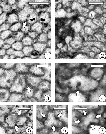
Thin-section photomicrographs showing transverse (2, 3, 5–7) and both transverse and longitudinal (1, 4) views of Reptamsassia divergens n. gen. n. sp. from localities B86 (4, 6, 7) and B97 (1–3, 5). (1) Skeleton showing phacelocerioid structure and variable growth directions of modules, ranging from upward to lateral and reptant; some small areas of sediment accumulation or microbial encrustation (left of center, lower right of center) seem to have been accommodated by diverted growth of modules without mortality; note module undergoing bipartite fission (arrow); holotype GSC 142679; scale bar = 5 mm. (2) Skeleton showing variable growth directions of modules; paratype GSC 142680; scale bar = 5 mm. (3) Skeleton showing partial growth interruptions associated with peloids and micritic clots likely of microbial origin (arrows); GSC 142681; scale bar = 5 mm. (4) An intraclast consisting of skeleton encrusted by microbialite; longitudinal fission resulting in pairs of modules (arrows); deep calices with concave-up bottoms (outlined area; enlarged in Fig. 8.3); GSC 142682; scale bar = 2 mm. (5) Cerioid area showing low size and shape variability of densely packed, polygonal to subpolygonal modules; GSC 142683; scale bar = 1 mm. (6) Loosely packed area showing high size variability of subcircular to circular modules, some flanked by micrite (dark colored); outlined area enlarged in 7; GSC 142684; scale bar = 0.5 mm. (7) Enlargement of outlined area in 6, showing back-to-back walls between adjoining modules (pairs of white arrows); GSC 142684; scale bar = 0.5 mm.

Diagnosis
Reptamsassia n. gen. with maximum module diameter 0.53 mm.
Description
Growth form massive, shape predominantly irregular; structure phacelocerioid (Figs. 6.1–6.4, 7.1). At locality B97, complete skeletons not observed due to development of stylolites and partial diagenetic dissolution; many apparently in growth orientation, some fragmentary and abraded skeletons evidently transported; size over 80 × 80 mm across and 55 mm high. Many skeletons at locality B97, predominantly in boundstone, were overgrown by and intergrown with comparatively small-module species Reptamsassia minuta n. gen. n. sp. (Figs. 4, 5), and contain various amounts of peloids and micritic clots likely of in situ microbial origin (arrows in Fig. 6.3). At locality B86, most skeletons occur as heavily abraded, transported grains in rudstone; original form and size unknown (Fig. 6.4).
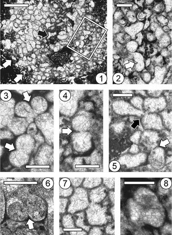
Thin-section photomicrographs showing transverse views of Reptamsassia divergens n. gen. n. sp. from localities B86 (1, 2, 6) and B97 (3–5, 7, 8). (1) Modules irregularly shaped where they encrust substrate during initial stage of skeletal growth (black arrow) and at base of skeleton during subsequent lateral expansion (white arrows); outlined area enlarged in 2; GSC 142685; scale bar = 2 mm. (2) Enlargement of outlined area in 1, showing variably sized modules, some flanked by micrite, in loosely packed area; module shapes subcircular to circular, or irregular due to interference from adjacent modules; note module undergoing bipartite fission involving infolding of wall (arrow); GSC 142685; scale bar = 0.5 mm. (3–6) Modules undergoing bipartite fission (white arrows) and probable quadripartite fission (black arrow in 5) involving infolding(s) of wall; GSC 142686a, GSC 142687, GSC 142686b, and GSC 142688, respectively; scale bars = 0.5 mm. (7) Module undergoing tripartite fission; GSC 142686a; scale bar = 0.5 mm. (8) Module probably undergoing tripartite fission involving infoldings of wall; GSC 142689; scale bar = 0.5 mm.

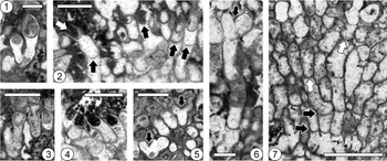
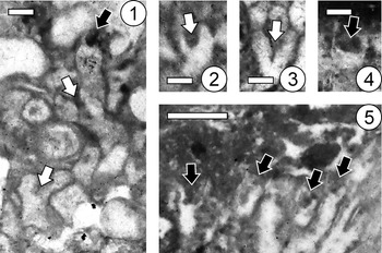
In transverse section, module shape extremely variable: polygonal to subpolygonal in densely packed areas (Fig. 6.5); subcircular to circular in loosely packed areas where partially or completely separated by micrite (Figs. 6.6, 7.2); shape irregular where modules encrust substrate, including initial stage of skeletal growth (black arrow in Fig. 7.1) and base of skeleton during subsequent lateral expansion (white arrows in Fig. 7.1); irregular due to highly variable growth directions of modules, ranging from upward to lateral and reptant (Figs. 3.4, 6.1–6.3); irregular due to interference from adjacent modules (Figs. 6.6, 7.2). Adjoining modules have back-to-back walls (Fig. 6.7). Module size extremely variable throughout skeletal growth, especially in loosely packed areas with modules partially or completely separated by micrite (Figs. 6.6, 7.2); diameter of largest 10% of modules 0.40–0.53 mm (Table 1). Module increase by longitudinal fission (arrows in Fig. 6.4) involving infolding(s) of wall (white arrows in Figs. 6.1, 7.2–7.6), which extend(s) to divide module; predominantly bipartite (white arrows in Figs. 6.1, 7.2–7.6), rarely tripartite (Fig. 7.7, 7.8) and probably quadripartite (black arrow in Fig. 7.5). In oblique or longitudinal section, some modules resulting from fission separate slightly (arrows in Fig. 6.4; Fig. 8.1, 8.3) or diverge from one another in opposite directions with an inter-module angle up to 90° (arrows in Fig. 8.5, 8.6). In longitudinal section, module walls usually undulate (Fig. 8.6, 8.7). Tabulae absent. Regeneration or rejuvenation, usually following constriction of modules (white arrows in Fig. 8.7), recorded by typically concave-up tabula-like structures forming bases of subsequent module growth, structurally continuous with vertical module wall, commonly developed at same levels in adjacent modules (black arrows in Fig. 8.7). Calices of modules deep with concave-up bottoms (white arrow in Fig. 8.2; Fig. 8.3, 8.4), similar in shape to the tabula-like structures (black arrows in Fig. 8.2).
Thin-section photomicrographs showing longitudinal views of Reptamsassia divergens n. gen. n. sp. from localities B86 (3, 4) and B97 (1, 2, 5–7). (1) Longitudinal fission resulting in modules that separate slightly; GSC 142690; scale bar = 0.5 mm. (2) Deep calice with concave-up bottom (white arrow) similar in shape to tabula-like structures within modules (black arrows); GSC 142691; scale bar = 1 mm. (3) Enlargement of outlined area in Figure 6.4, showing longitudinal fission resulting in modules that separate slightly (left side); note deep calices of modules; GSC 142682; scale bar = 1 mm. (4) Deep calices with concave-up bottoms; GSC 142692; scale bar = 1 mm. (5, 6) Longitudinal fission resulting in modules that diverge (arrows); GSC 142693 and GSC 142694, respectively; scale bars = 1 mm and 0.5 mm, respectively. (7) Tabula-like structures forming bases of regenerated modules, usually following constriction of modules (e.g., white arrows), commonly developed at same levels in adjacent modules (black arrows); GSC 142695; scale bar = 2 mm.

Diameter (mm) of modules of Reptamsassia divergens n. gen. n. sp., based on 12 skeletons from locality B86 and 11 (including holotype and paratype) from locality B97. In transverse thin-sections, diameter was measured internally in the largest 10% of modules; module diameter = (long axis + short axis) ÷ 2 (see Scrutton, Reference Scrutton1981). Abbreviations: Min. = minimum; Max. = maximum; Avg. = average; S.D. = standard deviation; N = number of modules.

Etymology
The name of the new species refers to the divergent growth patterns of modules following longitudinal fission.
Materials
In addition to holotype and paratype (see above), thin sections of the following 16 skeletons are figured. GSC 142682 (Figs. 6.4, 8.3), GSC 142684 (Fig. 6.6, 6.7), GSC 142685 (Fig. 7.1, 7.2), GSC 142688 (Fig. 7.6), and GSC 142692 (Fig. 8.4) occur in rudstone, Barbace Cove Member (Floian; Tulean) of Boat Harbour Formation, locality B86 in Isthmus Bay, eastern Port au Port Peninsula, Newfoundland, Canada. GSC 142673 (Fig. 3.4), GSC 142686a (Fig. 7.3, 7.7) and 142686b (Fig. 7.5), GSC 142690 (Fig. 8.1), GSC 142694 (Fig. 8.6), and GSC 142695 (Fig. 8.7) occur in floatstone/rudstone, GSC 142681 (Fig. 6.3), GSC 142683 (Fig. 6.5), GSC 142687 (Fig. 7.4), GSC 142689 (Fig. 7.8), GSC 142691 (Fig. 8.2), and GSC 142693 (Fig. 8.5) occur in boundstone, Barbace Cove Member (Floian; Tulean) of Boat Harbour Formation, locality B97 in Isthmus Bay, eastern Port au Port Peninsula, Newfoundland, Canada.
Remarks
The most distinctive feature of Reptamsassia divergens n. gen. n. sp. is the high degree of variability in shape and size of modules, related in part to the encrusting base of the skeleton and the highly variable growth directions of modules, ranging from upward to lateral and reptant. Reptamsassia divergens n. gen. n. sp. is the large-module species of this genus; diameters of the largest 10% of modules are 0.40–0.53 mm for R. divergens n. gen. n. sp. versus 0.19–0.30 mm for R. minuta n. gen. n. sp. Although diameters of subcircular to circular modules in R. divergens n. gen. n. sp. tend to be smaller at locality B86 than at locality B97, the ranges of values overlap (Table 1). Many skeletons of R. divergens n. gen. n. sp. were overgrown by and intergrown with R. minuta n. gen. n. sp. at locality B97, predominantly in the boundstone.
Types
Holotype GSC 142675a (thin section; Figs. 4.1, 5.1, 9.1, 10.4), 142675b (thin section; Figs. 4.2, 9.3, 9.6), and 142675c (thin section; Fig. 9.2, 9.4), and paratype GSC 142677a (thin section; Figs. 4.4, 10.5) and 142677b (thin section; Fig. 4.5) occur in boundstone, Barbace Cove Member (Floian; Tulean) of Boat Harbour Formation, locality B97 in Isthmus Bay, eastern Port au Port Peninsula, Newfoundland, Canada.
Thin-section photomicrographs showing transverse views of Reptamsassia minuta n. gen. n. sp. from locality B97. (1) Mostly polygonal to subpolygonal modules showing variable shapes and sizes, with one module undergoing bipartite fission involving infolding of wall (white arrow); adjoining modules have back-to-back walls (black arrows); holotype GSC 142675a; scale bar = 0.5 mm. (2) Shape and size variability of subcircular to circular and irregularly shaped modules, some flanked by sediment (dark area left of center); holotype GSC 142675c; scale bar = 0.5 mm. (3) Module undergoing bipartite fission (arrow) where modules are relatively densely packed; holotype GSC 142675b; scale bar = 0.2 mm. (4, 5) Modules undergoing bipartite fission involving infolding(s) of wall (arrows); holotype GSC 142675c and GSC 142696a, respectively; scale bars = 0.2 mm. (6) Module undergoing tripartite fission involving infoldings of wall (arrows); holotype GSC 142675b; scale bar = 0.2 mm. (7) Module probably undergoing quadripartite fission involving infoldings of wall (arrows); GSC 142697; scale bar = 0.2 mm.

Thin-section photomicrographs showing longitudinal views of Reptamsassia minuta n. gen. n. sp. from locality B97. (1–3) Longitudinal fission resulting in modules that separate slightly or diverge (white arrows); note calice of a module (black arrow in 1); GSC 142696b (1, 2) and GSC 142698 (3); scale bars = 0.2 mm. (4, 5) Modules having moderately deep calices (arrows); holotype GSC 142675a and paratype GSC 142677a, respectively; scale bars = 0.2 mm and 0.5 mm, respectively.

Diagnosis
Reptamsassia n. gen. with maximum module diameter 0.30 mm.
Description
Growth form massive, shape irregular; overall structure phacelocerioid, predominantly cerioid with inconspicuous phaceloid areas. Size (difficult to determine in most cases) up to 10 × 8 mm across and 4 mm high. All observed skeletons grew in direct association with R. divergens n. gen. n. sp. (Figs. 3.4, 4, 5); occasional partial mortality was followed by rejuvenation/regeneration (Fig. 4.5). In transverse section, modules predominantly polygonal to subpolygonal in densely packed areas (Fig. 9.1), subcircular to circular in more loosely packed areas (Fig. 9.2); shape of modules typically irregular, especially where in contact with R. divergens n. gen. n. sp. (Figs. 3.4, 4.2, 5.1). Growth direction of modules variable, ranging from upward to lateral and reptant (Fig. 4.1). Adjoining modules have back-to-back walls (black arrows in Fig. 9.1). Module size highly variable throughout skeletal growth; diameter of largest 10% of modules 0.19–0.30 mm (Table 2). Module increase by longitudinal fission (white arrows in Fig. 10.1–10.3); predominantly bipartite (white arrows in Fig. 9.1, 9.3–9.5), rarely tripartite (Fig. 9.6) and probably quadripartite (Fig. 9.7), involving infolding(s) of wall seen where module is not constrained by tight packing (white arrows in Fig. 9.1, 9.3–9.7). In oblique or longitudinal section, some modules resulting from fission separate slightly or diverge from one another in opposite directions with an inter-module angle up to 60° (white arrows in Fig. 10.1–10.3). In longitudinal section, module walls highly undulate (Fig. 4.4) and conform with modules of R. divergens n. gen. n. sp. where in contact (Figs. 4.3, 5.2). Tabulae absent. Regeneration or rejuvenation, usually following constriction of modules, recorded by typically concave-up tabula-like structures forming bases of subsequent module growth, structurally continuous with vertical module wall, commonly developed at same levels in adjacent modules (between black arrows in Fig. 4.4). Calices of modules moderately deep with concave-up bottoms (black arrows in Fig. 10.1, 10.4, 10.5).
Diameter (mm) of modules of Reptamsassia minuta n. gen. n. sp., based on five skeletons (including holotype). In transverse thin-sections, diameter was measured internally in the largest 10% of modules; module diameter = (long axis + short axis) ÷ 2 (see Scrutton, Reference Scrutton1981). Abbreviations: Min. = minimum; Max. = maximum; Avg. = average; S.D. = standard deviation; N = number of modules.

Etymology
The name of the new species refers to the small size of its modules.
Materials
In addition to holotype and paratype (see above), thin sections of the following six skeletons are figured. GSC 142674 (Fig. 3.4) occurs in floatstone/rudstone, GSC 142676 (Fig. 4.3), GSC 142678 (Fig. 5.2), GSC 142696a (Fig. 9.5) and 142696b (Fig. 10.1, 10.2), GSC 142697 (Fig. 9.7), and GSC 142698 (Fig. 10.3) occur in boundstone, Barbace Cove Member (Floian; Tulean) of Boat Harbour Formation, locality B97 in Isthmus Bay, eastern Port au Port Peninsula, Newfoundland, Canada.
Remarks
Reptamsassia minuta n. gen. n. sp. is the small-module species of this genus; diameters of the largest 10% of modules are 0.19–0.30 mm for R. minuta n. gen. n. sp. versus 0.40–0.53 mm for R. divergens n. gen. n. sp. Reptamsassia minuta n. gen. n. sp. is only known to occur in direct association with R. divergens n. gen. n. sp., overgrowing and intergrown with the latter species.
Acknowledgments
This study was supported by funding from the National Research Foundation of Korea (grant 2018R1A2B2005578) to D.-J. Lee, and the Natural Sciences and Engineering Research Council of Canada to B.R. Pratt. Pratt is grateful to N.P. James for supervising his M.Sc. thesis work, during which specimens from western Newfoundland were discovered, and to S.P. Robson for help in the field when further collections were made. We thank H.M. Park for assistance in preparing thin sections. The reviewers (M.G. Carrera, O. Vinn) and Associate Editor (S.T. LoDuca) provided helpful comments on the manuscript.