INTRODUCTION
An intraerythrocytic protozoon, Babesia bovis, is one of the major causative agents of babesiosis in cattle (Sam-Yellowe, 1996; Homer et al. 2000). This parasite causes severe symptoms such as fever, anaemia, and cerebral dysfunctions during its erythrocytic stage, and thereby has a significant impact in the cattle industry worldwide (Kuttler, 1988). The host-parasite interaction between B. bovis and the host is complex. A better understanding of the biological processes involved in the growth cycle at the erythrocytic stage is required in order to develop effective therapeutic tools against bovine babesiosis (Bork et al. 2004a).
Protein kinases play essential roles in various signalling pathways of eukaryotic cells by catalysing the transfer of phosphate from ATP to an amino acid side-chain of a protein (Karin, 1991). They are vital for cell growth and survival (Bray, 1990). Any irregularity in their enzymatic activities may result in serious diseases, such as cancer, diabetes, and cardiovascular disorders (Komuro, 2001; Fulop, Larbi and Douziech, 2003; Russello and Shore, 2004). Since such protein phosphorylations are also essential in protozoa, notable advances have been made in determining the growth inhibitory activities of specific kinase inhibitors and clarifying the structure and function of the target protein kinases (Wiser and Schweiger, 1985; Flawia, Tellez-Inon and Torres, 1997; Doerig, 2004).
A serine/threonine kinase inhibitor, staurosporine (Becker and Jaffe, 1997), has broad biological activity ranging from anti-fungal (Sancelme, Fabre and Prudhomme, 1994) to anti-hypertensive (Berg, 2003). Additionally, it inhibits host cell invasion or growth of several Plasmodium species (Ward et al. 1994; Dluzewski and Garcia, 1996; Gazarini and Garcia, 2003), the epimastigotes of Trypanosoma cruzi (Vieira, de Carvalho and de Souza, 1994; Malaquias and Oliveira, 1999), and the promastigotes of Leishmania major and L. amazonensis (Becker and Jaffe, 1997). A selective protein kinase C (PKC) inhibitor, Ro-31-7549 (Hashimoto et al. 1997), is a potent down-regulator of several PKC-subtypes in human cancer cells (Turner et al. 1996), but study of its effect on protozoa is still lacking. Finally, a Ca2+/calmodulin (CaM)-dependent protein (calcium-modulin) kinase inhibitor, KN-93 (Sumi et al. 1991), inhibited the growth of the epimastigotes of T. cruzi (Malaquias and Oliveira, 1999) and hampered the morphological development of the P. gallinaceum zygote to ookinete in mosquitoes (Silva-Neto, Atella and Shahabuddin, 2002). In the present work, we investigated the inhibitory activities of these protein kinase inhibitors on the growth of B. bovis in vitro.
MATERIALS AND METHODS
In vitro cultivation of B. bovis
The Texas strain of B. bovis was maintained in purified bovine erythrocytes in a serum-free GIT growth medium (Wako Pure Chemical Industrial Ltd., Osaka, Japan) as described previously (Bork et al. 2003a, 2004b, 2005).
Chemicals
Indolcarbazole staurosporine was purchased from Wako, and bisindolylmaleimide-VIII-acetate, Ro-31-7549, and the methoxybenzenesulfonamide KN-93 were obtained from Calbiochem (Darmstadt, Germany). Stock solutions of 1 M of the chemicals were prepared in dimethylsulfoxide (DMSO; Wako) and stored at −30 °C until use. Different drug concentrations were prepared by diluting the stock solution with the GIT medium. The concentrations of DMSO used in this study did not exert any abnormally inhibitory effects on the growth, as described previously (Bork et al. 2003a).
Growth inhibition assays
In vitro growth inhibition assays were performed as described previously (Bork et al. 2003a,b). In brief, 100 μl of infected bovine erythrocytes were diluted with non-infected erythrocytes to obtain 1% parasitaemia in a 0·1 ml volume, and the mixture was subsequently suspended in 0·9 ml of the growth medium supplemented with the indicated concentrations of chemicals. The suspension was added to 24-well culture plates (Nunk, Roskilde, Denmark), and the plates were incubated in a humidified multigas water-jacketed incubator at 37 °C for 4 days. During the incubation period, the overlaid culture medium was replaced daily with 0·9 ml of fresh growth medium containing the chemicals at the indicated concentrations. In parallel, chemical-free cultures were prepared as controls. Giemsa-stained thin blood smears were prepared daily, and the parasitic growth was monitored as parasitaemia by evaluating at least 1000 erythrocytes per well using light microscopy (Nikon, Tokyo, Japan). The percentage of extraerythrocytic (free) merozoites was calculated on the first day of culture as the ratio of free merozoites to the entire (intra- and extraerythrocytic) parasite population in approximately 250 parasites. The percentage of ring-shaped parasites was determined as the ratio of ring-shaped intraerythrocytic parasites to the entire amount of intraerythrocytic parasites in approximately 250 parasites. Viability tests were also conducted following the method described by Bork et al. (2004a). In brief, after 4 days of chemical exposure, 30 μl of each of the infected and treated erythrocytes were mixed with 70 μl of non-infected erythrocytes and suspended in fresh growth medium without chemical supplementation. The plates were incubated for the next 10 days. The culture medium was replaced daily, and parasite recrudescence was determined by light microscopy to evaluate the parasite viability.
Statistical analysis
The 50% inhibition (IC50) values of the drugs were determined in triplicate on the fourth day of in vitro culture after curve-fitting using Cricket Graph Software (Malvern, Pennsylvania, USA). Differences in the percentage parasitaemia and other percentages were statistically analysed using an independent Student's t-test at P<0·01 and P<0·05 as the values representing significant differences.
RESULTS
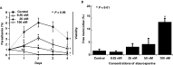
A concentration of 100 μM staurosporine irreversibly arrested the growth of B. bovis as early as the first day of in vitro culture, as shown in light microscopy and a subsequent viability test (Fig. 1A). During the time-course of the growth inhibition assay, concentrations from 6·25 to 50 μM significantly suppressed the parasitaemia but did not destroy the parasites. The IC50 value was determined as 5·12 μM. On the first day of culture, 25, 50, and 100 μM staurosporine resulted in substantial increases in the percentages of extraerythrocytic (free) merozoites, i.e. 3·2%±2·8, 4·8%±4·3, and 14·5%±4·1 (P<0·01), respectively, as compared to the non-treated control group (1·1%±1·1) (Fig. 1B). In the subsequent time-course with staurosporine, almost all of the free merozoites became pycnotic and died (data not shown).

Fig. 1. (A) In vitro growth curves of Babesia bovis in the presence of different concentrations of staurosporine, based on the dynamics of parasitaemia. Parasitic viability: viable (+), dead (−). (B) Percentage of free merozoites in in vitro cultures of B. bovis exposed to different concentrations of staurosporine on the first day of culture. Each value represents the mean±standard deviation (S.D.) in 3 separate trials carried out in triplicate. Asterisks indicate significant differences (P<0·05 in (A) and P<0·01 in (B)) between the drug-treated and control groups.
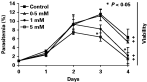
Concentrations of 1 and 5 mM Ro-31-7549 reduced the parasitaemia, starting from the second and third day of culture, respectively. However, full destruction of the parasite was not observed and, additionally, a concentration below 1 mM did not exert any growth inhibitory effects on B. bovis (Fig. 2). The IC50 value was estimated as 4·83 mM.

Fig. 2. In vitro growth curves of Babesia bovis in the presence of different concentrations of Ro-31-7549. Parasitic viability: viable (+), dead (−). Asterisks indicate significant differences (P<0·05) between the drug-treated and control groups. Each value represents the mean±S.D. in 3 separate trials conducted in triplicate.
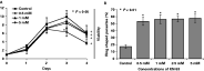
Treatment with 5 mM KN-93 slightly but significantly hampered the parasitic growth as early as the third day of culture, but failed to completely clear the parasites from the culture (Fig. 3A). The IC50 value was estimated as 11·75 mM. Interestingly, on the second day of culture, the percentages of ring-shaped parasites within the infected erythrocytes increased (P<0·01) in the cultures treated with 0·5 mM (53·3%±5·8), 1 mM (56·3%±6·4), 2·5 mM (56·7%±4·2), and 5 mM (57·7%±6·1) of KN-93, as compared to the control (17·1%±2·5) (Fig. 3B).

Fig. 3. (A) In vitro growth curves of Babesia bovis in the presence of different concentrations of KN-93. Parasitic viability: viable (+), dead (−). (B) Percentage of ring-shaped intraerythrocytic parasites in in vitro cultures of B. bovis exposed to different concentrations of KN-93 on the second day of culture. Each value represents the mean±S.D. in 3 separate trials conducted in triplicate. Asterisks indicate significant differences (P<0·05 in (A) and P<0·01 in (B)) between the drug-treated and control groups.
DISCUSSION
In this study, 3 kinds of protein kinase inhibitors were shown to have substantial inhibitory effects on the in vitro growth of B. bovis. Among them, the serine/threonine kinase inhibitor, staurosporine, showed the strongest activity of growth inhibition. The significant increase in the percentage of free merozoites in the staurosporine-treated cultures might be attributed to the inhibition of the parasite's invasion into host erythrocytes. Our findings correspond to previous publications reporting that staurosporine significantly inhibited the erythrocyte invasions by P. knowlesi (Ward et al. 1994), P. falciparum (Dluzewski and Garcia, 1996), and P. chabaudi (Gazarini and Garcia, 2003) in their cultures. The inhibitory mechanism, however, remains unclear yet, because the significant increase of free merozoites was accomplished only at the highest concentration of staurosporine that affected the parasite viability in this study. Further study will be required to understand the inhibitory mechanism of staurosporine on B. bovis.
The PKC-inhibitor, Ro-31-7549, has not been studied in any protozoa. Thus, our work is the first study of its possible inhibitory effect on the propagation of B. bovis. Previously, Ray et al. (1990) demonstrated that B. bovis possesses a powerful PKC activity in the parasitic membrane. However, the growth inhibition activity of Ro-31-7549 was not as high as we initially expected. Whether the result in our growth inhibition assay was due to insufficient drug concentrations used in our study or the possibilities that Ro-31-7549 may not be a specific inhibitor of PKC activity in B. bovis or that PKC activity may not be essential for in vitro growth, remains to be elucidated.
Although treatment with a Ca2+/CaM-dependent protein kinase inhibitor, KN-93, failed to effectively interrupt the parasitic propagation, it caused a significant increase in the percentage of ring-shaped parasites, which are the early forms after erythrocyte invasion that follow the subsequent division to 2 elongated parasites (Levine, 1988). This morphological phenomenon was also observed in the Plasmodium species (Silva-Neto et al. 2002). Ray et al. (1990) demonstrated the existence of a Ca2+/CaM environment in B. bovis and mentioned a possible correlation between the growth of B. bovis and Ca2+-dependent protein phosphorylation by kinases. For understanding the biological effect of KN-93 on B. bovis, it is necessary to identify the Ca2+/CaM-dependent protein kinase of B. bovis and investigate its relationship with the morphological shape changes from ring-shaped to elongated Babesia parasites.
Our endeavour to work with these protein kinase inhibitors will contribute to the understanding of the biological mechanisms of erythrocyte invasion and development of B. bovis in future. In particular, the identification of the target protein kinases, which possibly exist in the parasite or host erythrocyte, is of major interest. A more detailed exploration of the biological roles of protein kinases involved in the asexual growth of Babesia parasites will pave the way for a better understanding and control of bovine babesiosis.
This work was supported by Grants-in-Aid for Scientific Research from the Japanese Society for the Promotion of Science, by grants from the Special Coordination Funds for Science and Technology from the Science and Technology Agency and from The 21st Century COE Program (A-1), Ministry of Education, Culture, Sports, Science, and Technology, Japan, and by the Japan International Cooperation Agency (JICA), F.Y. 2003.