
Introduction
The family Cryptogonimidae is a cosmopolitan group of trematodes that infect fish, reptiles and, rarely, amphibians (Miller and Cribb, Reference Miller, Cribb, Bray, Gibson and Jones2008). Cryptogonimid adult worms from Exorchis Kobayashi, 1921 are intestinal parasites of the order Siluriformes Cuvier, 1816, members of which are widespread throughout East and Southeast Asia (Gibson et al., Reference Gibson, Bray and Jones2002). Exorchis oviformis Kobayashi, Reference Kobayashi1921, a type species of Exorchis, was described based on samples from Japan collected from the intestine of Silurus asotus Linnaeus, 1758 (Kobayashi, Reference Kobayashi1921). Subsequently, the species was recorded in Korea, China, Vietnam and in the Russian southern Far East (Wang, Reference Wang and Wu1991; Sohn and Choi, Reference Sohn and Choi1997; Besprozvannykh et al., Reference Besprozvannykh, Ermolenko and Shedko2000; Nguyen et al., Reference Nguyen, Dalsgaard, Ly and Murrell2007). Later, three other species of the genus were described from Siluridae in China: E. multivitellarius (Pan, Reference Pan1984), E. ovariolobularis Cao, 1990, and E. dongtinghuensis Zhang et al. Reference Zhang, Zuo, Zheng, Liu and Zhou1993. In addition, Pan (Reference Pan1984) described the new genus, Parametadena Pan, 1984, when Parametadena macrobursae (Pan, Reference Pan1984) was collected from the freshwater siluriform Tachysurus fulvidraco (J. Richardson, 1846) in China. Thereafter, Miller and Cribb (Reference Miller, Cribb, Bray, Gibson and Jones2008) synonymized Parametadena with Exorchis, and transferred P. macrobursae to Exorchis as E. marobursae (Pan, Reference Pan1984). As a result, the genus Exorchis currently includes five valid species. Previous study of Exorchis representatives established similarities with respect to life cycles in different species of this genus: Stenothyridae Tryon, 1866, Bithyniidae Gray, 1857, and Pomatiopsidae Stimpson, 1865 molluscs act as the first intermediate hosts of Exorchis, while their second intermediate hosts consist of different species of cyprinid fish, and Siluriformes are definitive hosts (Chung et al., Reference Chung, Joo and Choi1980; Zhang et al., Reference Zhang, Zuo, Zheng, Liu and Zhou1993; Besprozvannykh et al., Reference Besprozvannykh, Ermolenko and Shedko2000; Shimazu, Reference Shimazu2017). Published molecular studies involving Exorchis are non-existent.
During a survey of intestinal parasites of Siluridae, adult Exorchis were identified in Japan and the Russian southern Far East. Morphological and genetic (complete ITS2 rDNA region (ITS2), partial 28S rRNA gene (28S) and cox1 mtDNA gene (cox1)) data were used for taxonomic identification of these samples. In addition, genetic data of Exorchis metacercariae from naturally infected fish in Japan were analysed. The population and intrageneric relationships were also assessed for Exorchis representatives and the phylogenetic relationships determined within the family Cryptogonimidae.
Materials and methods
Sample collection and slide preparation
Adult Exorchis spp. removed from the intestines of naturally infected S. asotus were used in this study. Nets were used for fishing. Fish were caught from three localities: Lebedinoe Lake, located in the wellhead region of the Karasik River, Russia (42°58–23′N, 130°68–57′E); the Razdolnaya River, Russia (43°51–79′N, 131°90–40′E); and the Tara River, Tara Town, Saga, Kyushu Island, Japan (33°01–76′N, 130°10–00′E). Three S. asotus were observed in Russia and one was observed in Japan. Following the removal from the intestine, worms are rinsed in distilled water, killed in hot distilled water and preserved in 70% ethanol. After fixation, they are placed in 96% ethanol. Whole mounts were prepared for microscopic examination by staining with alum carmine, then dehydrated using a concentration gradient of ethanol, cleared using xylene, and mounted on a slide using Canada balsam under the coverslip. All measurements are given in micrometres (μm).
In the analysis, Exorchis spp. metacercariae from naturally infected fish from Japan were also used. Fish were caught from two localities: a small stream on Aomori, Honshu Island (Gymnogobius urotaenia (Hilgendorf, 1879), 40°74–61′N, 141°20–95′E), and a small stream on Saga, Kyushu Island (Tanakia lanceolata (Temminck and Schlegel, 1846) and Carassius sp. Nilsson, 1832, 33°29–05′N, 130°30–34′E). Metacercariae were preserved in 70% ethanol.
DNA extraction, amplification and sequencing
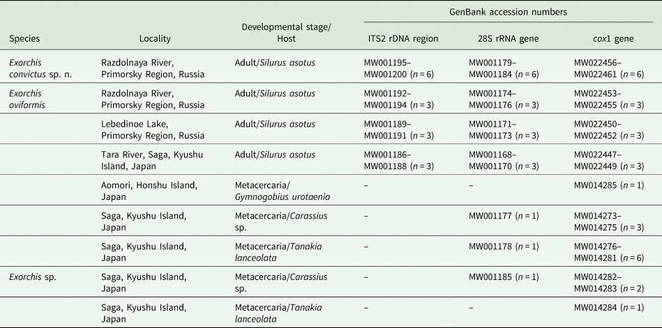
Genomic DNA was extracted from adult worms using the HotSHOT technique (Truett et al., Reference Truett, Heeger, Mynatt, Walker and Warman2000). The number of samples isolated, their developmental stages, hosts and localities are presented in Table 1. Complete ITS2, and partial 28S and cox1 sequences are amplified using the following primer pairs: 1/F (5′-CAC-ACC-GCC-CGT-CG-3′) (Krieger and Fuerst, Reference Krieger and Fuerst2002) and BD2 (5′-TAT-GCT-TAA-G(A)T-TCA-GCG-GGT-3′) (Morgan and Blair, Reference Morgan and Blair1995) for ITS2; DIGl2 (5′-AAG-CAT-ATC-ACT-AAG-CGG-3′) and 1500R (5′-GCT-ATC-CTG-AGG-GAA-ACT-TCG-3′) for 28S (Tkach et al., Reference Tkach, Littlewood, Olson, Kinsella and Swiderski2003), and Co1-Fw (5′-GGG-CAT-CCT-GAG-GTT-TAT-G-3′) and Co1-Rv (5′-AAC-AAA-TCA-TGA-TGC-AAA-AGG-TA-3′) for cox1 (Katokhin et al., Reference Katokhin, Shekhovtsov, Konkow, Yurlova, Serbina, Vodianitskai, Fedorov, Loktev, Muratov, Ohyama, Makhnev, Peltek and Mordvinov2008). The reaction mixture (10 μL of total volume) contains 0.25 mm of each primer, 5 μL GoTaq® Green Master Mix (2 × ) (Promega Corporation, USA), and 3 μL DNA. Polymerase chain reaction (PCR) was performed using a GeneAmp 9700 Thermocycler (Applied Biosystems, USA) with the following reaction conditions: initial denaturation (1 min, 94 °C) followed by 35 cycles: denaturation (15 s, 94 °C), annealing (30 s, 55 °C and 52 °C for nuclear and mitochondrial markers, respectively) and extension (2 min, 72 °C); final extension (5 min, 72 °C). Amplicons were sequenced using an ABI 3500 Genetic Analyser (Applied Biosystems; at the Federal Scientific Center of the East Asia Terrestrial Biodiversity FEB RAS, Russia). For sequencing, external and internal primers are used: BD2 for ITS2; DIGl2, 900F (5′-CCG-TCT-TGA-AAC-ACG-GAC-CAA-G-3′, forward, Tkach et al., Reference Tkach, Littlewood, Olson, Kinsella and Swiderski2003), 1200R (5′-CTT-GGT-CCG-TGT-TTC-AAG-ACG-GG-3′, reverse, Tkach et al., Reference Tkach, Littlewood, Olson, Kinsella and Swiderski2003), 28S4R (5′-TAT-TTA-GCC-TTG-GAT-GGA-GTT-TAC-C-3′, reverse, Besprozvannykh et al., Reference Besprozvannykh, Tatonova and Shumenko2019) for 28S, and Co1-Fw, Co1-Rv for cox1. The cox1 gene of metacercariae in Japan is amplified and sequenced using the following primers: JB3 (5′-TTT-TTT-GGG-CAT-CCT-GAG-GTT-TAT-3′, forward) and CO1-R trema (5′-CAA-CAA-ATC-ATG-ATG-CAA-AAG-G-3′, reverse) (Miura et al., Reference Miura, Kuris, Torchin, Hechinger, Dunham and Chiba2005). The 28S sequences for metacercariae were obtained using the same primers as for adult worms.
List of Exorchis samples determined in this study

n, number of determined sequences.
Analysis of genetic data
Obtained sequences were assembled manually and aligned using the ClustalW option within MEGA version 5.03 (Tamura et al., Reference Tamura, Peterson, Peterson, Stecher, Nei and Kumar2011). Complete ITS2 and partial 28S and partial cox1 were deposited to GenBank (Table 1). For all markers, p-distances between and within species were analysed using MEGA software. Using Arlequin version 3.11 (Excoffier et al., Reference Excoffier, Laval and Schneider2005), a minimum spanning tree (MST) was reconstructed based on cox1 sequences.
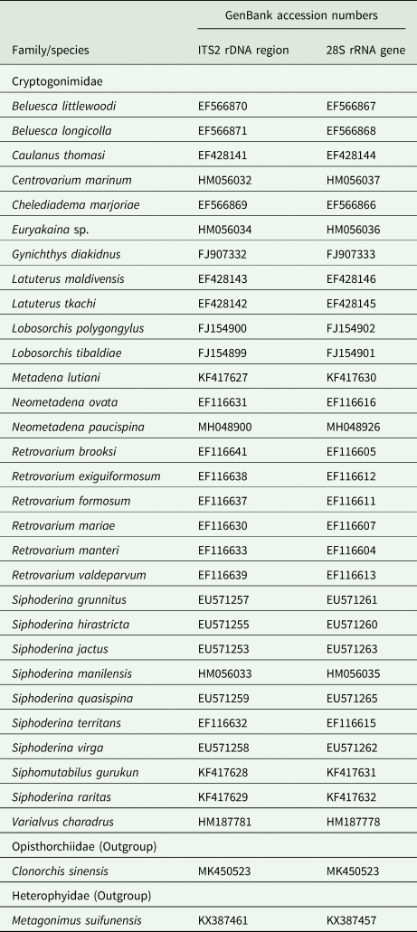
The phylogeny within the genus Exorchis was constructed based on partial cox1 data (759 bp) with Tabascotrema verai Lamothe-Argumedo & Pineda-Lopez, 1990 (JX023291) as an outgroup. Phylogenetic relationships within Cryptogonimidae were assessed using combined complete ITS2 and partial 28S sequencing data (with total length 1157 bp) for cryptogonimid species included from GenBank (Table 2). To align the sequences of ribosomal cluster, MAFFT online service was used (Katoh et al., Reference Katoh, Rozewicki and Yamada2019). Both phylogenetic relationships are reconstructed via the Bayesian inference (BI) using MrBayes version 3.1.2 (Ronquist et al., Reference Ronquist, Huelsenbeck and Teslenko2011). In accordance with the Akaike information criterion in jModeltest version 2.1.10 (Darriba et al., Reference Darriba, Taboada, Doallo and Posada2012), the GTR + I and TVM + I + G models were determined to be optimal for cox1 and ITS2-28S, respectively. BI analysis was performed using 600 000 and 400 000 Markov chain Monte Carlo generations, respectively. The number of generations is determined to sufficient, since the s.d. value calculated is <0.01. The chain was sampled every 100 generations. The first 25% of trees produced were discarded as burn-in to permit the construction of consensus trees.
List of sequences from GenBank used in phylogenetic analysis of the family Cryptogonimidae

Results
Exorchis convictus sp. n.
Definitive host: Silurus asotus.
Site: intestine.
Intensity of infection: 15 specimens.
Infection indices: One fish of three examined.
Type locality: the Razdolnaya River, Primorsky Region, the Russian southern Far East, 43°51–79′N, 131°90–40′E.
Type-deposition: Holotype No. 154–Tr, paratypes No. 155–158–Tr.
Materials are deposited in the parasitological collection of the Zoological Museum (Federal Scientific Center of the East Asia Terrestrial Biodiversity, Far East Branch of the Russian Academy of Sciences, Vladivostok, Russia); e-mail: petrova@biosoil.ru. Deposited 2018.11.20.
Etymology: Species name refers to shared dwelling and feeding alongside E. oviformis in the definitive host.
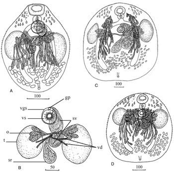
Adult worm (material examined: 5 specimens) (Fig. 1A; Table 3)
Adult worms of Exorchis. Exorchis convictus sp. n.: A, adult worm, B, reproductive complex; Exorchis oviformis (Kobayashi, Reference Kobayashi1921): C, from Japan, D, E. oviformis from Russia. gp, genital pore; o, ovary; sr, seminal receptacle; sv, seminal vesicle; t, testis; vd, vitelline duct; vgs, ventrogenital sac; vs, ventral sucker.

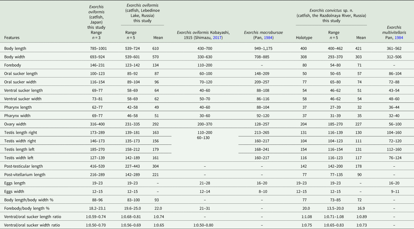
Morphometric parameters of Exorchis spp. (μm)

Body wide, oval, narrowed on anterior end. Tegument with fine spines. Forebody short. Two eye-spots, pigment diffuse, on dorsal side of forebody, on either side of ventral sucker. Numerous diffuse gland cells in forebody. Ducts of gland cells open on dorsal surface of body. Oral sucker transversally oval to transversally rectangular. Prepharynx inconspicuous. Pharynx spherical, adjacent to oral sucker. Oesophagus short. Caecal bifurcation at level of ventral sucker. Branches of intestine reach middle of posterior quarter of body. Ventral sucker round, smaller than oral one, located in middle of anterior half of body. Testes two, oval, large, opposite, lateral, medial, symmetrical, posterior to ventral sucker. Seminal vesicle bipartite, both parts large, sinistral to median line. Anterior part of seminal vesicle anterior to testes, posterior part of seminal vesicle at level of testes. Ventrogenital sac overlaps ventral sucker. Gonotyl inconspicuous. Genital pore at level of anterior margin of ventral sucker. Ovary transversely elongated, with two lobes on either side of midline of body and partially overlaps testes. Each lobe consists of transversely elongated lobes forming clusters. Seminal receptacle large, saccular, usually located in middle of body, at level of middle of testes. Uterus occupies posterior part of body. Eggs small, numerous. Vitelline fields located from level of anterior edge of testes to level of middle of posterior half of body. Vitellarium consists of longitudinally elongated follicles. Excretory vesicle Y-shaped.
Genetic data
Six complete ITS2, partial 28S and partial cox1 sequences were obtained (288, 1234 and 753 bp in length, respectively) and analysed. No genetic variability within the species was observed using these markers, with the exception of the ITS2 sequence of one specimen (MW001196), which contains an intragenomic polymorphism (T ↔ C) at the position 283.
Exorchis oviformis (Kobayashi, Reference Kobayashi1921)
Definitive host: Silurus asotus.
Site: intestine.
Intensity of infection: 8 specimens from Japan and 25–45 specimens from Russia.
Infection indices: One fish of one examined from Japan; three fish of three examined from Russia.
Localities: Tara River, Tara Town, Saga Kyushu, Japan, 33°01–76′N, 130°10–00′E; Lebedinoe Lake, Primorsky Region, the Russian southern Far East, 42°58–23′N, 130°68–57′E; the Razdolnaya River, Primorsky Region, the Russian southern Far East, 43°51–79′N, 131°90–40′E.
Voucher deposition: No. 159.1–159.3 (Japan) and No. 159.4–159.8 (Russia) in the parasitological collection of the Zoological Museum (Federal Scientific Center of the East Asia Terrestrial Biodiversity, Far East Branch of the Russian Academy of Sciences, Vladivostok, Russia); e-mail: petrova@biosoil.ru. Deposited 2019.11.20.
Adult worm (material examined: 3 specimens from Japan and 5 from Russia) (Fig. 1B, C; Table 3)
Body wide, oval or round. Tegument with fine spines. Forebody short. Two eye-spots, pigment diffuse, on dorsal side of forebody, on either side of ventral sucker. Numerous diffuse gland cells in forebody. Ducts of gland cells open on dorsal surface of forebody. Oral sucker subterminal, round. Prepharynx inconspicuous. Pharynx spherical, adjacent to oral sucker. Oesophagus short. Caecal bifurcation anterior to or at level of ventral sucker. Branches of intestine reach middle of posterior quarter of body. Ventral sucker globular, smaller than oral one, located in middle of anterior half of body. Testes two, oval or round, large, opposite, lateral symmetrical, at level of ventral sucker. Seminal vesicle bipartite, both parts large, sinistral to ventral sucker, at level of testes or slightly posterior to posterior level of testes. Ventrogenital sac overlaps ventral sucker, contains two gonotyls. Genital pore at level of anterior margin of ventral sucker. Ovary transversely elongated, with two lobes on either side of midline of body and partially overlaps testes. Each lobe consists of transversely elongated lobes forming clusters. Seminal receptacle large, saccular, usually located in middle of body, near posterior part of seminal vesicle. Uterus occupies posterior part of body. Eggs small, numerous. Vitelline fields located from level of pharynx or from level of ventral sucker to level of middle of posterior half of body. Anterior part of vitellarium from pharynx to level of anterior third of testes, consists of drop-shaped follicles; posterior part of vitellarium consists of elongated follicles. Excretory vesicle Y-shaped.
Genetic data
Nine, 11 and 19 complete ITS2, partial 28S and partial cox1 sequences with length 288, 1234 and 753 bp, respectively, were obtained. Sequences from both Russia and Japan were generated from both adult and metacercaria stages (Table 1).
All ITS2 sequences of E. oviformis were identical, with the exception of an intragenomic polymorphism (C ↔ T) that is observed at position 52 in two Russian samples (MW001191, MW001193).
For 28S data, intraspecific variations occurred. At position 770, variability was obtained: two sequences of Japanese samples (adult worm MW001170, metacercaria MW001177) have A, while two other specimens from Japan (MW001168, MW001169) have an intragenomic polymorphism (G ↔ A), the remaining seven sequences from both countries have G. This site has no effect on the value of intraspecific distances for E. oviformis due to existence of intragenomic polymorphism. Intragenomic polymorphisms are also found in the following positions: 514 bp (G ↔ A, MW001175, Russia), 719 bp (C ↔ T, MW001170, Japan) and 815 bp (G ↔ A, MW001170, Japan). This type of variability in 28S marker is only observed for mature individuals.
Based on сох1 data, sequences of E. oviformis vary from 0 to 1.6% (with average 0.6 ± 0.2%).
Exorchis sp. (metacercariae)
Second intermediate hosts: Tanakia lanceolata and Carassius sp.
Locality: Saga, Kyushu Island, Japan, 33o29–05′N, 130o30–34′E.
Site: Scale.
Genetic data
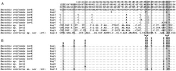
One 28S and three cox1 sequences (1234 and 753 bp in length, respectively) were generated for Exorchis sp. isolated from Japan. ITS2 data were not obtained. Genetic distances between сох1 sequences vary from 0.3 to 1.1% (with average 0.8 ± 0.3%). One sample differs from the other two by eight nucleotide substitutions, and one nucleotide substitution identified produces an amino acid substitution. The other more similar sequences vary by two nucleotide substitutions (Fig. 2).
Variable sites of nucleotide (A) and amino acid (B) sequences of partial cox1 gene for Exorchis spp. n – number of sequences in a haplotype; Hap designation relates to haplotypes corresponding to Fig. 4. Nucleotide substitutions corresponding to amino acid substitutions are highlighted in grey. Curly brace indicates the effect of two nucleotide substitutions in codon on the appearance of an amino acid substitution in different haplotypes.

Discussion
Based on the data presented in Table 3, all known representatives of Exorchis can be divided into two groups, one of which includes ‘large’ worms, which can reach over 700 μm in length, and another that is composed of ‘small’ worms with a maximum body length of <700 μm. The group of ‘large’ worms comprises E. oviformis and E. macrobursae. The ‘small’ worms include E. dongtinghuensis, E. ovariolobularis and E. multivitellaris.
E. convictus sp. n. belongs to the ‘small’ group (the maximum body length is 462 μm). We determined adult worms of E. convictus sp. n. are most similar to E. multivitellaris based on morphological metrics. Between individuals of the two species, there are only slight differences in body width, oral sucker and pharynx length and egg size (Table 3). The main difference of E. convictus sp. n. from E. multivitellaris, E. dongtinghuensis and E. ovariolobularis is within its two-lobed ovary, which is located laterally on both sides of the midline of the body. The other three species have a compact ovary with a median or submedian location. Exorchis dongtinghuensis and E. ovariolobularis differ very minimally morphologically and with respect to metric characters, with the exception of egg sizes: 24–26 × 14–15 vs 11–14 × 7–9. To determine whether these worms belong to the same or different species, additional research will be required using both morphological and molecular features.
Regarding E. oviformis, Kobayashi described mature worms from S. asotus in Japan (Kobayashi, Reference Kobayashi1921). Yamaguti (Reference Yamaguti1938) subsequently made adjustments to the description of worms of this species. Besprozvannykh et al. (Reference Besprozvannykh, Ermolenko and Shedko2000) provided morphological data for this species from S. asotus in the Russian southern Far East. Analysis of the morphology of E. oviformis from published data and additional material, as well as a large amount of material studied from collections in Japan, allowed Shimazu (Reference Shimazu2017) to identify inaccuracies in previous descriptions of mature individuals of this species, in particular, the morphology of the ovary and vitellarium. Based on analysis, Shimazu provided a high-quality detailed description of E. oviformis. In the current study, the worms from Japan and Russia are similar in morphological parameters but have some differences in body and organ sizes. They mainly differ in the metric indicators of the body and organs (Table 3). Of note, there are no gaps in metric indicators between the specimens of E. oviformis from Russia and the samples presented by Shimazu (Reference Shimazu2017). The presently studied worms from Japan are larger than both the parasites presented by Shimazu (Reference Shimazu2017) and the worms found in Russia. However, the difference in size may be due to the period of growth and development of worms, which takes place from the moment of infection of the final host with metacercariae until the moment the worms are removed from this host. In accordance with the above, based on the morphological characteristics of the discussed worms, we consider them all E. oviformis.
All designated worms of E. oviformis from Japan and Russia also differ from another ‘large’ worm, namely E. macrobursae from China. In contrast to E. macrobursae, the Japanese and Russian worms have smaller oral and ventral suckers and a smaller pharynx (Table 3). Further, they have a two-lobed vitellarium, while E. macrobursae has a compact vitelline mass with a median or submedian location.
For all morphological and metric criteria, E. oviformis differs from E. convictus sp. n.; molecular data have confirmed these differences. Using 28S data, adult worms and metacercariae of E. oviformis (from Japan and Russia) and metacercariae of Exorchis sp. differ by 0.1%, and each differs from E. convictus sp. n. by 0.3 and 0.4%, respectively. Indicators of the variability of this marker for Exorchis spp. correspond to those between species of other genera of Cryptogonimidae. The distance between Lobosorchis polygongylus (Miller et al. Reference Miller, Downie and Cribb2009) and Lobosorchis tibaldiae Miller & Cribb, 2005 is 0.1% (Miller et al., Reference Miller, Downie and Cribb2009), and the genetic distance between Retrovarium exiguiformosum (Miller and Cribb, Reference Miller and Cribb2007) and Retrovarium formosum (Miller and Cribb, Reference Miller and Cribb2007) is 0.2% (Miller and Cribb, Reference Miller and Cribb2007). Thus, based on the 28S marker, both species, E. convictus sp. n. and E. oviformis, are valid. At the same time, the ITS2 rDNA region was more conserved for Exorchis spp. compared with the 28S marker. Moreover, ITS2 sequences are identical for all samples of E. oviformis and E. convictus sp. n. and did not divide these species. For other cryptogonimids, there are no data for species that have differences based on the 28S marker, but are 100% identical based on the ITS2 rDNA region. The smallest difference for the latter marker was no less than 0.4% for species within the genus Retrovarium (Miller and Cribb, Reference Miller and Cribb2007). However, we cannot exclude that conservatism of ITS2 is a feature for Exorchis, and species within this genus actually do not have variability in this spacer region. In any case, additional data for other species are needed to confirm this hypothesis.
Along with confirming the validity of the worms, we analysed intraspecific variability based on nuclear markers. Only a single intragenomic polymorphism has been identified both in the ITS2 rDNA sequences isolated from two E. oviformis (at position 52) and one E. convictus sp. n. (at position 283) samples from Russia. Intragenomic polymorphisms have also been identified within 28S sequences of E. oviformis from Japan and Russia, but most sites are in specimens from Japan. There is a possibility that worms from Japan, as a result of the geographic isolation of island populations, have developed special adaptation mechanisms to a wider range of hosts or other environmental factors, which are due to expressing various tandem repeats of the ribosomal cluster. This type of variability has previously been described for other species of trematodes including representatives of the superfamily Opisthorchioidea Braun, 1901 (Tatonova et al., Reference Katokhin, Shekhovtsov, Konkow, Yurlova, Serbina, Vodianitskai, Fedorov, Loktev, Muratov, Ohyama, Makhnev, Peltek and Mordvinov2017). However, more data are needed to determine how common intragenomic polymorphism in populations of Exorchis spp. and other cryptogonimids are,
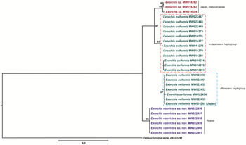
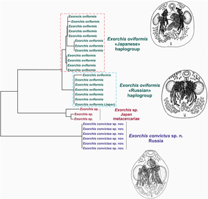
Analysis of mitochondrial cox1 revealed differences between the Exorchis spp. assessed. Three species of Exorchis presented in this study have been divided on a BI reconstruction based on this marker with high statistical support (Fig. 3). In the present analysis, E. oviformis and Exorchis sp. are closer to each other (5.3%) than to E. convictus sp. n. (7.1–7.4%), which is located at a basal position of the tree. These values are comparable to interspecies genetic distances between representatives of related families, namely Opisthorchiidae Looss, 1899 and Heterophyidae Leiper, 1909 (Tatonova et al., Reference Tatonova, Shumenko and Besprozvannykh2018; Besprozvannykh et al., Reference Besprozvannykh, Tatonova and Shumenko2019). Fixed amino acid substitutions have also been identified between analysed species (Fig. 2), which have led to 1.2–2.6% differences between the sequences of studied species and confirm their validities.
BI-reconstruction for Exorchis spp. based on sequences of the partial cox1 gene. Tabascotrema verai (JX023291) was used as an outgroup. Exorchis oviformis haplogroups are showed by frames.

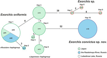
In addition to the analysis of interspecies differences, MST-reconstruction based on cox1 data has revealed that E. oviformis sequences formed ‘Japanese’ and ‘Russian’ haplogroups, which differ by six fixed nucleotide substitutions (Fig. 4). One nucleotide substitution has identified has been determined to be nonsynonymous (Fig. 2). The genetic distance between these haplogroups is 1.1%, and distances within them are 0.3 and 0.1% for ‘Japanese’ and ‘Russian’ flukes, respectively. These values are 5–7 times lower than the differences between species of Exorchis (5.3–7.4%). There are similar values for another cryptogonimid representative, T. verai, for which based on cox1 data, there are three independent groups with a range of distances of 0–2.32% and 8.3–11.9% within and between these groups, respectively (Razo-Mendivil et al., Reference Razo-Mendivil, Rosas-Valdez, Rubio-Godoy and Pérez-Ponce de León2015). Using results of genetic data, the authors concluded that the groups obtained are a complex of cryptic species. Analysis of Oligogonotylus Watson, 1976 using morphological and molecular data made it possible to determine that, based on cox1, interspecies differences were from 9.55 to 10.39%, while the genetic divergence within species of this genus varied from 0.28 to 3.65% (Razo-Mendivil et al., Reference Razo-Mendivil, Rosas-Valdez and Pérez-Ponce de León2008). The intraspecies and interspecies distances within Neocladocystis Manter & Ritchard, 1696 based on same mitochondrial marker are 1.3–4.2% and 15.5–16.2%, respectively (Kmentová et al., Reference Kmentová, Bray, Koblmüller, Artois, De Keyzer, Gelnar, Vanhove and Georgieva2020). Thus, we confirmed that all E. oviformis in this study are the same species.
MST for the cox1 haplotypes of Exorchis spp. n – number specimens in a haplotype (Hap); numbers of substitutions are shown on the branches.

Value of distance between haplogroups of E. oviformis indicates isolation between populations from different countries, which, as shown above, has led to the presence of fixed substitutions, including nonsynonymous ones. However, this value does not reach the level of interspecies differences.
The ‘Japanese’ E. oviformis haplogroup comprises samples from Kyushu Island. At the same time, one sample (MW014285) from another island of Japan (the north part of Honshu) is included in the ‘Russian’ haplogroup and is identical to samples from Russia, despite the fact that the island and continental populations of E. oviformis are separated by seawater. This sea barrier cannot be overcome for all known freshwater hosts of E. oviformis. The anadromous fish Tribolodon Sauvage, 1883 have been identified among the second intermediate hosts of Exorchis (Nagasawa and Katahira, Reference Nagasawa and Katahira2013), but they only carried out offshore migrations in coastal waters, a fact confirmed by analysis of genetic data for four members of the genus Tribolodon from the Sea of Japan (Watanabe et al., Reference Watanabe, Sakai, Sanada and Nishida2018). Based on our study, it was shown that Tribolodon spp., distributed throughout the Sea of Japan, have no common haplotypes between continental southwestern parts and the Japanese archipelago. This indicates the absence of migration of these species across the sea at the current time. The presence of specimens of the same haplogroup both in the island (Japan) and continental (Russia) populations of worms may be associated with the participation of currently unknown hosts (fish) that can perform long offshore migrations, providing a connection between these populations. Regarding the continental haplogroup in Russia, there is a common haplotype in separate river basins. This may be also related to migrations of Tribolodon spp., which are an intermediate host of E. oviformis, between completely separated rivers connected via the sea.
Within the ‘Russian’ haplogroup of E. oviformis, there are two haplotypes. One of them includes six samples (Hap 1; Fig. 4) while the other (Hap 2) comprises one individual that differed from Hap 1 by three nucleotide substitutions. Two of the aforementioned substitutions produce amino acid changes. By contrast, the ‘Japanese’ haplogroup unites four haplotypes. this finding may indicate that the centre of the species formation occurred in Japan, which is characterized by its increased degree of variability.
For metacercariae of Exorchis sp. from the territory of Saga Kyushu (Japan) and for E. oviformis from both countries, there are two haplogroups based on cox1 data, which are separated by a distance of 1.1%. This value is comparable to genetic distances found between ‘Japanese’ and ‘Russian’ haplogroups of E. oviformis, that is, it also indicates the intraspecific level of variability. There are different haplogroups of Exorchis spp. are found within different fish: Carassius sp. and T. lanceolata. In this case, parasitizing in different hosts may have affected the formation of isolation between specimens of the same species. Additional studies that cover most of the populations of both species and other representatives of the genus are needed to confirm this assumption and to clarify their relationships in the parasite–host system.
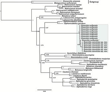
Despite the unresolved topology of most parts of the phylogenetic reconstruction for the family Cryptogonimidae based on the combined sequences of ribosomal markers (Fig. 5), the genus Exorchis is monophyletic and differs from other representatives of the family Cryptogonimidae with high posterior probability. Exorchis spp. are clustered with representatives of the genus Siphomutabilus, from which they differ by 6.1%, which corresponds to the range of intergeneric differences within the family (6.1–9.1%).
The phylogeny of Cryptogonimidae based on combined sequences of complete ITS2 rDNA region and the partial 28S rRNA gene using the BI method. Bayesian posterior probabilities of ⩾0.50 are shown. Genus Exorchis is presented in frame. The accession numbers are listed in Tables 1 and 2. Asterisk marks the combined sequences MW001188/MW001170.

The results of this study confirm the validity of the genus Exorchis and three species: E. convictus sp. n. (Russia), E. oviformis (Russia and Japan) and Exorchis sp. (Japan). We identified two haplogroups of E. oviformis, and their presence is associated with the geographic isolation of populations within the continental part of Russia and islands of Japan. There was also a representative in the island population of E. oviformis that is identical to individuals from the haplogroup of the continental population of this species. In addition, we revealed an intragenomic polymorphism, the significance of which for Exorchis spp. requires additional research.
Data
Materials are deposited in the parasitological collection of the Zoological Museum (Federal Scientific Center of the East Asia Terrestrial Biodiversity, Far East Branch of the Russian Academy of Sciences, Vladivostok, Russia); e-mail: petrova@biosoil.ru.
Acknowledgments
We thank Yusuke Hibino (Kitakyushu Museum of Natural History & Human History) and Hiromi Ikadai (Kitasato University) for providing freshwater fish from Saga and Aomori, Japan.
Financial support
This research was supported by the Russian government basic research programme (grant number 0267–2019–0018).
Conflict of interest
The authors declare there are no conflicts of interest.
Ethical standards
Euthanasia of fish was carried out in accordance with the Committee on the Ethics of Animal Experiments of the Federal Scientific Center of the East Asia Terrestrial Biodiversity, FEB RAS, Russia and according to ethical standards of the national guides.