Duchenne muscular dystrophies are the most common form of muscular dystrophy in children and is caused by mutations in the DMD gene, located on chromosome Xp21.2. DMD encodes the dystrophin protein, an important component of the dystrophin–glycoprotein complex that links the intracellular cytoskeleton to the extracellular matrix in skeletal muscle cells. Mutations in the DMD gene lead to a reduced production of dystrophin, causing increases in muscle ragility and leading to contraction-induced injury.Reference Bushby, Finkel and Birnkrant1–Reference Evans, Miayak and Robertson3 Duchenne muscular dystrophies is a life-limiting disorder with an incidence estimated at 1 per 3600 to 6000 live male birthsReference Romitti, Zhu and Puzhankara27 and is characterised by progressive skeletal and cardiac muscle weakness usually leading to death by 30 years of age.Reference Wong and Christopher4 Symptoms typically appear in early childhood, with loss of ambulation by age 12 years if untreated with corticosteroids.Reference Dubowitz5
Duchenne muscular dystrophies causes progressive respiratory muscle weakness and respiratory failure.Reference Tandon, Villa and Hor6 Since the advent of home respiratory support in the early 1990s, the life expectancy for Duchenne muscular dystrophies patients has increased from 19.3 to 25.3 years.Reference Eagle, Baudouin and Chandler7, Reference Gomez-Merino and Bach8, Reference Birnkrant, Ashwath and Noritz23–Reference Humbertclaude, Hamroun and Bezzou25 Numerous studies have shown that both non-invasive positive pressure ventilation and cough-assist ventilation have improved survival for these patientsReference Dubowitz5, Reference Eagle, Baudouin and Chandler7–Reference Toussaint, Soudon and Kinnear11, Reference Duiverman, Wempe and Bladder28 by reducing respiratory failure which, prior to non-invasive positive pressure ventilation, was a prominent cause of death in this patient population. As a consequence of extending life through use of non-invasive positive pressure ventilation, left ventricular dysfunction has become a more clinically significant feature of Duchenne muscular dystrophies.Reference Eagle, Baudouin and Chandler7–Reference Ishikawa, Miura and Ishikawa9, Reference Hamada, Ishikawa and Aoyagi12 However, the impact of non-invasive positive pressure ventilation on progression of left ventricular dysfunction in Duchenne muscular dystrophies is unknown.
The loss of dystrophin protein in the cardiac myocytes produces fibrofatty infiltration that begins in the posterobasal wall of the left ventricle. This infiltration leads to fibrosis, thinning of the ventricular wall, dilation of the ventricle, and progressive decline in ejection fraction.Reference Tandon, Villa and Hor6, Reference Judge, Kass, Thompson and Wagner13–Reference Spurney16 The goal of this study was to determine the rate of decline in left ventricle function in patients with Duchenne muscular dystrophies and echo data and to compare the rate of decline in left ventricle function between patients who initiated non-invasive positive pressure ventilation and those who did not. We hypothesised that patients who initiated non-invasive positive pressure ventilation would be associated with a slower decline of left ventricle function when compared with Duchenne muscular dystrophies patients who did not initiate non-invasive positive pressure ventilation.
Methods
The Muscular Dystrophy Surveillance, Tracking, and Research Network (MD STARnet) is a population-based surveillance program that retrospectively identifies and longitudinally follows individuals diagnosed with childhood-onset muscular dystrophy who were diagnosed by the age of 21 years. Information is gathered on individuals born between 1 January, 1982 and 31 December, 2011 and residing in Arizona, Colorado, Georgia, Hawaii, Iowa, and the western 12 counties of New York. Surveillance started in 2004 for Arizona, Colorado, Iowa, and western New York, 2005 for Georgia, and 2008 for Hawaii. Each site maintains permission for medical record abstraction through Institutional Review Board approval or state-mandated public health authority.
Multiple methods were used to identify potential cases. All retrospectively identified, and newly diagnosed cases had annual medical record abstraction through 31 December, 2011 or until death or migration out of a MD STARnet site. A detailed description of the MD STARnet abstraction and identification methodology has been previously published.Reference Miller, Romitti and Cunniff17 Key clinical and diagnostic data, including family history, first signs and symptoms, and muscle biopsy, were used to assign a MD STARnet case status (definite, probable, possible, female, asymptomatic, or not Duchenne muscular dystrophies). Final case status was validated by consensus of a committee of neuromuscular clinicians from the MD STARnet sites.Reference Matthews, Cunniff and Kantamnent18
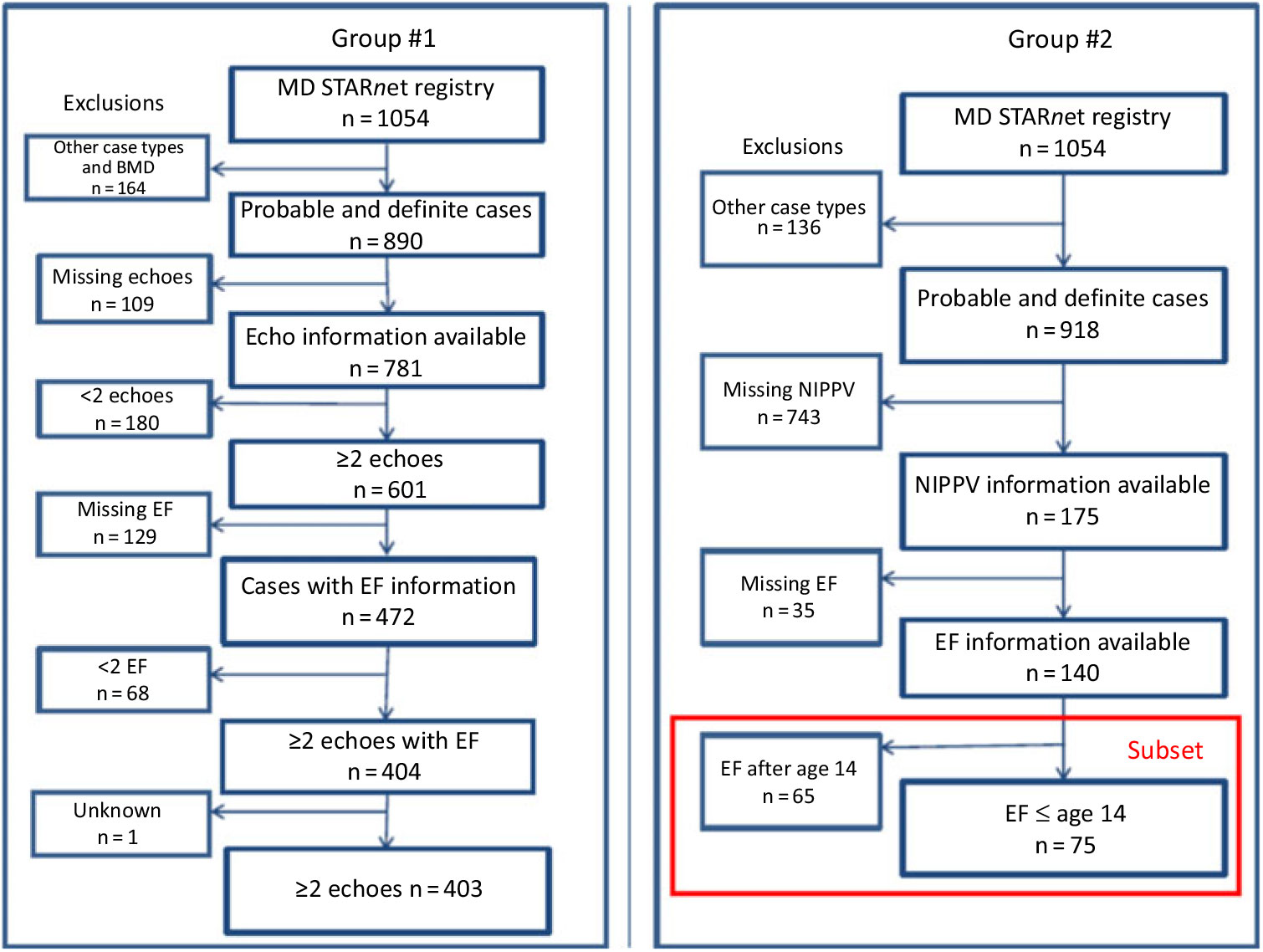
The study cohort, derived from the MD STARnet surveillance system, included individuals with a case status of “definite” or “probable” (n = 918) and were not Becker Muscular Dystrophy (n = 28) Individuals from this study cohort (n = 890) were eligible for the analytic cohorts based on the available cardiac and non-invasive positive pressure ventilation data (Fig 1).
Selection of analytic cohorts from the MD STARnet Surveillance System, 1982–2011.

The first cohort, Cohort 1, was utilised to determine the rate of decline in left ventricle function in individuals and included all individuals from the study cohort with two or more echo measurements and two or more ejection fraction measurements (n = 403) (Fig 1). The second cohort, Cohort 2, was utilised to evaluate the association between non-invasive positive pressure ventilation initiation and the decline of left ventricle function in individuals and included all individuals from the study cohort with at least one ejection fraction and had full/complete documented non-invasive positive pressure ventilation compliance (n = 140) (Fig 1). Date of initiation was based on clinical indication abstracted from individual medical records. Since the average age of onset of left ventricular dysfunction has been shown to be 14 years in previous studies, a secondary analysis was completed on Cohort 2 excluding patients with their first ejection fraction after age 14 years based on Paediatric Cardiomyopathy Registry data (n = 75) (Cohort 2a).Reference Barber, Andrews and Lu20, Reference Silva, Mahalhaes and Alves Meira21 Each analytic cohort was analysed separately and was not compared to the other. Linear mixed effect modelling was performed to control for patients with only one ejection fraction time point (see statistical section below).Reference Fitzmaurice and Ravidchandran30
For the quantitative analysis, left ventricular dysfunction was defined as an ejection fraction of <55% or a shortening fraction of <28%. In a separate qualitative analysis, left ventricular dysfunction was defined from categorical data as normal, mildly depressed, moderately depressed, or severely depressed. Since the abstracted echo date only includes month and year, the missing day of the month for the echo was set to 15. Echoes falling on the same month and year were assumed to be duplicates. In case of echo duplication, the data selected for inclusion were the one indicating the highest ejection fraction and best qualitative function for that day or hospitalisation. The highest value was taken because multiple echoes in 1 month were typically seen in the setting of hospitalisations, in which transient decrease in systolic function is clinically common and reversible.
Non-invasive positive pressure ventilation was defined as medical record evidence of any of the following procedures: bilevel positive airway pressure (Bi-PAP), continuous positive airway pressure or mechanical ventilator with sip (mouthpiece) or a mask/nasal ventilator mode. For those using non-invasive positive pressure ventilation, frequency within the database is categorised as >16 hours a day, night and <16 hours a day, and night. For any given patient, the date of ejection fractions was coded as “prior” or “post” non-invasive positive pressure ventilation initiation.
Medications were classified as corticosteroid, angiotensin-converting enzyme inhibitor, and beta blocker. Medication use was categorised as “yes” or “no.” Variables were created to indicate if patients used a combination of corticosteroids and angiotensin-converting enzyme inhibitors or corticosteroids and beta blockers. Corticosteroid time period of use was defined as cumulative use for more than 6 months as determined from the MD Starnet calculated variable for corticosteroid use. Time of use for angiotensin-converting enzyme inhibitors and beta blockers were recorded in the dataset as year and not exact dates. For this analysis, start dates were the middle of the first year of documented use (July 1st) and stop dates were the middle of the year before the first year of undocumented use. For corticosteroids, angiotensin-converting enzyme inhibitors, and beta blockers, use was categorised as “prior” or “post” when the echo was performed.
Statistical methods
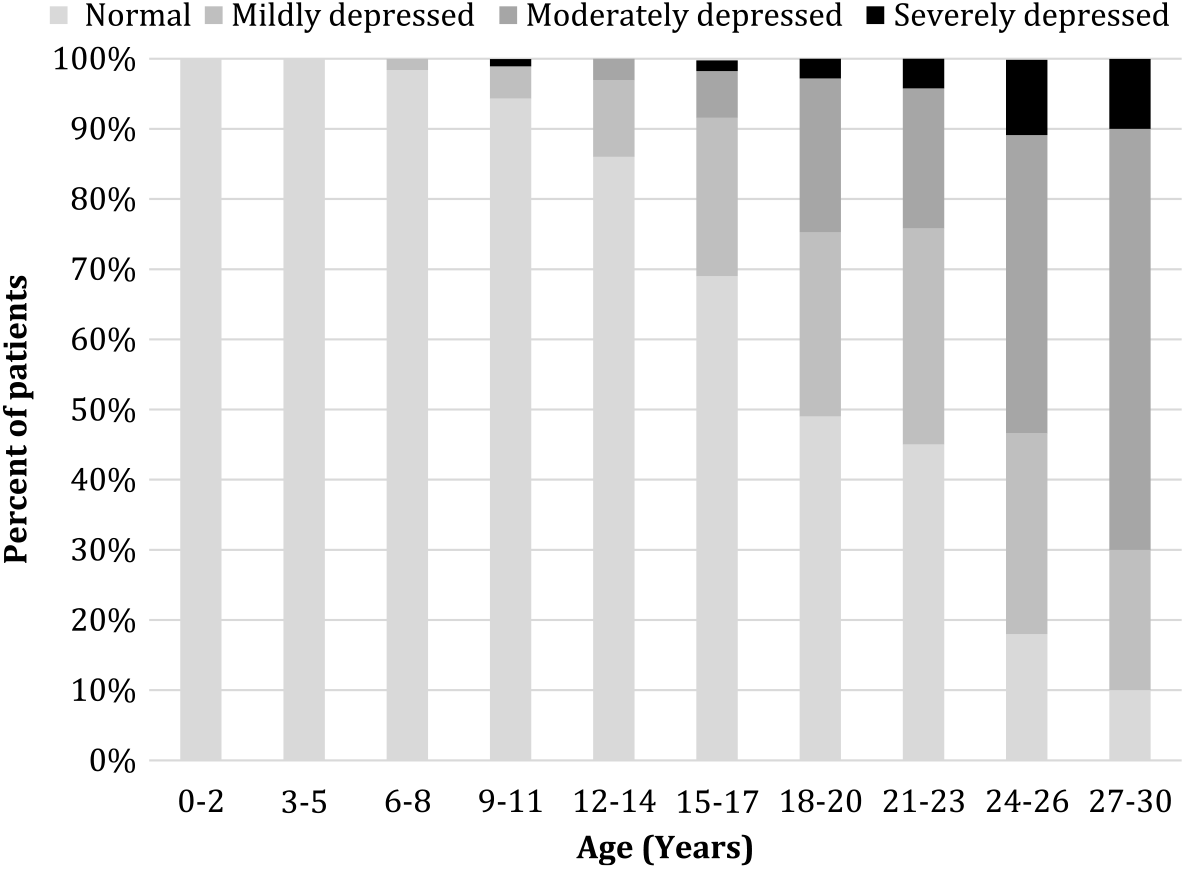
Descriptive statistics were calculated for all independent variables. Mean age and its standard deviations were calculated for first signs and symptoms, loss of ambulation, first echo, first echo with ejection fraction <55%, and initiation of medication. Using Cohort 1, a linear mixed effect model was used to estimate the rate of decline in left ventricle function over time based on repeated ejection fraction measurements starting from the first recorded ejection fraction. A linear mixed effect model was used to accommodate fixed and random effects within individuals with repeated ejection fraction measurements, while adjusting for missing data and medication use (each independently corticosteroid, angiotensin-converting enzyme inhibitors, and beta blockers).Reference Tandon, Jefferies and Villa29 In a separate analysis, in Cohort 1, echo results with only a qualitative assessment documented (normal, mildly depressed, moderately depressed, or severely depressed) at each age (n = 403) were plotted according to the number of echoes in that age group and qualitative category.
An additional analysis was performed to predict the change in mean ejection fraction over time in Cohort 2. A linear mixed effects model evaluated the progression of left ventricular dysfunction over time using repeated ejection fraction measurements. This analysis was similar to the analysis of change in ejection fraction over time in Cohort 1, except that it was limited to a cohort with non-invasive positive pressure ventilation initiation to determine if non-invasive positive pressure ventilation slows the progression of left ventricular dysfunction. Models were adjusted for missing data, corticosteroid, angiotensin-converting enzyme inhibitors, and beta blocker use (independently), as previous studies have shown slower progression of disease in Duchenne muscular dystrophies patients taking these medications.Reference Schram, Fournier and Leduc31, Reference Duboc, Meune and Pierre32 Linear mixed models were used to evaluate within-individual progression of left ventricular dysfunction and whether the progression (slope) changed with initiation of non-invasive positive pressure ventilation. This analysis was replicated in Cohort 2a limiting to those from Cohort 2 with ejection fraction prior to or at 14 years to evaluate whether earlier respiratory intervention slows progression of left ventricular dysfunction. All analyses were performed using SAS Version 9.3 (SAS Institute Inc., Cary, North California, United States of America) and validated by investigators from a different MD STARnet site.
Results
MD STARnet data included 1054 cases with a mean age of first signs and symptoms of 3.7 ± 3.0 years and a mean age of loss of ambulation of 11.1 ± 2.6 years. In Cohort 1, the mean age of first signs and symptoms of 3.2 ± 2.3 years and the mean age of loss of ambulation was 10.4 ± 2.2 years. The distribution of birth year in Cohort 1 was normal with most births occurring between 1986 and 2003. Patients were categorised by year of birth in 5-year increments (1982–1986, 1987–1991, 1992–1996, 2997–2001, and >2002) due to expected changes in clinical practice over time. In a descriptive analysis, there was temporal decline in age of medication use (corticosteroid, angiotensin-converting enzyme inhibitors, and beta blockers) and age of first echo. The mean age at first echo was 10.8 ± 4.6 years, with an average first ejection fraction of 63.1 ± 12.6%. The average age at first echo with an ejection fraction <55% was 15.2 ± 3.9 years. Eighty percent of Cohort 1 patients were treated with corticosteroids. Of those treated with corticosteroids, 42% were treated with a combination of corticosteroids and angiotensin-converting enzyme inhibitors, and 14.5% were treated with corticosteroids and beta blockers. The mean age at initiation of non-invasive positive pressure ventilation was 15.8 ± 3.7 years. Bilevel positive airway pressure was the most common non-invasive positive pressure ventilation used (n = 153, 37.9%) occurring most frequently at night. There were a total of 998 (n = 403) echo measurements with ejection fraction recorded in Cohort 1. Each patient had at least two ejection fractions with a maximum of 13 ejection fractions. Approximately, 75% of the patients had ≥ 3 ejection fractions (n = 302).
The rate of decline in ejection fraction over time from first recorded ejection fraction was 1.6% per year, adjusted for age, birth cohort, corticosteroid use, and angiotensin-converting enzyme inhibitor and beta blocker treatment.
Cohort 2 included 140 patients with ejection fraction data who used non-invasive positive pressure ventilation. The mean age of first signs and symptoms was 3.4 ± 2.6 years, and the mean age of loss of ambulation was 10.9 ± 2.3 years. The mean age at first echo was 13.9 ± 4.6 years with an average first ejection fraction of 60.5 ± 14.4%. The mean age at first echo with an ejection fraction <55% was 17.6 ± 3.4 years and 77% of Cohort 2 were treated with corticosteroids (n = 108). Of those treated with corticosteroids, 22.9% were treated with a combination of corticosteroids and angiotensin-converting enzyme inhibitors, and 11.4% were treated with corticosteroids and beta blockers. The mean age of initiation of non-invasive positive pressure ventilation was 17.3 ± 3.7 years. Bilevel positive airway pressure was the most common non-invasive positive pressure ventilation used (n = 103, 73.6%). More than half of the Cohort 2 patients used non-invasive positive pressure ventilation at night only (n = 94, 67.1%).
Among those who had an ejection fraction at or before age 14 years (n = 75), the mean age of first signs and symptoms was 3.0 ± 2.4 years and the mean age of loss of ambulation was 10.8 ± 2.4 years. The median number of echoes for Cohort 2a was 4 with a range from 2 to 13. The mean age at first echo was 10.6 ± 2.7 years with an average first ejection fraction of 66.9 ± 9.8%. The mean age at first echo with an ejection fraction <55% was 11.9 ± 1.1 years. Almost 80% of Cohort 2a were treated with corticosteroids. Of those treated with corticosteroids, 13.3% were treated with a combination of corticosteroids and angiotensin-converting enzyme inhibitors, and 4.0% were treated with corticosteroids and beta blockers. The mean age of initiation of non-invasive positive pressure ventilation for Cohort 2a was 15.9 ± 3.1 years, with bilevel positive airway pressure being the most common non-invasive positive pressure ventilation used (n = 62, 82.7%). More than half of the Cohort 2 patients used non-invasive positive pressure ventilation at night (n = 56, 74.7%).
Figure 2 shows the categorical distribution of the 998 qualitative echo measurements for 403 cases (Cohort 1). Left ventricular function was categorised by type (normal, mild dysfunction, moderate dysfunction, and severe dysfunction) and age. Subjectively normal function was found in nearly 100% of patients less than 8 years old, and the percentage of patients with left ventricular dysfunction increased with age. By age 18 years, nearly 60% of patients had some degree of left ventricular dysfunction.
Qualitative assessment of left ventricular dysfunction (subjective echo function), MD STARnet 1982–2011 (n = 998 echoes in 403 individuals).

Using mixed modeling methods, an association between non-invasive positive pressure ventilation initiation and the change in the rate of decline of left ventricle function in Cohort 2 (140 patients) was not statistically significant (F-test, p = 0.60) while adjusting for age, birth cohort, corticosteroid use, and angiotensin-converting enzyme inhibitor and beta blocker treatment. When this model was limited to patients with their first echo before age 14 (Cohort 2a, n = 75), there was no significant association between initiation of non-invasive positive pressure ventilation and change in the rate of progression of left ventricular dysfunction adjusting for age, birth cohort, corticosteroid use, and angiotensin-converting enzyme inhibitor and beta blocker treatment (F-test, p = 0.36).
Discussion
Duchenne muscular dystrophy is a progressive neuromuscular disorder associated with an increasing proportion of deaths attributable to cardiac failure.Reference Gomez-Merino and Bach8–Reference Ishikawa, Miura and Ishikawa9, Reference Hamada, Ishikawa and Aoyagi12, Reference Barber, Andrews and Lu20 The goals of our study were to determine the overall rate of decline in left ventricle function in the MD STARnet surveillance system patient cohort and to determine if initiation of non-invasive positive pressure ventilation use was associated with a change in the rate of decline in left ventricle function in patients with Duchenne muscular dystrophies. In this cohort, ejection fraction declined over time at a rate of 1.6% per year, while the proportion of patients with Duchenne muscular dystrophy with qualitatively depressed left ventricle function increased with age. The quantitative assessment of left ventricle function by ejection fraction in Cohort 1 showed that the mean age of onset of the decline in left ventricle function (ejection fraction <55% or shortened fraction <28%) was 15.2 years. This is only slightly older than the mean age of left ventricle dysfunction of 14.4 years previously found by the Paediatric Cardiomyopathy Registry and 14.3 years in a prior MD STARnet study.Reference Connuck, Sleeper and Colan19, Reference Barber, Andrews and Lu20 This difference in age at onset of left ventricular dysfunction may be due to variation in the inclusion criteria between studies or variation in entry into the surveillance system for patients across sites within MD STARnet. Additionally, our study cohorts had more than one ejection fraction measurement and larger number of patients compared to the Paediatric Cardiomyopathy Registry study. When all MD STARnet participants with historical recorded echoes (results available and unavailable) are included (n = 809), the mean age of onset for quantitative left ventricular dysfunction was 15.1 years. Lastly, older age and left ventricular dysfunction onset may be due to potential beneficial effect from early corticosteroid or cardiac medication use since that time.Reference Barber, Andrews and Lu20, Reference Silva, Mahalhaes and Alves Meira21
Left ventricle function data were reviewed for this study. Some patients had only qualitative left ventricle function assessments due to the difficulty in obtaining usable left ventricle volumes in patients with Duchenne muscular dystrophies due to poor acoustic windows. It is well described that an experienced echocardiographer is more accurate at estimating left ventricle volumes than quantitative methods, such as the Biplane Simpson’s method.Reference Gudmundsson, Rydberg, Winter and Willenheimer22 Therefore, poor acoustic windows due to inaccurate tracing of the endocardial surface could lead to inaccurate estimates of ejection fractions. Knowledge of the natural history of decline can aid physicians in counselling patients and their families as well as to guide interventions.
We did not find a significant change in the progression of left ventricular dysfunction due to the initiation of non-invasive positive pressure ventilation independent of age, corticosteroid use, and angiotensin-converting enzyme inhibitor and beta blocker treatment. There are several possible explanations. While positive pressure ventilation is beneficial in the setting of acute heart failure by decreasing left ventricle afterload, there are few studies demonstrating a long-term benefit from its use.Reference Gomez-Merino and Bach8, Reference Ishikawa, Miura and Ishikawa9 The benefit of decreased afterload through non-invasive positive pressure ventilation may require use of non-invasive positive pressure ventilation around the clock. Since the vast majority of patients in this study only used non-invasive positive pressure ventilation at night during sleep, the full benefit of non-invasive positive pressure ventilation on reduced afterload may not have been achieved. Another possibility is that age of non-invasive positive pressure ventilation initiation could have affected the effectiveness in slowing the rate of left ventricular dysfunction. In Cohorts 1 and 2, age at non-invasive positive pressure ventilation initiation was 15.8 and 17.3 years, respectively. Comparatively, in Cohort 2a, age at initiation of non-invasive positive pressure ventilation was 15.9 years, 4 years after left ventricular dysfunction was diagnosed. Perhaps, in order to obtain the maximal benefit of non-invasive positive pressure ventilation in slowing the rate of left ventricular dysfunction, non-invasive positive pressure ventilation needs to be initiated prior to the onset of left ventricular dysfunction. Finally, it is difficult to ascertain whether cardiac and pulmonary functions decline together. However, non-invasive positive pressure ventilation is likely prescribed as pulmonary status declines. If so, then patients who require non-invasive positive pressure ventilation may be “sicker” than other patients.
Strengths of this study include the large sample size and longitudinal surveillance data. MD STARnet is the largest database of males with MD in the United States and is a population-based surveillance cohort with reasonable generalisability to other male MD populations. However, there are several limitations to this study. Since this database records data over multiple decades, there may be changes in practice over time that may affect outcomes. For example, as of now most patients with both Duchenne muscular dystrophy are on a treatment plan that includes both an annual echocardiogram after diagnosis,Reference Birnkrant, Bushby and Bann33 and some kind of cardiac medication by age 10.Reference McNally, Kaltman and Benson34 The proportion of participants in this study taking cardiac medication was likely related to time of diagnosis and the accepted medical practices at time of diagnosis.Reference Wittlieb-Weber, Pantea and Krikov35 In a sensitivity analysis looking at a potential birth cohort effect, we did not find a change in the association between non-invasive positive pressure ventilation use and left ventricle function decline in Cohorts 2 or 2a. Studies using more current data are likely to see a greater proportion of participants overall who are actively taking cardiac medications than was seen in this study.
Echo outcomes included both those obtained from routine out-patient visits and from hospital admissions. Although the highest recorded values for suspected hospital admissions were used, those values still may not have represented the true baseline level for those patients, as recovery may not have been completed at that point. Echoes are also difficult to perform on patients who are unable to lie flat and/or have large barrel chests due to poor acoustic windows. This may limit collection of ejection fraction or qualitative function data and may explain why there is a significant amount of missing echo data. In a study by Tandon et al 2015, the decline of ejection fraction was similar to our findings with results validated by cardiac MRI which is considered a more accurate method of volume measurement.Reference Tandon, Jefferies and Villa29 Additionally, participants in this cohort are from multiple sites over a long period of time, which could induce variation in the ejection fraction values both across and within participants. This limitation would be non-differential by cardiac status and expected to bias the findings towards the null hypothesis.
Finally, this research is an observational study of changes in the progression of left ventricular dysfunction within patients after initiation of non-invasive positive pressure ventilation. While an analysis with between-patient (comparison group) analysis could address whether non-invasive positive pressure ventilation initiation causes a change in the progression of left ventricular dysfunction, a within-patients analysis in this surveillance cohort was practical given the smaller numbers and observational nature of the study. Our findings do not suggest a causal relationship between non-invasive positive pressure ventilation and left ventricular dysfunction, further research in a randomised control trial is suggested to further explore the hypothesis that non-invasive positive pressure ventilation slows the progress of left ventricular dysfunction. Additionally, non-invasive positive pressure ventilation may have prevented episodes of acute respiratory distress with respiratory acidosis. Such episodes of respiratory acidosis further depress left ventricle function in patients who already have left ventricular dysfunction, resulting in subsequent metabolic acidosis, low cardiac output, and, without intervention, cardiac death. It is unknown how many acute deaths by such cardiopulmonary interactions were prevented by non-invasive positive pressure ventilation use. The relatively small numbers are also a limitation of this study; even though this is the largest surveillance-based analytic cohort, it had limited statistical power. This could be why an association was not detected in this analysis.
Conclusion
Using the largest longitudinal surveillance system of U.S. patients with Duchenne muscular dystrophy, the rate of left ventricle ejection fraction decline was 1.6% per year via quantitative evaluation, and in a separate qualitative analysis, the percentage of patients with left ventricular dysfunction increased with age. By age 18 years, nearly 60% of patients had some degree of left ventricular dysfunction. While we did not find a change in the progression of left ventricular dysfunction with initiation of non-invasive positive pressure ventilation, it is possible that the benefit of non-invasive positive pressure ventilation with respect to left ventricular dysfunction may still exist. Other study designs such as randomised control trials may be needed to examine the impact of respiratory support on cardiac function.
Clinical perspectives
We found the mean age of left ventricle dysfunction by ejection fraction to be 15.2 years, but left ventricle dysfunction can also be seen in early childhood. The average decline in ejection fraction was 1.6% per year. While we did not find a significant change in the decline in ejection fraction over time based on the use of non-invasive positive pressure ventilation, our findings are descriptive in nature and do not suggest a lack of benefit of non-invasive positive pressure ventilation with respect to cardiopulmonary interactions. Given the clear respiratory benefit Duchenne muscular dystrophy patients receive from support with non-invasive positive pressure ventilation, Duchenne muscular dystrophy patients should continue to receive non-invasive positive pressure ventilation when indicated and research should continue towards therapies that have the potential to preserve ventricular function.
Acknowledgements
We would like to acknowledge all patients with muscular dystrophy including those included in the surveillance cohort. We would also like to acknowledge all of the MDStarNet staff, abstractors, and data managers.
Financial Support
The authors disclosed receipt of the following financial support for the research, authorship, and/or publication of this article: This research was supported by Cooperative Agreement 5 U01 DD000191 from the Centers for Disease Control and Prevention and by site Arizona – DD000187Colorado: DD001126, Iowa: DD001119, Western New York: DD001123.
Conflicts of Interest
The authors declared no potential conflicts of interest with respect to the research, authorship, and/or publication of this article.
Ethical Standards
Each site has different approval numbers and the institutional review board approval was maintained at each site. The findings and conclusions in this publication are those of the authors and do not necessarily represent the official position of the Centers for Disease Control and Prevention.