Intrapericardial teratomas are rare tumours usually diagnosed antenatally or during the neonatal period. If not correctly or timely managed, it may be associated with large pericardial effusions, tamponade, fetal hydrops, or even death. Reference Pachy, Raiffort, Mechler, Zilberman and Mandelbrot1 The treatment of fetal intrapericardial teratoma can be challenging. Prenatal management most often includes observation with intervening measures of pericardiocentesis and delivery once the fetus reaches a viable gestational age for postnatal surgical resection. Reference Benatar, Vaughan, Nicolini, Trotter, Corrin and Lincoln2 We described a case of immature intrapericardial teratoma diagnosed at 25 weeks of gestation but required multiple foetal pericardiocentesis and premature delivery due to massive pericardial effusion. The importance of making early diagnosis and a multidisciplinary team approach to ensure success in the management will be highlighted in this case report.
Case
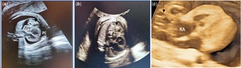
A 31-year-old gravida lady with underlying gestational diabetes presented at 25 weeks of gestation with a foetal ultrasound scan that demonstrated pericardial effusion and extracardiac mass. The cystic mass size was 10 × 14 mm and located near the right atrium. The tumour mass rapidly increases in size associated with the formation of massive pericardial effusion requiring multiple foetal pericardiocentesis. The first pericardiocentesis was performed at 28 weeks of gestation when there was evidence of right atrial diastolic collapse consistent with pericardial tamponade and pleural effusion, resulting in foetal hydrops (Fig 1). About 45 ml of haemorrhagic pericardial fluid was aspirated, which caused an improvement of the foetal hydrops after the procedure. Microscopic examination of the pericardial fluid revealed scattered mesothelial cells with small lymphocytes and red blood cells but without any atypical cells. The foetal pericardial fluid for tumour makers showed elevated α-fetoprotein levels of 84,066 U/ml and normal β-human chorionic gonadotrophin of 0.3 ng/ml. The pregnancy was subsequently closely monitored with weekly follow-ups. Repeated foetal pericardiocentesis was performed at 30 and 32 weeks of gestation due to the reaccumulating of pericardial fluid. The tumour was rapidly increasing in size to 55 × 50 mm at 33 weeks of gestation with the persistence of the pericardial effusion.
( a ) Four-chamber view fetal echocardiography demonstrates an intrapericardial teratoma (black arrow), described as a 10 × 14 mm cystic mass near the right atrium (RA) with global pericardial effusion (PE). ( b ) A repeat fetal scan shows an increasing tumor size, ( c ) cystic heterogenous mass from 3D scan view.

A multidisciplinary team discussion was conducted between a fetomaternal specialist, paediatric cardiologist, cardiothoracic surgeon, anaesthesiologist, and neonatologist, and the consensus was on early delivery. Following dexamethasone doses completion, elective lower segment caesarian section was performed at 33 weeks and 5 days and delivered a male baby with a birth weight of 2.8 kg. The baby required immediate intubation and resuscitation for poor oxygenation and perfusion. Intratracheal surfactants for severe respiratory distress and pericardial drainage were performed in the operation theatre. His initial chest radiograph showed marked cardiomegaly and homogenous lung fields with very minimal aeration. He required a high ventilator setting to maintain saturation and received multiple fluid boluses and a high dose of dopamine and dobutamine initially to maintain his blood pressure. The haemodynamic parameters improved after a few days, and he became more stable to allow surgery to be performed. Postnatal echocardiography confirmed the antenatal scan findings, and other than a small patent ductus arteriosus, the intracardiac structure and function were otherwise normal. His CT thorax revealed a large solid-cystic mediastinal mass measuring 55 × 35 × 43 mm with calcification and fat components, causing mass effect and compression onto the mediastinal structures.
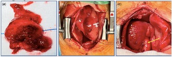
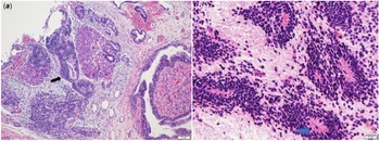
He underwent surgical tumour resection via median sternotomy approach without cardiopulmonary bypass. A large extracardiac tumour was resected from the ascending aorta’s adventitia layer, extending anteriorly, near the right ventricle and behind the ascending aorta. The tumour mass was described as a large lobulated greyish mass with multiple cystic components (Fig 2). The pericardial lining was clear from the mass except near the right ventricle, probably related to previous intrauterine puncture for foetal pericardiocentesis. The blood supply to the mass was via a pedicle from the descending aorta. The operation was uneventful, and he was extubated to continuous positive airway pressure on a post-operative day 4 without any inotropic support. Histopathological examination revealed a high-grade (Grade 3) immature teratoma containing an immature and variable number of mature tissues with three germ cell layers without yolk sac components. The immature tissues showed neuroectodermal tubules and rosettes displaying mitotically active hyperchromatic cells (Fig 3). An oncologist was consulted, who advised closed surveillance of the tumour with imaging scan and tumour marker monitoring. He was discharged well from the neonatal intensive care unit at 2 months of life with close regular follow-ups. His repeated echocardiography and tumour marker readings suggest no evidence of recurrence. His alpha-fetoprotein was reduced from >80,000 ng/ml to 32,682 ng/ml at 1 month of life, decreasing slowly to less than 100 ng/ml by 4 months and normalised by 6 months of life (normal range 0–8.78 ng/ml). At his latest follow-up at 1 year and 3 months, he remained well with no evidence of tumour recurrence.
( a ) En-bloc excision of a large lobulated greyish mass 63 × 37 × 15 mm. The solid area shows pale yellowish or greyish tissue as well bony component. ( b ) tumor in-situ, lies anterior to the cardiac mass with no attachments to the pericardial lining, ( c ) Cardiac mass after clear resection to the tumor. Yellow arrow was site of the pedunculated stump arising from the ascending aorta.

( a ) The immature teratoma consists of neuroectodermal tubules (black arrow), ( b ) Under high power field slide shows neuroectodermal tubules and rosettes displaying mitotically active hyperchromatic cells (blue arrow).

Discussion
Teratoma is the most common type of germ cells tumour occurring in the perinatal period. Histologically, most germ cell tumours presented during this period are benign and classified as mature or immature teratomas. Reference Isaacs3 According to Gonzalez-Crussi histopathological grading system, mature teratomas (Grade 0) are more frequent (54.5%) than immature teratomas (Grades 1–3) seen in 45.5% of the cases. Grade 3 immature teratoma, as seen in our case, accounts for 7.8% of all teratomas. Reference Gonzalez‐Crussi4 In the congenital form of teratomas, the grade of immaturity is not predictive of malignant behaviour; instead, the presence of a yolk sac component can adversely affect the prognosis. Complete tumour resection is an effective treatment for children with immature teratomas, with or without admixed yolk sac tumours. In contrast, incomplete surgical resection is associated with tumour recurrences. Reference Isaacs3
Intrapericardial teratomas are frequently diagnosed in utero or the neonatal period. Usually, the tumour appears as a single, nonhomogeneous, lobular, cystic, calcified, intrapericardial mass, often associated with pericardial effusion on echocardiography. Reference Rychik, Khalek and Gaynor5 Frequently, it is a right-sided, well-encapsulated intrapericardial mass arising from the region of the pericardial reflection at the junction of the ascending aorta and right atrial appendage. Reference Rychik, Khalek and Gaynor5 Sometimes, the tumour capsule can be firmly attached to the aorta, as seen in our case but can also be connected to the pulmonary artery adventitia. The feeding vessels are usually from the aortic vasa vasorum with potential massive bleeding from the aorta during dissection. Reference Manoly, Viola, Fowler, Roman and Haw6 Another essential feature is that the tumour can grow rapidly increasing in size over a few weeks. Reference Rychik, Khalek and Gaynor5 This tumour mass effect can be associated with large pericardial effusions and tamponade with physiological consequences of diminished cardiac output. The reduction in cardiac output is directly related to a large tumour size that can cause fetal hydrops and death if left untreated. An early prenatal diagnosis of intrapericardial teratoma was made in our case based on the presentation and echocardiographic findings. The significance of elevated alpha-fetoprotein levels in the fetus’s pericardial fluid needs to be carefully interpreted. It can also be produced by immature fetal liver cells other than germ cell tumours. Reference Meuris, Gewillig and Meyns7 Another factor that may explain raised level in our case was prematurity. A study demonstrated a significant negative correlation between the alpha-fetoprotein level and gestational age. Reference Blohm, Vesterling-Hörner, Calaminus and Göbel8
The treatment of fetal intrapericardial teratoma can be challenging. Prenatal management most often includes observation with intervening measures of pericardiocentesis and delivery once the fetus reaches a viable gestational age for postnatal surgical resection. Reference Rychik, Khalek and Gaynor5,Reference Manoly, Viola, Fowler, Roman and Haw6 Our patient required multiple fetal pericardiocentesis due to reaccumulation of pericardial fluid that helped to stabilise the haemodynamic parameters and prevent the progression to severe hydrops. In the current era of improved prenatal imaging and cardiovascular surveillance utilising fetal echocardiography and the potential for in utero treatment through foetal surgery techniques, survival with a good outcome may be possible. To date, in utero surgical resection can be an option in experienced foetal heart centres with reported successful prenatal resection in a selected foetus with intrapericardial teratoma. Reference Rychik, Khalek and Gaynor5
A carefully planned and coordinated caesarean section followed by immediate surgical resection is lifesaving and curative because most tumours are benign. The operation ideally should be performed without delay once an ultrasound confirms the presence of an intrapericardial teratoma with pericardial effusion. In our case, the baby needed to be stabilised for a few days, mainly due to severe respiratory distress requiring surfactant and high-frequency oscillatory ventilation. His haemodynamic parameters were more stable with the transcatheter pericardial drainage, allowing an optimised surgery condition. The operative procedure for removal of intrapericardial teratoma is usually uncomplicated and does not necessitate cardiopulmonary bypass unless complicated by a difficult surgery or a high risk of significant bleeding from tumours adherence to other tissues during dissection.
In conclusion, a multidisciplinary team approach is important to ensure the success of managing intrapericardial teratoma that usually presents with hydrops foetalis. Urgent intervention for haemodynamic stabilisation is usually required. The prognosis after complete surgical clearance is good, but it needs a long-term follow-up to monitor for recurrences.
Acknowledgements
We would like to acknowledge all the people involved in managing this patient, the Pediatric cardiology unit, Neonatal Intensive Care Unit, Dr Zuraini Mohd Noor from the Anesthesiology Department and Norren Haneezah Sahak from the Pathology Department, Serdang Hospital.
Conflicts of interest
None.