Introduction
Anurans represent one of the most diverse and ecologically relevant groups among vertebrates, playing highly important roles in trophic chains, both aquatic and terrestrial, since they present a life cycle divided into an aquatic larval stage and a terrestrial adult stage (Campião et al Reference Campião, Ribas, Morais, Silva and Tavares2015; Hüsken et al Reference Hüsken, Schwelm, Rückert and Sures2025; Koprivnikar et al Reference Koprivnikar, Marcogliese and Rohr2012). Tadpoles, the aquatic larval stage, are important ecological indicators, as they exhibit high sensitivity to environmental changes; these animals can also act as sensitive intermediate hosts to helminth infections (Delgado et al Reference Delgado, de Souza Queiroz, Pinto, Pezutti and Anjos2026; Johnson and McKenzie Reference Johnson, McKenzie, Fried and Toledo2008; Svinin et al Reference Svinin, Bashinskiy, Ermakov and Litvinchuk2023). The high diversity of anurans in Neotropical regions like Brazil reinforces the ecological relevance of these organisms (Duellman Reference Duellman1999; Haddad et al Reference Haddad, Lopes, Becker, da Silva and Lyra2022; Penhacek et al Reference Penhacek, Souza, Santos, Guerra, Castro-Souza and de Jesus Rodrigues2024). Combined with diverse parasitic interactions (Campião et al Reference Campião, Ribas, Morais, Silva and Tavares2015; Neves et al Reference Neves, Cardoso, Rebêlo, Félix, Machado, Costa-Campos, Santos and Melo2024), this biodiversity highlights the urgent need for studies addressing the impacts of helminth infections across different host life stages.
Among the group of helminth parasites that infect anurans, trematodes stand out, presenting a complex life cycle and the ability to cause significant harmful effects in their hosts (Hernandez-Caballero et al Reference Hernandez-Caballero, Garcia-Longoria, Gomez-Mestre and Marzal2022; Hüsken et al Reference Hüsken, Schwelm, Rückert and Sures2025). Several trematode genera, including Ribeiroia, Echinostoma, Clinostomum, and Neodiplostomum, become established as metacercariae in different host tissues and organs, with or without cyst formation (Achatz et al Reference Achatz, Kostadinova, Georgieva, Fecchio and Tkach2025; Aguiar et al Reference Aguiar, Morais, Silva, Anjos, Foster and Da Silva2021; Billet et al Reference Billet, Wuerthner, Hua, Relyea and Hoverman2020; Calhoun et al Reference Calhoun, Leslie, Riepe, Achatz, McDevitt-Galles, Tkach and Johnson2020, Reference Calhoun, Groves, Schaffer, Achatz, Greiman and Johnson2025; Cavataio and Johnson Reference Cavataio and Johnson2025; Johnson et al Reference Johnson, Calhoun, Achatz, Greiman, Gestos and Keeley2024; Orlofske et al Reference Orlofske, Belden and Hopkins2017; Rosa et al Reference Rosa, de Souza Queiroz, Pelinson, Anjos and Rossa-Feres2024; Schotthoefer et al Reference Schotthoefer, Cole and Beasley2003). Among the effects of trematode presence in tissues are physiological stress, tissue deformities, behavioural alterations, and even mortality (Holland Reference Holland2010; Koprivnikar et al Reference Koprivnikar, Hoye, Urichuk and Johnson2019; Rosa et al Reference Rosa, de Souza Queiroz, Pelinson, Anjos and Rossa-Feres2024; Svinin et al Reference Svinin, Bashinskiy, Ermakov and Litvinchuk2023). Both the occurrence and intensity of trematode infections are strongly associated with environmental factors such as eutrophication, which increases the abundance of aquatic molluscs serving as intermediate hosts (Johnson and McKenzie Reference Johnson, McKenzie, Fried and Toledo2008; Rohr et al Reference Rohr, Schotthoefer, Raffel, Carrick, Halstead, Hoverman, Johnson, Johnson, Lieske, Piwoni, Schoff and Beasley2008). In addition, anthropogenic disturbances, including increasing temperatures and other human-induced environmental changes, can indirectly influence amphibian populations by altering their physiological condition and their ability to respond to environmental stressors (Chen et al Reference Chen, Cao, Dong and Li2025; Marcogliese Reference Marcogliese, Smit and Sures2025).
In Brazil, trematode infections have been reported in a wide variety of amphibian species across different biomes, including the Atlantic Forest, Amazon, Pantanal, and Cerrado, demonstrating the high diversity of host–parasite associations in the country (Aguiar et al Reference Aguiar, Morais, Silva, Anjos, Foster and Da Silva2021; Campião et al Reference Campião, Ribas, Morais, Silva and Tavares2015; Queiroz et al Reference Queiroz, Delgado, Pinto and Anjos2025; Rosa et al Reference Rosa, de Souza Queiroz, Pelinson, Anjos and Rossa-Feres2024). Most available studies have focused on parasite diversity, host records, and ecological patterns of infection, whereas investigations addressing the pathological consequences of these infections, particularly in tadpoles from natural populations, remain scarce.
Despite the increasing interest in the ecological and morphological relationships of trematode parasitism in tadpoles, few studies have investigated in detail the histopathological effects of these infections in internal organs. Some studies have evaluated the effects of trematode infection in different tissues of tadpoles, demonstrating that the location of the parasite throughout larval development is associated with different types of physiological and morphological alterations, with potential consequences for host survival and development (Calhoun et al Reference Calhoun, Schaffer, Gregory, Hardy and Johnson2015). However, although these studies correlate the presence of the parasite with the affected tissue structures, studies that describe in depth the histological alterations after parasite establishment are still scarce, with histology being predominantly addressed based on the location of trematodes in organ systems and the structural and functional alterations they induce in host tissues (Johnson et al Reference Johnson, Lunde, Ritchie and Launer1999; Schotthoefer et al Reference Schotthoefer, Cole and Beasley2003).
A significant gap in the literature remains regarding the histopathological characterisation of trematode infections in tadpoles from different families, such as the family Hylidae, especially in populations from the Brazilian Cerrado, a highly threatened biome (Soares et al Reference Soares, Sano, Aquino and Td2025). Most studies focus on ecological aspects, prevalence, or external malformations (Castro-Bastidas et al Reference Castro-Bastidas, Bucio-Pacheco, Serrano and Aguillón-Gutiérrez2025; Henle and Dubois Reference Henle and Dubois2017; Johnson et al Reference Johnson, Lunde, Ritchie and Launer1999, Reference Johnson, Calhoun, Achatz, Greiman, Gestos and Keeley2024; Koprivnikar et al Reference Koprivnikar, Marcogliese and Rohr2012; Presswell and Bennett Reference Presswell and Bennett2025). These investigations often overlook the cellular and tissue impacts within vital internal organs. However, metacercariae can establish in tissues like the liver, kidneys, and musculature, causing inflammation, necrosis, hyperplasia, and other significant structural alterations (Amaral et al Reference Amaral, Silva, Dias, Malta, Rosa, Costa-Neto, Gentile and Melo2017; Campião et al Reference Campião, Ribas, Morais, Silva and Tavares2015; Martinez et al Reference Martinez, Gilardoni, Medina, Cremonte and Etchegoin2024; Regueira et al Reference Regueira, O’Donohoe, Pavon Novarin, Michou Etcheverria, Tropea and Hermida2022; Shareef and Abidi Reference Shareef and Abidi2015).
Therefore, the present study aimed to describe the histopathological alterations caused by trematode metacercariae in the kidneys and musculature of tadpoles collected in a natural environment, contributing to the understanding of host–parasite interactions in the early stages of larval development.
Materials and methods
Sampling of biological material
The specimens were collected using a rectangular sieve (50 cm × 75 cm, 2 mm mesh), through sweeps along the water body with a sampling effort of 1 hour during both morning and night periods, according to Queiroz et al (Reference Queiroz, Alves, López-Hernández, Anjos and Pinto2021). Collections were carried out monthly during two consecutive rainy seasons, from October to March (2023–2024 and 2024–2025), in a Cerrado fragment located in the municipality of Selvíria, Mato Grosso do Sul State, Brazil (20.380010° S, 51.375447° W). The specimens were individually placed in plastic bags containing water from the collection site and transported to the laboratory for classification of the developmental stage according to Gosner (Reference Gosner1960) and taxonomic identification based on Rossa-Feres and Nomura (Reference Rossa-Feres and Nomura2006), Schulze et al (Reference Schulze, Jansen and Köhler2015), and Rossa-Feres (Reference Rossa-Feres2024).
The specimens, after euthanasia with a solution of eugenol and distilled water (50 μL/L) (protocol SISBIO (Biodiversity Authorization and Information) number 91997-1 and CEUA (Animal Use Ethics Committee) no. 218773/2025), were dissected to verify the presence of trematodes. The parasitised organs were extracted and fixed in a solution of 2% glutaraldehyde and 4% paraformaldehyde in Sorensen phosphate buffer (pH 7.2) for histological analyses following standard light microscopy protocols. All 39 infected individuals were included in the histopathological evaluation. Likewise, organs from non-parasitised tadpoles representing all developmental stages in which parasitised individuals were found were also collected and processed under the same conditions. These specimens were used as controls to establish a comparative baseline between healthy and parasitised tissues.
Parasite characterisation
Trematodes collected in vivo were analysed on temporary slides stained with the vital dye neutral red, especially in the case of encysted metacercariae. The parasites collected from the tissues were mounted on permanent slides stained with carmine and Delafield’s haematoxylin, with morphometric and morphological records and photomicrographs obtained using the LAS v3 system (Leica Application Suite), coupled to a Leica DM 2500 microscope with phase interference contrast. Taxonomic identification of the trematodes was performed based on Travassos et al (Reference Travassos, Freitas and Kohn1969), Thatcher (Reference Thatcher1993), and Fernandes and Kohn (Reference Fernandes and Kohn2014).
Tissue processing for light microscopy
After fixation, the parasitised portions of the tegument were processed in historesin (glycol methacrylate), following the steps described below: storage in 70% alcohol for at least 24 h, followed by immersion in 95% alcohol for 4 h for dehydration. The material was progressively infiltrated in an equimolar solution of resin + alcohol for 4 h and subsequently in pure resin for 12 h. Embedding was performed using the JB-4 Embedding Kit (Polysciences), composed of resin monomer, hardener, accelerator, and plasticiser.
The blocks were sectioned into 3-μm histological sections using a Leica RM 2245 microtome equipped with a steel blade. The sections were stained with haematoxylin and eosin (H&E) and Masson’s trichrome (MT) according to standard protocols. The slides were analysed and photographed using a Leica DM 2500 microscope.
Results
A total of 207 tadpoles belonging to eight anuran species were analysed, distributed among the families Hylidae and Leptodactylidae: Boana raniceps (Cope, 1862) (n = 20), Boana albopunctata (Spix, 1824) (n = 106), Dendropsophus nanus (Boulenger, 1889) (n = 31), Dendropsophus cerradensis (Napoli and Caramaschi, 1998) (n = 2), and Scinax fuscovarius (Lutz, 1925) (n = 30) (Hylidae); and Leptodactylus fuscus (Schneider, 1799) (n = 11), Leptodactylus labyrinthicus (Spix, 1824) (n = 4), and Physalaemus cuvieri (Fitzinger, 1826) (n = 3) (Leptodactylidae).
Among the investigated animals, 39 (P = 18.84%) were found to be naturally parasitised. In the kidneys, metacercariae of Neodiplostomum sp. (15) were observed, and in the tail muscle tissue, encysted Strigeidae gen. sp. parasites (10) were recorded, whose infections were associated with the structural alterations observed in the tissues, resulting in evident histological modifications. Other animals presented parasites in the intestine (5) and in the body cavity (20); however, in these cases, the parasites were not encysted, and no histopathological alterations were observed. Some individuals presented parasitism in more than one site (kidneys and musculature = 2; Gyrinicola sp. in the cavity + trematode in the intestine = 1).
Despite the difficulties inherent to the analysis and identification of trematodes in tissues, the observed characteristics allow the recognition of the presence of a Strigeidae gen. sp. parasite and Neodiplostomum sp., previously recorded for the region and already described parasitising the same species analysed here, both in larval and adult stages, located in the kidneys (Queiroz et al Reference Queiroz, Pontes, Neto, Campião and Anjos2020, Reference Queiroz, Alves, López-Hernández, Anjos and Pinto2021; Rosa et al Reference Rosa, de Souza Queiroz, Pelinson, Anjos and Rossa-Feres2024). Thus, although identification was limited to the family level in some cases, the results presented consistently demonstrate the effects of parasitism on the host organism.
Parasitised kidney tissue
The region formed by fibrous tissue is composed of connective tissue and presents an abundant extracellular matrix, with fibroblasts sparsely distributed (Fig. 1). Together with the fibrous tissue layer, the layer that gives rise to the capsule surrounding the parasite, Neodiplostomum sp., is observed; this capsule is formed by a single layer of simple squamous epithelial tissue (Fig. 1), which once formed the renal tubules where the parasites are now established.
Micrograph of the kidney of a parasitised Dendropsophus nanus tadpole (Gosner 27), showing the presence of the parasite (*) located within the renal tissue. The formation of connective tissue (CT) surrounding the parasite can be observed, in which fibroblasts (f) are identified. The connective tissue induced by the parasite within a previously intact renal tubule (rt) now presents a simple squamous epithelium (sse), resulting from structural modification of the tubule. Staining: H&E.

The infection foci, together with fibrotic and inflammatory areas, displaced the functional renal tissue. As a result, the remaining functional structures occupied smaller areas and became restricted to peripheral or central regions (Fig. 2a, b). This displacement may compromise renal function in tadpoles.
A – Micrograph of the kidney of a healthy Boana raniceps tadpole (Gosner 31), showing the typical organisation of renal tissue. B – Micrograph of the kidney of a parasitised Boana raniceps tadpole (Gosner 31), presented as a digitally coloured image, in which the displacement of functional renal tissue to the peripheral regions can be observed, resulting in a reduction in its proportion and in the available space within the organ. Green areas represent functional tissue (rft); red areas correspond to the space occupied by encapsulation and parasite establishment; uncoloured regions indicate connective tissue without renal function. m – muscle tissue. A and B – Histological staining: H&E.

Figure 2. Long description
Panel A is a micrograph of healthy kidney tissue. The tissue is densely packed with purple and pink stained structures. The label r f t is positioned in the center of the renal functional tissue. Below the main kidney mass is a section of muscle tissue labeled m. A scale bar representing 200 micrometers is located in the bottom-right corner.
Panel B is a digitally colored micrograph of parasitized kidney tissue. Large, irregular red-colored regions dominate the center and right side, representing parasite encapsulation. Green-colored areas representing functional renal tissue are displaced to the peripheral edges and are significantly reduced in volume compared to Panel A. Uncolored white and light purple regions indicate connective tissue. A scale bar representing 200 micrometers is located in the bottom-right corner.
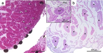
Among the renal structures regularly observed, the glomeruli stand out, presenting a rounded shape and a capsule composed of simple squamous epithelial cells, which delimit the urinary space; the central region is formed by a cluster of fenestrated capillaries and mesangial cells (Fig. 3a, cap). The glomerular structure, which was previously abundantly distributed (Fig. 3a), is now less widespread in the infected tissue (Figs. 2b and 3b).
Micrographs of the kidneys of Boana raniceps tadpoles. A – Kidney of a non-infected tadpole (Gosner 30), showing the regular and healthy distribution of glomeruli (arrowheads) throughout the organ. In cap – structure of an intact glomerulus. B – Kidney of a parasitised tadpole (Gosner 31), showing an apparent reduction in the number of glomeruli in areas affected by the presence of parasites (*). cr – central region of the glomerulus containing fenestrated capillaries; mc – mesangial cells. Histological staining: H&E.

Figure 3. Long description
A two-panel histological photo series.
Panel A on the left shows a healthy kidney at Gosner stage 30. The tissue is densely packed with pink-stained structures. White arrowheads point to several healthy glomeruli distributed regularly throughout the organ. A scale bar at the bottom right indicates 100 micrometers.
An inset at the top center of panel A provides a high-magnification view of a single intact glomerulus. An arrow labeled m c points to the mesangial cells on the outer edge of the structure. The label c r identifies the central region containing fenestrated capillaries. A scale bar in the inset indicates 50 micrometers.
Panel B on the right shows a parasitized kidney at Gosner stage 31. The tissue appears significantly more fragmented with large white gaps. Asterisks mark multiple areas affected by parasites, showing a clear reduction in the number of healthy glomeruli compared to panel A. A single white arrowhead at the bottom left points to one remaining glomerulus. A scale bar at the bottom right indicates 200 micrometers.
The renal ducts in healthy animals (Fig. 4a) present a tubular structure lined by simple cuboidal epithelium (Fig. 4b). The epithelial cells present clear cytoplasm, well-defined cell boundaries, and central nuclei (Figs. 4a, b); between the ducts, interstitial connective tissue can be observed. Infected renal tubules exhibited cytoplasmic degeneration of epithelial cells (Fig. 4c). These alterations included reduced cytoplasmic basophilia, cytoplasmic vacuolation, oedema, irregular cell contours, and condensed or displaced nuclei. Together, these changes indicate cellular damage and loss of tubular epithelial integrity.
Micrographs of the kidneys of Boana albopunctata tadpoles (Gosner 25) highlighting structural and physiological damage associated with trematode infection, with emphasis on alterations in the renal tubules. A and B – Kidneys of non-infected tadpoles, showing the integrity of tubular architecture; renal tubules (rt) formed by simple cuboidal epithelium (sce) are visible. C – Degeneration of the duct epithelium (ed) observed in infected animals. D – Ectasia of the renal ducts (area delimited in red), characterised by an increase in tubular lumen due to the presence of trematodes in the tissue. E – Micrograph of a chromaffin cell (cc) identified in healthy tissue of a Boana albopunctata tadpole (Gosner 25), a structure not observed in infected animals. Histological staining: H&E.

Figure 4. Long description
A multi-panel histological photo series.
Panel A (top-left): A 50 micrometer scale view of healthy kidney tissue. Multiple renal tubules, labeled r t, are clustered together, showing organized circular and elongated structures with distinct cellular borders.
Panel B (top-middle): A 20 micrometer close-up of healthy tissue. Three arrows point to the simple cuboidal epithelium, labeled s c e, which forms the lining of the tubules.
Panel C (top-right): A 50 micrometer view of infected tissue. The duct epithelium, labeled e d, appears dense and dark purple, indicating degeneration and loss of clear cellular architecture.
Panel D (bottom-center): A 100 micrometer view showing a large, irregular area outlined by a thick red line. Inside this boundary, the renal ducts exhibit ectasia, appearing as large, empty circular voids where trematodes have displaced or dilated the tissue.
Panel E (bottom-right): A 20 micrometer high-magnification view of healthy tissue. An arrow points to a single, pale, circular chromaffin cell, labeled c c, nestled among the darker stained renal cells.
With regard to the ducts, ectasia of the renal ducts was observed here in infected renal tissues. Ductal ectasia (Fig. 4d) is characterised by a widening of the lumen of the renal ducts and a thinning of the epithelial cell layer, with flattening of the cuboidal cells; nuclei that were previously centralised and rounded now appear displaced or condensed.
Chromaffin cells (Fig. 4e), which play a fundamental endocrine role in tadpole development, were observed here only in healthy animals, presenting clear to slightly eosinophilic cytoplasm, granular due to the presence of secretory granules, round-to-oval basophilic nuclei, and were arranged individually. Chromaffin cells were not observed in animals infected by trematodes, suggesting a possible impairment or alteration of the endocrine tissue associated with the infection.
It is important to emphasise that the histopathological alterations observed in the kidneys were consistent across all infected host species, with no species-specific differences in tissue response. Instead, parasite load was the main factor influencing the severity of the lesions. Lightly infected individuals exhibited more discrete changes (Fig. 5a), whereas tadpoles with higher parasite burdens presented more pronounced histological alterations, including cellular degeneration, ductal ectasia, and functional impairment (Figs. 5b, c). These findings indicate that the severity of tissue damage is associated with infection intensity rather than with host species.
Micrographs of the kidneys of Boana albopunctata tadpoles infected by trematodes (*), showing the different structural and morphological impacts on renal tissue according to infection intensity. A – Animal with low infection intensity (Gosner 25), in which most of the renal tissue is composed of healthy renal tissue (hrt). B – Animal with moderate to high infection intensity (Gosner 27). C – Animal with high infection intensity (Gosner 30). m – muscle. Histological staining: H&E.

Figure 5. Long description
The three-panel micrograph series displays histological sections stained with H and E.
Panel A at the top shows a low infection intensity in a Gosner 25 tadpole. The top portion contains muscle tissue labeled m. Below this is a large area of healthy renal tissue labeled h r t, characterized by dense, organized tubular structures. A single trematode, marked with an asterisk, is visible at the far right edge. A 200 micrometer scale bar is at the bottom right.
Panel B in the middle shows moderate to high infection intensity in a Gosner 27 tadpole. The healthy renal tissue h r t is reduced and fragmented on the left. The right side is dominated by at least five distinct trematode parasites, each marked with an asterisk, embedded within the tissue. A 200 micrometer scale bar is at the bottom right.
Panel C at the bottom shows high infection intensity in a Gosner 30 tadpole. Only a small patch of healthy renal tissue h r t remains at the top left. The rest of the section is heavily infiltrated by approximately nine trematode parasites, marked with asterisks, which appear as large, elongated, or curved structures displacing the native renal architecture. A 200 micrometer scale bar is at the bottom right.
Parasitised muscle tissue
Healthy animals presented tail musculature organised into well-defined myomeres, composed of long, multinucleated striated skeletal muscle fibres surrounded by a basal membrane. The fibres were arranged in dorsal and ventral layers, delimited by loose connective tissue and adjacent to the epidermis (Figs. 6a, b); however, once parasitised, the tail musculature also presented a new morphology and organisation.
Tail muscle tissue of Dendropsophus nanus tadpoles (Gosner 30). A and B – Micrographs of healthy tissue, showing the structural layers of the tail: m – muscle tissue; ct – connective tissue; e – epidermis, whose organisation may vary from simple to stratified depending on species and developmental stage, together with the underlying dermis. C, D, and E – Trematode cysts (*) located in muscle tissue, showing structural alterations, including modifications in collagen fibres (cf). The fibrous capsule formed during encystment (parasite tissue) (c) is surrounded by an epithelium produced by the host (cp), composed of multiple epithelial layers indicated by 1, 2, and 3 and delimited by the red dotted line. Histological staining: H&E.

Figure 6. Long description
A multi-panel histological study using H and E staining.
Panel A and B show healthy tissue. Panel A is a wide view with a 200 micrometer scale bar. A dashed box indicates the zoomed area for Panel B, which has a 50 micrometer scale bar. From the top surface moving inward, the layers are labeled E for epidermis, C T for connective tissue, and m for muscle tissue. In Panel B, an arrow points to c f for collagen fibers within the connective tissue.
Panels C, D, and E show trematode infection.
Panel C (100 micrometer scale) shows two dark trematode cysts marked with asterisks embedded in the C T. They are surrounded by a host-produced epithelium labeled c p.
Panel D (20 micrometer scale) provides a closer view of the interface. From left to right, the asterisk marks the parasite, c points to the fibrous capsule of the parasite, and c p points to the host epithelium.
Panel E (10 micrometer scale) shows a high-magnification cross-section of the host epithelium. Red dotted lines delineate three distinct epithelial layers numbered 1, 2, and 3, moving outward from the parasite capsule c.
Unlike the kidney, in the muscle tissue, parasites identified as Strigeidae gen. sp. were found; these parasites formed, in addition to the fibrous capsule, an epithelial layer that is part of cyst formation. In the muscle tissue, it was observed that the fibrous capsules presented an irregular shape, with significant variations in thickness and less alignment of collagen fibres than those found in the renal tissue (Fig. 6c). The presence of fibrous capsules presents a similar effect in both tissues, in which capsule formation ultimately alters tail morphology, displacing healthy and functional tissue to other regions, although there is a lower presence of parasites in a tissue of greater dimension.
In addition to the connective-tissue characteristics, capsule and cyst formation involved another difference: the epithelial tissue layer became stratified; in this epithelium that surrounds the entire cyst in which the parasite is established, it is possible to observe the formation of three layers of juxtaposed squamous cells (Figs. 6d, e). Another difference between parasitised renal tissue and muscle tissue is the formation of a cyst itself, between the parasite and the host tissue (Figs. 6d, e); to form part of or the entire cyst wall, cystogenic material is released by the cystogenic glands in the parasite; once formed within a host, the tissue of the cyst wall may present secretions or tissues from the host itself.
Local inflammatory response
In the tissue adjacent to the region surrounding the parasites, it is possible to identify different types of immune defence cells, such as neutrophils, eosinophils, mast cells, macrophages, and fibroblasts (Fig. 7a–e). The presence of these cell types indicates an inflammatory process associated with the immune response in the infected tissue; blood cells extravasated into the parenchyma are also observed (Fig. 7a, b), which may indicate the occurrence of a haemorrhagic process or even a congestive process; these immunological, inflammatory, and haemorrhagic responses are compatible with a chronic response to helminths.
Micrographs showing inflammatory foci in infected tissues of a parasitised Dendropsophus nanus tadpole (Gosner 30). A – Kidney; B – Tail muscle tissue. In A and B, inflammatory foci are observed, characterised by the presence of blood cells extravasated into the tissue (black arrows). In A, the area delimited in red indicates a region with accumulation of immune cells. C, D, and E – Detail of immune cells present in infected tissues: n – neutrophil; mt – mast cell; mc – macrophage; eo – eosinophil; m – muscle; ed – duct ectasia. Histological staining: H&E.

Figure 7. Long description
Panel A shows kidney tissue with multiple black arrows pointing to extravasated blood cells. A red dashed line outlines a dense cluster of immune cells. The label e d is located in the bottom right near a 50 micrometer scale bar. Panel B shows tail muscle tissue labeled m, with black arrows indicating inflammatory foci. A 50 micrometer scale bar is at the bottom. Panel C is a high-magnification view showing a neutrophil labeled n and a mast cell labeled m t, with a 10 micrometer scale bar. Panel D shows a macrophage labeled m c with a 10 micrometer scale bar. Panel E shows an eosinophil labeled e o and a region of duct ectasia labeled e d, with a 10 micrometer scale bar. All panels use H and E staining, appearing in shades of pink and purple.
Discussion
The results of this study allow the discussion of the histopathological effects of trematode metacercarial infection in the kidneys and musculature of Hylidae tadpoles in a natural environment. The observed alterations indicate that the presence and intensity of infection are associated with the progression of tissue damage, highlighting specific host responses at different infection sites. These findings provide the basis for discussion, in light of the literature, on the structural patterns of cysts and the dynamics of host–parasite interaction in the early stages of larval development.
Several studies have investigated the structure and development of metacercarial cysts in different trematode families, demonstrating that cyst formation is a dynamic process that begins after parasite establishment in the second intermediate host and culminates in the formation of encapsulated structures (Calhoun et al Reference Calhoun, Curtis, Hassan and Johnson2023; Faltýnková et al Reference Faltýnková, Karvonen and Valtonen2011; Galaktionov et al Reference Galaktionov, Malkova, Irwin, Saville and Maguire1997). The morphology of these cysts varies considerably among species, particularly in wall thickness, the number of capsule layers, and the participation of host tissues in cyst formation. While some trematodes develop thick, highly organised cysts, others form loose cysts that allow continued parasite growth within the host. These differences likely reflect distinct adaptive strategies and host–parasite interactions.
Once metacercariae become established within host tissues, they stimulate the formation of a fibrous capsule. This capsule is produced by the host rather than by the parasite. Similar host responses have been reported in kidneys, intestines, and muscle tissues of amphibians (Hamann et al Reference Hamann, Fernández and Gonzalez2019; Hong et al Reference Hong, Hong, Lee, Seo and Chi1982; Svinin et al Reference Svinin, Bashinskiy, Ermakov and Litvinchuk2023). Comparable reactions have also been described in birds, fish, and snails, indicating that fibrous encapsulation represents a widespread defence mechanism against trematode infections (Asok Kumar et al Reference Asok Kumar, Kumar, Palanivelu, Annamalai, Mathesh, Singh, Sharma and Dhama2018; Gérard et al Reference Gérard, De Tombeur, Dahirel and Ansart2023; Matos et al Reference Matos, Meira-Filho, Lorenzetti, Lavorente, Caldart, Bizari, Matos, Domit and Bracarense2021; Santi et al Reference Santi, André, Lux Hoppe and Werther2018; Skorobrekhova Reference Skorobrekhova2024). Recent studies also indicate that infectious processes act in a similar way by inducing fibrosis and cellular infiltration in amphibian kidneys, leading to the loss of tissue architecture observed here and consequent functional impairment (Hossainey et al Reference Hossainey, Yaparla, Hauser, Moore and Grayfer2021).
The remodelling observed in the kidneys, characterised by fibrotic areas and inflammatory infiltrates, promoted the displacement of functional structures. This tissue arrangement may directly compromise the renal physiology of tadpoles (Martin and Conn Reference Martin and Conn1990). This process of alteration of tissue architecture is closely associated with a decrease in nephron efficiency, resulting in impairments to the organism’s filtration capacity and water homeostasis (Mahmoud et al Reference Mahmoud, Gadel-Rab, Said, Saber, ElSalkh, Said and Atia2022). In developing organisms, such as anurans in the larval stage, such alterations may be even more critical, since osmotic balance and the elimination of metabolites are fundamental for metamorphosis and survival (Blaustein et al Reference Blaustein, Gervasi, Johnson, Hoverman, Belden, Bradley and Xie2012; Warburg Reference Warburg1995).
Previous studies have shown that cellular infiltration and fibrosis resulting from infectious or parasitic processes can cause substantial structural disorganisation of the kidneys, potentially impairing renal function (Hossainey et al Reference Hossainey, Yaparla, Hauser, Moore and Grayfer2021; Martin and Conn Reference Martin and Conn1990). Similar alterations have been reported in tadpoles infected with echinostomatid metacercariae, in which parasite establishment was associated with renal damage in Rana pipiens and Rana clamitans (Martin and Conn Reference Martin and Conn1990). Comparable pathological responses have also been described in ranavirus infections, which induce viral persistence in the kidneys, leukocyte infiltration, fibrosis, and chronic immunosuppressive conditions (Hossainey et al Reference Hossainey, Yaparla, Hauser, Moore and Grayfer2021). Therefore, the presence of renal remodelling in the analysed tadpoles suggests a tissue response to parasitism and highlights the potential pathological consequences of trematode infection during the larval stage. As the severity of renal alterations increased with parasite infection intensity, these findings indicate that heavily infected individuals may experience greater impairment of renal tissue integrity than lightly infected tadpoles.
In infected tissues, the glomerular structure was less widespread than the regular distribution seen in healthy controls. This pattern suggests a direct impairment of renal organisation due to the infection. Such a reduction in glomerular density and distribution may reflect cellular degeneration and tissue remodelling, both of which are driven by inflammatory or fibrotic processes in response to parasitism (Christensen and Verroust Reference Christensen and Verroust2008; van Velthuysen Reference van Velthuysen1996). The structural rearrangement of glomeruli observed in infected animals appears to be an already established effect of renal infection by trematodes (Holland Reference Holland2010) and is particularly critical, since glomeruli are essential functional units for plasma filtration and the maintenance of osmotic homeostasis; consequently, the reduction in the area occupied by glomeruli may compromise the filtering capacity of the kidney, affecting water regulation, the elimination of metabolites, and, in developing organisms such as tadpoles, the proper progression of metamorphosis (Fox Reference Fox1963; Holland Reference Holland2010; Schotthoefer Reference Schotthoefer, Cole and Beasley2003).
Studies in amphibians and other vertebrates have demonstrated that renal infections or inflammatory processes frequently lead to the loss of glomerular architecture, with direct implications for renal function. Chronic glomerular lesions may result in corresponding tubular atrophy, degeneration of specific nephrons, and, ultimately, interstitial and tubular fibrosis, highlighting the close interdependence between glomeruli and renal tubules in the pathogenesis of kidney diseases (Christensen and Verroust Reference Christensen and Verroust2008; Holland Reference Holland2010; Schotthoefer et al Reference Schotthoefer, Cole and Beasley2003). This same pattern can be observed in the present study in association with the presence of Neodiplostomum sp., since the glomerular structure and its distribution were affected, indicating that parasitism compromises glomerular functionality and, consequently, renal function.
Among the differences observed in tissue infections, the formation of cysts by parasites of Strigeidae gen. sp. stands out, in contrast to the absence of this structure in Neodiplostomum sp., indicating that different species exhibit distinct infection dynamics and, consequently, histopathological variations. The differences in fibrous capsule formation between renal and muscular tissues may stem from their distinct structural characteristics. Unlike the dense renal tissue, muscle tissue is contractile and has a lower structural density. These traits allow the fibrotic response to adapt locally to mechanical conditions and tissue dynamics (Koprivnikar et al Reference Koprivnikar, Hoye, Urichuk and Johnson2019; Martin and Conn Reference Martin and Conn1990).
The comparison between the two tissue types highlights that the formation of fibrous capsules is a defence mechanism present in anurans, capable of limiting the contact of the parasite with host cells and reducing the direct damage caused by infection, which is evident from the inflammatory foci and the activity of immune cells in the tissue. However, this response is modulated by the morphology and function of the affected tissue, indicating that the host adjusts its local immune response according to the structural, physiological, and immunological characteristics of each organ (Koprivnikar et al Reference Koprivnikar, Hoye, Urichuk and Johnson2019; Martin and Conn Reference Martin and Conn1990; Stopper et al Reference Stopper, Hecker, Franssen and Sessions2002). Despite its protective function against the parasite, encapsulation represents a cost to the host; in the kidney, the presence of a dense capsule and the displacement of functional structures may partially compromise filtration and osmotic homeostasis (Martin and Conn Reference Martin and Conn1990).
In muscle tissue, irregular fibrosis may limit contractile capacity and reduce tadpole mobility. Thus, the fibrotic response represents a trade-off between immune defence and the maintenance of tissue function, with important implications for the physiology, performance, and survival of infected anurans (Johnson et al Reference Johnson, Lunde, Thurman, Ritchie, Wray, Sutherland, Kapfer, Frest, Bowerman and Blaustein2002; Perpiñán et al Reference Perpiñán, Garner, Trupkiewicz, Malarchik, Armstrong, Lucio-Forster and Bowman2010). It is also important to highlight the problems associated with limb malformations, known as anomaly P, resulting from the encystment of trematodes in strategically important muscular regions. This anomaly has already been recorded in several species and represents a significant problem associated with trematode infections (Svinin et al Reference Svinin, Bashinskiy, Litvinchuk, Ermakov, Ivanov, Neymark, Vedernikov, Osipov, Drobot and Dubois2020).
In the tissue surrounding the fibrous capsule formed around trematode cysts, the presence of an inflammatory infiltrate composed of different cell types can be observed, demonstrating the complexity of the local immune response (Buchmann Reference Buchmann2022; Kiesecker Reference Kiesecker2002; Ramos et al Reference Ramos, Discipio and Ferreira2006). Neutrophils are present in the analysed tissues, characterising an early inflammatory response and indicating their protective role against parasites, which has been demonstrated in different contexts, such as in vitro studies with trematodes of the genus Diplostomum, in which neutrophils were able to colonise and eliminate metacercariae at early developmental stages (Buchmann Reference Buchmann2022). Eosinophils are characteristic immune cells in helminth infections and are vital for host resistance (Kiesecker Reference Kiesecker2002). They actively release toxic proteins and organise the fibrous capsule around cysts. This process reduces contact between host cells and the parasite, while also contributing to cyst damage (Ramos et al Reference Ramos, Discipio and Ferreira2006).
Mast cells can be observed at the periphery of the region surrounding the parasites; the role of these cells in cellular recruitment through the release of inflammatory mediators, such as histamine, is well known and highlights the participation of adaptive immunity in the chronic response to the parasite present in the tissue (Dezfuli et al Reference Dezfuli, Manera, Lorenzoni, Pironi, Shinn and Giari2015; Varga et al Reference Varga, Bui-Marinos and Katzenback2019). In addition, macrophages identified here are commonly recruited to the site of infection and play a central role in the phagocytosis of parasitic remnants during inflammatory processes that modulate fibrosis. Activated fibroblasts, also observed in the analysed tissues, act in the deposition of extracellular matrix; this cell type has already been recorded in this type of infection and contributes to the formation of the fibrous capsule and to the modulation of the tissue response (Dezfuli et al Reference Dezfuli, Manera, Lorenzoni, Pironi, Shinn and Giari2015; Perpiñán et al Reference Perpiñán, Garner, Trupkiewicz, Malarchik, Armstrong, Lucio-Forster and Bowman2010; Varga et al Reference Varga, Bui-Marinos and Katzenback2019), which has been reported for several vertebrate groups in response to helminth infections (Dezfuli et al Reference Dezfuli, Manera, Lorenzoni, Pironi, Shinn and Giari2015). Thus, the presence of both innate and adaptive immune cells indicates that the host adjusts its response based on tissue characteristics and infection intensity. While this modulation helps contain the parasite, an exacerbated inflammatory response can also lead to pathological tissue damage (Koprivnikar et al Reference Koprivnikar, Hoye, Urichuk and Johnson2019; Martin and Conn Reference Martin and Conn1990).
Considering that trematodes may exert direct and often aggressive effects on their hosts, and that their occurrence and prevalence are influenced by anthropogenic activities, it becomes essential to advance studies that clarify these interactions and their impacts on organisms. In addition to the ecological perspective, physiological approaches are necessary to evaluate the stress associated with these infections and to understand how the host response is modulated in this context. These physiological data are highly relevant for clarifying host–parasite dynamics. Furthermore, they help integrate this knowledge with other global threats, such as infection by Batrachochytrium dendrobatidis (Bd). Bd is responsible for global amphibian declines and varies in its impact depending on ecological contexts, host species, and health conditions (Humphries et al Reference Humphries, Lanctot, Robert, McCallum, Newell and Grogan2022; Searle et al Reference Searle, Gervasi, Hua, Hammond, Relyea, Olson and Blaustein2011). In this sense, the integration of morphological, ecological, and physiological studies represents a promising path for consolidating advances and supporting conservation strategies.
Our findings demonstrate that the histopathological consequences of trematode infections in tadpoles depend primarily on parasite infection intensity rather than host species, highlighting the importance of considering infection burden when evaluating host–parasite interactions. By providing a detailed characterisation of tissue alterations associated with natural infections, this study expands current knowledge of the pathological effects of trematode metacercariae during amphibian larval development. Such information provides a valuable foundation for future studies investigating the physiological and ecological consequences of parasitism in natural amphibian populations, particularly under ongoing environmental change.
Acknowledgements
The authors would like to thank the Coordenação de Aperfeiçoamento de Pessoal de Nível Superior (CAPES, Brazil) for the financial support (student scholarship to MRLD).
Author contribution
M.L.R.D.: Conceptualization, methodology, formal analysis, data curation, and writing – original draft. C.S.R.: Formal analysis, conceptualization, and writing – review and editing. L.A.A.: Conceptualization, writing – review and editing, and supervision.
Financial support
The work was funded by the Coordenação de Aperfeiçoamento de Pessoal de Nível Superior (CAPES) (process number 88887.966100/2024-00).
Competing interests
The authors declare no competing interests.
Ethical standards
All international, national, and institutional guidelines regarding the care and use of animals were strictly observed (SISBIO number 91997-1). The experimental procedures complied with the ethical standards of the institution where the study was conducted (CEUA no. 218773/2025). This work did not involve any research with human participants carried out by the authors.