




No CrossRef data available.
Article contents
Characterization of a mitochondrion-like organelle in Cryptosporidium parvum
Published online by Cambridge University Press: 10 June 2004
Abstract
Cryptosporidium parvum is a protozoan parasite that causes widespread diarrhoeal disease in humans and other animals and is responsible for large waterborne outbreaks of cryptosporidiosis. Unlike many organisms belonging to the phylum Apicomplexa, such as Plasmodium spp. and Toxoplasma gondii, there is no clinically proven drug treatment against this parasite. Aspects of the basic biology of C. parvum remain poorly understood, including a detailed knowledge of key metabolic pathways, its genome organization and organellar complement. Previous studies have proposed that C. parvum lacks a relic plastid organelle, or ‘apicoplast’, but that it may possess a mitochondrion. Here we characterize a mitochondrion-like organelle in C. parvum by (i) ultrastructural and morphological description (ii) localization of heterologous mitochondrial chaperonin antibody probes (iii) phylogenetic analysis of genes encoding mitochondrial transport proteins (iv) identification and analysis of mitochondrion-associated gene sequences. Our descriptive morphological analysis was performed by energy-filtering transmission electron microscopy (EFTEM) of C. hominis and C. parvum. The ‘mitochondrion-like’ organelle was characterized by labelling the structure with a heterologous mitochondrial chaperonin probe (hsp60) both in immunoelectron microscopy (IMEM) and immunofluorescence (IMF). Phylogenetic analysis of the mitochondrial import system and housekeeping components (hsp60 and hsp70-dnaK) suggested that the C. parvum mitochondrion-like organelle is likely to have descended from a common ancestral apicomplexan mitochondrion. We also identified a partial cDNA sequence coding for an alternative oxidase (AOX) gene, a component of the electron transport chain which can act as an alternative to the terminal mitochondrial respiratory complexes III and IV, which has not yet been reported in any other member of this phylum. Degenerate primers developed to identify selected mitochondrial genes failed to identify either cytochrome oxidase subunit I, or cytochrome b. Taken together, our data aim to provide new insights into the characterization of this Cryptosporidium organelle and a logical framework for future functional investigation.
Keywords
Information
- Type
- Research Article
- Information
- Copyright
- © 2004 Cambridge University Press
INTRODUCTION
Many aspects of the cell biology and biochemistry of Cryptosporidium parvum are still poorly understood (Coombs, 1999) and little is known about its organelle complement, the nature of key metabolic pathways, or its extranuclear genome organization and evolution (Andersson et al. 1998). Preliminary evidence suggests the absence of plastid-like structures (Zhu, Marchewka & Keithly, 2000), but the presence of a relic mitochondrion with some associated functions (Riordan et al. 1999). Karyotype analysis (Blunt et al. 1997) has shown the presence of an extranuclear genome, similar to those found in other Apicomplexa (Feagin, 1994; Wilson & Williamson, 1997).
To investigate the organelle complement of Cryptosporidium, we analysed sporozoites using nucleic acid stains in order to demonstrate the existence of extranuclear DNA staining elements. We also undertook an ultrastructural investigation to identify organelles both in C. hominis (Morgan-Ryan et al. 2002) and C. parvum sporozoites and detected a double-membrane bound structure closely associated with the nucleus at the posterior end of sporozoites. This structure was further characterized by immunolocalization with heterologous hsp60 antibodies both by electron and fluorescence microscopy. Taken together, these observations suggest that there is a mitochondrion-like organelle in Cryptosporidium.
Screening of recent sequence data from C. parvum (Liu et al. 1999; Strong & Nelson, 2000; Widmer et al. 2000; http://medsfgh.ucsf.edu/id/CpTags/home.html, http://www.parvum.mic.vcu.edu/) revealed the presence of some mitochondrion-related sequences which are encoded by the nuclear genome in other organisms, but failed to identify any associated with extranuclear organellar genomes.
To help understand the function and evolution of the putative mitochondrion-like organelle, we amplified and sequenced genes coding for C. parvum hsp60 and hsp70 from a cDNA library and undertook phylogenetic analysis of the predicted polypeptide sequences. We also used a degenerate primer PCR approach to test for the presence of a mitochondrial genome by amplification of markers for the respiratory chain components cytochrome oxidase I and cytochrome b, but we failed to identify either of these components despite recent work suggesting that some form of aerobic metabolism occurs in C. parvum (Riordan et al. 1999; Kayser et al. 2001, 2002). We therefore adopted a further gene cloning strategy in which we demonstrated the presence of an alternative oxidase (AOX) gene, a terminal oxidase which can replace complex III and IV in the alternative respiratory pathways of plants, fungi and kinetoplastida (Sluse & Jarmuszkiewicz, 1998; Chaudhuri, Ajayi & Hill, 1998; Maxwell, Wang & McIntosh, 1999). We also used a similar approach to identify a superoxide dismutase gene (SOD), a component of the oxygen radical scavenging system (ROS) (Kabiri & Steverding, 2001). Our molecular and phylogenetic analysis, combined with the ultrastructural characterization, has led us to formulate a working hypothesis as to the possible biochemical pathways present in this organelle of C. parvum.
MATERIALS AND METHODS
Parasites
Purified C. parvum (Iowa isolate) and C. hominis (Morgan-Ryan et al. 2002) oocysts were obtained from the Scottish Parasite Diagnostic Laboratory (SPDL) Cryptosporidium oocyst bank (C. hominis isolates 1141 and 986). C. hominis oocysts were purified by water–ether sedimentation (Bukhari & Smith, 1995) and stored in water at 4 °C until used. The Iowa isolate oocysts were a gift from M. M. Marshall, University of Arizona, Tucson, AZ, USA. Purified C. parvum (Moredun [MD] isolate) oocysts were purchased from Moredun Scientific Ltd, (Edinburgh, Scotland, UK). Entamoeba histolytica trophozoites (Human Mexico [HM]1 isolate) were provided by the SPDL.
Electron microscopy
Cryopreservation for morphology
Oocysts were excysted following incubation for 1·5 h at 37 °C in 1·5% taurocholic acid (Sigma) in phosphate-buffered saline (PBS 150 mM, pH 7·2). Sporozoites and oocysts were impact frozen and cryo-processed for embedding in Spurr's resin according to previously described methods (Tetley et al. 1998).
Immuno-electron microscopy
Pelleted (2500 g for 10 min) C. parvum and C. hominis sporozoites and E. histolytica trophozoites were fixed immediately in 2% paraformaldehyde/PBS (pH 7·4) containing 0·1% glutaraldehyde at 4 °C for 40 min. Sections were collected from pelleted sporozoites that had been embedded in ethanol/LR White resin at −20 °C and finally UV polymerized for 2 days at −20 °C and 2 days at +20 °C. Residual aldehyde groups were inactivated with 0·05 M glycine/PBS for 20 min and blocked for non-specific protein interactions with PBS/0·2% acetylated-BSA buffer (incubation buffer) for 30 min. Undiluted primary antibody hsp60 (Tovar, Fischer & Clark, 1999) was applied to sections in incubation buffer and incubated overnight at 4 °C followed by washes (6×5 min) in incubation buffer. This was followed by indirect gold labelling with goat-anti-mouse IgG/10 nm gold conjugate (Aurion, Netherlands) diluted 1 in 20 in incubation buffer for 2 h at 20 °C. After a final rinse in incubation buffer, 6 washes in PBS buffer and 3 washes in distilled water, sections were additionally fixed in 2% glutaraldehyde/PBS for 15 min, washed again in distilled water (3×5 min), and stained in uranyl acetate and lead citrate. Alternatively, silver enhancement of goat-anti-mouse IgG/1·4 nm gold conjugate was used. The conjugate was diluted 1 in 50 in incubation buffer, sections incubated for 2 h at 20 °C, subsequently washed (6×5 min) in incubation buffer, washed twice with PBS and fixed in 2% glutaraldehyde/PBS for 10 min and then washed (3×5 min) in distilled water prior to silver enhancement according to the manufacturer's instructions (Aurion, Netherlands). Extensive washing in distilled water was followed by section staining with uranyl and lead stains. As a negative control, some sections were not incubated with primary antibody but subjected to the same protocol. Samples were examined with a Zeiss 902 energy-filtering transmission electron microscope (Zeiss, Oberkochen, Germany) at 80 KV and zero energy loss. Digital images were captured with a 10242 Proscan CCD camera using a SIS EsiVision Pro software.
Fluorescence microscopy
C. parvum sporozoites (excysted from oocysts, as described above) were pelleted for 30 s at 10000 g, gently washed in PBS, pH 7·4, resuspended at a density of 105/μl in PBS and spotted onto poly-L-lysine coated slides. The samples were air dried, fixed by 4% paraformaldehyde (Sigma) in PBS for 10 min at 4 °C, rinsed in PBS and permeabilized using 0·2% Triton X-100 in PBS for 3 min at room temperature. Immunostaining was carried out using two anti-hsp60 polyclonal antibodies raised against E. histolytica (Tovar, Fischer & Clark, 1999) and Toxoplasma gondii (Toursel et al. 2000) chaperonin 60. Samples were incubated with a solution of 1% BSA in PBS for 1 h at 37 °C in a humid chamber. Slides were incubated for 1 h at 37 °C with anti-hsp60 antibody from E. histolytica or T. gondii diluted 1[ratio ]1 and 1[ratio ]10 respectively. As secondary antibody, a fluorescein isothiocyanate (FITC) conjugated anti-mouse IgG (Diagnostic Scotland) was used at a dilution 1[ratio ]50 and the slides were incubated at 37 °C for 30 min. Samples were added in a mounting medium containing DAPI H-1200 (Vectashield, Vector Laboratories, CA, USA), except for some oocyst excystation experiments which were stained with a PBS solution 1[ratio ]5000 (pH 7·2, 150 mM) of DAPI (Sigma), and fixed by methanol treatment at room temperature for 1 min. Oocyst DAPI staining was compared to Sytox® Green (Molecular Probes) staining using serial dilutions of dye at, 1, 0·1, 0·01 μM in PBS, pH 7·2 and formaldehyde fixation. All samples were viewed under oil immersion using an Olympus BH2 fluorescence microscope, equipped with Nomarski differential interference contrast (DIC). Images were acquired by a 3.3 MegaPixel SIS camera and processed by analySIS® software, 2001 edition.
DNA cloning and sequence analysis
C. parvum (MD isolate) DNA was isolated from sporozoite cDNA or genomic libraries (Spano et al. 1998; Ranucci et al. 1993). Oocysts from C. parvum (Iowa and MD) and C. hominis (isolate 986) were disrupted using the method described by Nichols, Campbell & Smith (2003) and lysed in Tris/HCl (50 mM, pH 8·0), EDTA (1 mM), 0·5% SDS, followed by digestion at 55 °C for 3 h with proteinase K (100 μg/ml) and then treated with Rnase A (50 μg/ml) at 37 °C for 1·5 h. DNA was extracted by using the ‘Genomic Prep™ Cells and Tissue DNA Isolation Kit’ (Pharmacia Biotech). Three combinations of cytochrome oxidase I degenerate primers (CX1S1 and CX1AS; CX1S1 and CX1CAS1; CX1BS1and CX1CAS1) (Inagaki et al. 1997) were used in an attempt to amplify the gene from C. parvum genomic DNA or cDNA; T. gondii and E. tenella genomic DNA were used as controls (prepared as described above). Amplification conditions were 40 cycles of 94 °C (5 min), 48 °C (1 min), 72 °C (1 min).
A set of degenerate primers for the cytochrome b gene (cytb1fA, (5′-T(AG) (GCT)AA(CT)IIIGGTTT(CT)(AT)T(AGCT)(GT)T(AGT)G(GC)T-3′); cytb 1fB, (5′-T(AG)(GCT)AA(CT)IIIGGTTT(CT)(AT)T(AGCT)(GT)T(AGT)-3′); cytb 2f, (5′-GC(AT)TT(CT)IIIGG(AT)TA(CT)GT(AT)(CT)T(AGT)CCA-3′); cyb 3rA, (5′-(AT)A(AT)(AT)A(AT)III(AT)A(AT)(AGT)A(CT)(AT)A(AT)ACC(AT)GA-3′); cyb 3rB, (5′-(AGCT)A(CT)(AGCT)A(AT)ACC(AT)GAIIICCA(AT)G(AC)-3′); cyt 4r (5′-(AG)AT(AT)AC(AT)GT(AT)GC(AT)CCCCA(AG)(AT)A(AT)GA-3′); cyt 5r (5′-ATG(CT)A(AG)(AGT)A(CT)(AG)(AT)A(AG)AATCT(CT)T(GT)AAT(CT)AA(AT)GT-3′); cyt 6r (5′-(CT)AA(AGCT)AA(AG)TACCA(CT)TCTGG(AGT)A(CT)AAT-3′) was designed, accounting for C. parvum codon bias (Char et al. 1996), on conserved regions inferred from a multiple alignment containing the orthologues from Plasmodium falciparum (AAP58252), P. gallinaceum (AAC39148), Theileria parva (CAA80800), Babesia bovis (AF053002), and T. gondii (AAC34138) and used to amplify the locus using 40 cycles of 94 °C (5 min), 42 °C (50 sec), 72 °C (1 min). E. tenella and T. gondii genomic DNA was included in the experiment as a control.
A second set of degenerate primers for the alternative oxidase gene (AOX1forward, (5′-(AGCT)T(AGCT)TT(CT)(CT)T(AGT)GA(AG)(AT)C(AT)(AG)T(ACT)GC(AT)-3′); AOX2forward (5′-(GG(AT)(GT)(GT)(ACT)CCAGG(AT)(AT)(CT)(AGT)GT(AT)G(GC)(AT)GG(AT)-3′); AOX3forward(5′-AA(CT)GA(AG)AGAATGCAT(CT)T(AGT)(ACT)T(AGCT)AC(AT)-3′); AOX3reverse (5′-(AT)GT(AGCT)A(AGT)(ACT)A(AG)ATGCATTCT(CT)(TC)(AG)TT-3′); AOX4reverse (5′-(AT)GC(CT)TC(CT)(TC)(CT)(TC)(ACT)A(AG)(AG)(AT)A(AT)CC(AT)AC-3′); AOX5reverse (5′-(AG)TGIII(AT)GC(CT)TC(AG)(TC)(AT)GCTCT(AGT)A(CT)-3′) was designed on conserved regions inferred from a multiple alignment including Q26710 (Trypanosoma brucei brucei), O74180 (Aspergillus niger), AB039832 (Emericella nidulans), L46869 (Neurospora crassa), AF031229 (Candida albicans), AF047832 (Chlamydomonas reinhardtii, AOX 1), AF314255 (Chlamydomonas reinhardtii, AOX 2) alternative oxidase homologues. The primers were used to amplify the AOX locus with 40 cycles of 94 °C (5 min), 48 °C (1 min), 72 °C (1 min) from C. parvum genomic DNA. The multiple products were cloned using the T-easy TOPO vector (Promega) and sequenced.
A partial superoxide dismutase fragment was also identified by a degenerate primer approach based on SOD sequences from P. falciparum (Z49819) and T. gondii (AF029915): the primers FCRP-SOD, (5′-CAGTTCCATCATGGCAAGCAT-3′) and RCRP-SOD, (5′-ACCCTCGTACGAATAATA-3′) were used to amplify SOD by using 32 cycles of 94 °C (5 min), 50 °C (1 min), 72 °C (1 min) from genomic DNA. Nested primers AOX Crypto forward (5′-ATGGTTGGTGCAATGCTTAG-3′), AOX Crypto reverse (5′-TGATTAACATCTCTGTGATG-3′), SOD forw (5′-AATAAAATCTACAGAATTTG-3′) and SOD rev (5′-ATGTAGTGTCCAATCTTGAC-3′), were designed on the amplicon sequence. Chaperonin 60 (hsp60) hsp60-forward 1 (5′-ATGTTGCTTCGGTCTGGTAT-3′), hsp60-forward 2 (5′-TTGGGTGCTCAGTTATTAAA-3′) and hsp60 reverse (5′-TGCATTTGAGAACTCTTCTT-3′) were based on the AF 082520 NCBI entry. DnaK forward (5′-GAGCCAAGTATTCTCCACTG-3′) and dnaK reverse (5′-GTGATTGCCCCAGAGGCCTG-3′) primers were designed on the AQ023533 EMBL entry (Liu et al. 1999). Specific primers ATP-synthase forward (5′-GAAATATTATAGGCGAAGAG-3′) and ATP-synthase reverse (5′-ACAAGTAGAATCCGGCTTTC-3′) amplifying the β component of the ATP-synthase F1 subunit, were designed on the entry AQ989070 (Widmer et al. 2000; http://www.parvum.mic.vcu.edu/). Both SOD and dnaK amplicons were used in a preliminary cDNA library screening experiment and two ORFs of 375 and 978 bp were respectively identified. All the products amplified from the cDNA library: AOX (434 bp), hsp60-1 (hsp60forward 1-hsp60 reverse, 1020 bp) and hsp60-2 (hsp60-forward 2-hsp60 reverse, 720 bp), ATP-synthase (341 bp) were obtained by 35 cycles of 92 °C (50 sec), 50 °C (1 min), 72 ° C (1 min) verified by sequencing and analysed using GeneDoc software (version 2.6.001). Analysis of sequences either published (Liu et al. 1999; Strong & Nelson, 2000) or available in public databases (http://medsfgh.ucsf.edu/id/CpTags/home.html; http://www.parvum.mic.vcu.edu/) was undertaken using the NCBI local programs (ORF finder, tBLASTx, BLASTx, BLASTp) and by the European Bioinformatics Institute (EMBL-EBI) FASTA program searching against non redundant (nr) and parasite databases (http://www.ebi.ac.uk/fasta33/, http://www.ebi.ac.uk/blast2/parasites.html). Putative signal sequences and transmembrane domains within predicted proteins (hsp60, hsp70, SOD, PNT and AOX) were detected by iPSORT prediction server (http://hypothesiscreator.net/iPSORT/) and TMpred (http://www.ch.embnet.org/software/TMPRED_form.html), TMHMM Server v. 2.0 (http://www.cbs.dtu.dk/services/TMHMM/) programs respectively. Alignments of translated cDNA library amplicons (Cpn60, AOX) were performed by using MultAlin program (http://prodes.toulouse.inra.fr/multalin.html), Clustal W (http://www.ebi.ac.uk/clustalw/) and DbClustal (http://igbmc.u-strasbg.fr:8080/DbClustal/dbclustal.html). For the AOX polypeptide, hydrophobicity patterns were computed by using the Kyte-Doolittle method and plotted by a 19 amino acid window size (http://bioinformatics.weizmann.ac.il/hydroph/plot_hydroph.html).
All sequences have been deposited in GenBank under Accession numbers AF529276 (hsp60), AF529277 (AOX), AF529278, AF529279 and AF529280 (SOD), AY191368 (hsp70, dnaK-like), AY192150 (ATP-synthase, F1 subunit β component), AY280715 (COXI, MD isolate sporozoite cDNA library), AY280716 (COXI, Iowa isolate genomic DNA).
Phylogenetic analysis
Predicted polypeptides from the C. parvum hsp60, hsp70 (dnaK-like), and AOX sequences were aligned using Clustal W and DbClustal and refined manually. Only unambiguously aligned regions were used for phylogenetic analyses, leaving datasets of 423 characters, 45 taxa (hsp60, AF 082520.3 updated version), 317 characters, 40 taxa (hsp70, AY191368), 138 characters, 39 taxa (AOX, AF529277). Phylogenetic analyses were performed using the program MrBayes v2.01 under the JTT-f substitution matrix with 1 invariable and 8 variable gamma rate categories. In total, 2000001 trees were sampled with sampling performed every 100 generations. The first 200 trees recovered by MrBayes were discarded as ‘burn-in’ before compilation of consensus topologies.
These trees were used to estimate both the alpha parameter and the proportion of invariable sites in TREE_PUZZLE 5.0. The program PUZZLEBOOT (http://hades.biochem.dal.ca/Rogerlab/Software/software.html#puzzleboot) was used to automate TREE_PUZZLE generation of distance matrices from 100 re-sampled datasets (generated using SEQBOOT, PHYLIP package, version 3.6) for each gene. The program FITCH (PHYLIP package, version 3.6) was used to calculate minimum evolution trees for each re-sampled dataset and bootstrap proportions were calculated with the program CONSENSE (PHYLIP package, version 3.6).
RESULTS
Morphological characterization
Cryo-preparations of embedded oocysts and free sporozoites from C. parvum (Iowa) and C. hominis (isolate 1141) were examined to produce a comparative ultrastructural analysis by energy-filtering transmission electron microscopy (EFTEM) (Fig. 1). We analysed both a human clinical isolate of C. hominis (Fig. 1A–D) and the C. parvum Iowa isolate (Fig. 1E). Sections from both species routinely revealed a round, organellar structure, bounded by a double membrane (Fig. 1A and inset in A) which was closely associated with the nucleus (Fig. 1A–E). No more than one such structure was found per cell and the structure was typically located at the posterior end of the sporozoite. This organelle was about one third the size of the nucleus. Sections through this organelle showed 2 internal features: a series of electron-dense granules (Fig. 1A) and membrane-like structures that resemble simple cristae (Fig. 1C and D).

Fig. 1. Ultrastructural morphology of the Cryptosporidium parvum and C. hominis mitochondrion organelle. Transverse (A and B) and longitudinal sections (C and D) show free sporozoites obtained from a C. hominis isolate, while a transverse section from the C. parvum Iowa isolate shows an oocyst with unexcysted sporozoites (E). The sections show a nuclear associated round structure (A–E), surrounded by a double membrane (inset in A), and characterized by electron-dense internal structures (A, C and D).
Immuno-electron microscopy (IMEM)
In order to characterize this organelle, we used an anti-hsp60 polyclonal antibody raised against the E. histolytica mitochondrial chaperonin 60 (Tovar et al. 1999) to determine whether the hsp60 protein was localized in the organelle. As a positive control, E. histolytica trophozoite sections were used in the experiment. In both C. parvum and in E. histolytica sections, the immuno-gold staining localized to a discrete region characterized by differential counter-staining (Fig. 2A, B and C). In C. parvum, the staining identified a compartmentalized round structure close to, but not part of, the nucleus (Fig. 2C). In the sections prepared using the cryo-preparation technique (Fig. 1) the structure was clearly characterized by a round, discrete region close to the nucleus. In the sections prepared using the LR-white resin embedding technique (Fig. 2) the separation between the nucleus and the antibody labelled area was less clear, although there was no evidence of antibody localization across the whole nucleus. Controls for both C. parvum and E. histolytica prepared without primary antibody incubation, showed no gold particles, demonstrating the specificity of the labelling (data not shown).

Fig. 2. Immunoelectron microscopy (IMEM) by hsp60 labelling of Cryptosporidium parvum sporozoites. The labelling was achieved by exploiting the heterologous reactivity of an anti-hsp60 polyclonal antibody raised against the Entamoeba histolytica chaperonin 60 (Tovar et al. 1999). C. parvum oocyst and free sporozoite cross-sections (A and C respectively) show a consistent compartmentalized labelling. In the free sporozoite the labelling is concentrated in an electron-dense region just beside the nuclear region in the same area in which the ultrastructure images indicate the double-membrane organelle presence (C). A section of an E. histolytica trophozoite, showing the labelling in a electron-dense structure within the cell, demonstrate the labelling specificity (B).
Immunofluorescence (IMF)
In immunofluorescence experiments, the E. histolytica anti-hsp60 polyclonal antibody (Tovar et al. 1999) localized to a structure at the posterior end of C. parvum sporozoites, showing labelling close to the nucleus (Fig. 3A). Identical results were obtained by using a T. gondii anti-hsp60 polyclonal antibody (Toursel et al. 2000) (Fig. 3D). Nuclear labelling by DAPI staining, observed by both normal and differential interference contrast light microscopy, confirmed the close proximity of the FITC-staining (Fig. 3B, C, E and F). These data support the results obtained using immuno-electron microscopy with the E. histolytica anti-hsp60 antibody and direct ultrastructural analysis. However, because of the close proximity of the organelle to the nucleus, the fluorescence microscopy lacks the resolution to discriminate completely between the DAPI staining of the nucleus and the fluorescence staining of the organelle.

Fig. 3. Immunofluorescence (IMF) by hsp60 labelling and combined DAPI and SYTOX® staining. Using as hsp60 probes, the heterologous Entamoeba histolytica and Toxoplasma gondii polyclonal antibodies (Tovar et al. 1999; Toursel et al. 2000) the labelling indicates a structure close, to the nucleus (A and D) as confirmed by the simultaneous DAPI staining under normal (B and E) and interference light emission (C and F). DAPI staining shows multiple dots for unexcysted sporozoites (G). Multiple dots with DAPI labelling are shown in a free sporozoite (I) with the corresponding interference images (L). The presence of multiple dots in unexcysted sporozoites is also confirmed by using SYTOX® Green staining (H).
DAPI and Sytox® green staining
In order to investigate the presence of an extranuclear genome, staining was performed using DAPI and SYTOX® green nucleic acid dyes. Oocysts were analysed during excystation and were stained using both dyes. DAPI staining revealed extranuclear staining which varied in intensity depending on the stage of excystation (Fig. 3G and I). Both DAPI and SYTOX® Green staining showed one or more dots that were clearly distinct from the nucleus (Fig. 3G, H and I).
Analysis of mitochondrion-related sequences: molecular cloning
With the aim of identifying genes that encode proteins involved in aerobic metabolism, we attempted to clone genes encoding key components of the oxidative-phosphorylation pathway. Primers were designed on the basis of C. parvum codon usage (Char et al. 1996) and E. tenella and T. gondii genomic DNA samples were included as positive controls. Despite repeated attempts using all primer combinations, no cytochrome b amplicons could be obtained from C. parvum, although amplicons of the predicted size were obtained using E. tenella or T. gondii DNA as a template.
In the case of cytochrome oxidase I, one primer combination (CX1S1 and CX1AS) gave an amplicon from both the C. parvum cDNA library (AY280715) and genomic Iowa DNA (AY280716). However, subsequent sequence analysis revealed 2 different bacterial-like COXI genes which showed 94% identity to Xanthomonas campestris (AE012504.1) and 96% identity to Pseudomonas putida (AE016774.1). Environmental contamination of the PCR reactions was ruled out by using an appropriate negative control, but it seems likely that these sequences were probably derived from bacterial contamination present in the original oocyst preparations.
The failure to detect a C. parvum cytochrome b or COXI gene using degenerate primers does not rule out the existence of these genes or necessarily imply the absence of a conventional respiratory chain in C. parvum. High sequence divergence or non-conventional gene structure may explain the lack of success using this approach. We extended our search for respiratory pathway genes to the terminal oxidase pathway which can act as an alternative to the cytochrome complexes III and IV (alternative oxidase, AOX). Degenerate primers were designed to highly conserved regions of AOX based on a multiple alignment using the Trypanosoma brucei AOX and some related homologues (for details see Materials and Methods section). The following primer combination (1) AOX1 and AOX3 rev (49 aa. fragment, expected amplicon size around 150 bp); (2) AOX2 and AOX3 rev (42 aa. fragment, expected amplicon size around 126 bp; (3) AOX1 and AOX4 rev (96 aa. fragment, expected amplicon size around 290 bp); (4) AOX2 and AOX4 rev (89 aa. fragment, expected amplicon size around 267 bp; (5) AOX1 and AOX5 rev (151 aa. fragment, expected amplicon size around 453 bp; (6) AOX2 and AOX5 rev (145 aa. fragment, expected amplicon size around 435 bp; (7) AOX3 and AOX4 rev (55 aa. fragment, expected amplicon size around 165 bp; (8) AOX3 and AOX5 rev (112 aa. fragment, expected amplicon size around 336 bp), were tested by using C. parvum Iowa genomic DNA. In contrast to attempts to amplify cytochrome b or COXI from C. parvum, an AOX gene fragment was readily amplified from both genomic DNA and the cDNA library. By a nested PCR approach, the same 434 bp fragment was amplified from both C. hominis and C. parvum templates and verified by sequencing. The fragment amplified from the C. parvum cDNA library was deposited in Genbank (AF529277). Figure 4 shows conserved domains of the C. parvum AOX sequence revealed by analysis of the predicted translated polypeptide against database entries and characterization by hydrophobicity and transmembrane prediction plots. The translated polypeptide shared the major features of other AOX genes (Vanlerberghe & McIntosh, 1997; Chaudhuri et al. 1998; Andersson & Nordlund, 1999). These characteristics include (i) typical highly conserved domains (H1, H2, H3), (ii) a transmembrane region (M) spanning the amino acid positions 50–70 (inferred from the TMHMM prediction and confirmed by the hydrophobic pocket revealed by plotting the hydrophilicity Kyte-Doolittle function) and (iii) an intermembrane surface-exposed helix S, (Vanlerberghe & McIntosh, 1997) overlapping the H1 region (Fig. 4). The alignment shows the hypothetical di-iron coordination motif E and EXXH (Fig. 4) buried in 3 of the 4 contiguous helices of this region (Chauduri et al. 1998; Nihei, Fukai & Kita, 2002) and the well-conserved residue Y in the transmembrane region M2 (Fig. 4) which is a component of the potential quinone-binding site (Sluse & Jarmuszkiewicz, 1998).

Fig. 4. DbClustal multiple alignment for the Cryptosporidium parvum alternative oxidase (AOX) partial coding sequence cloned from a sporozoite cDNA library and aligned, as translated polypeptide, against other AOX data-base entries. The alignment shows some typical features of the polypeptide: the presence of 3 highly conserved regions of the molecule (H1, H2, H3, boxed blocks), consistent with all AOX retrieved AOX entries, and containing the putative iron-binding motifs EXXH and E (in H1, H3 and H2 domains, marked with red asterisks at the top, Andersson & Nordlund, 1999). Membrane-spanning and surface-exposed regions are designated as M2 and S respectively (red boxed blocks). The symbol [utrif ] indicates the highly conserved residue Y in the transmembrane region M2, proposed to be a component of the potential quinone-binding site (Sluse & Jarmuszkiewicz, 1998). The cylinders under the sequences represent the four-helix bundle regions proposed for the AOX model (Andersson & Nordlund, 1999). Red and blue characters indicate, respectively, residue conservation over 90% and 50%.
To identify further genes encoding components of respiratory metabolism, the radical oxygen scavenging system (ROS) gene superoxide dismutase (SOD), was also amplified by degenerate primers which were designed from the iron-dependent Apicomplexa homologue entries Z49819 and AF029915. A partial SOD ORF of 375 bp (deposited in Genbank under AF529278; AF529279; AF529280) was cloned and sequenced from both C. parvum and C. hominis genomic DNA and also from the C. parvum cDNA library.
We also investigated mitochondrial housekeeping genes by further analysis of the C. parvum chaperonins hsp60 and hsp70 (dnaK-like). These molecules co-ordinate the translocation of proteins targeted to the mitochondrion and assist their correct folding once imported into the matrix. A 1017 bp fragment of C. parvum hsp60 (AF529276) was amplified from C. parvum genomic DNA and the resulting sequence confirmed the N-terminal, typical leader peptide. Analysis of the translated sequence indicated the presence of typical charged residues (Arg, His and Lys) of the mitochondrial targeting signals (Toursel et al. 2000) and the RXXS/A/T cleavage-site motif (Tovar et al. 1999; Sanchez et al. 1999). A partial sequence of 978 bp (Genbank AY191368) coding for the chaperonin hsp70 (dnaK-like) was also isolated by screening the C. parvum sporozoite cDNA library. Finally, the presence of a gene encoding a subunit of complex V, which is essential for mitochondrial ATP production and inner membrane proton gradient generation, was detected by amplifying a partial sequence (341 nt., AY192150) coding for the β component of the F1 subunit of this multicomplex enzyme from the C. parvum sporozoite cDNA library. This sequence is also present in the cp_a948 contig from the C. hominis sequencing project.
Analysis of mitochondrion-related sequences: in silico analysis
Based on our cloning experiments and analysis of previously published sequences, it is possible to identify genes that are key components of several mitochondrial functions (Table 1). We tested for the presence of genes that encode components of 7 pathways involved with (i) protein import and folding, (ii) complex assembly, (iii) a transport system, (iv) protein synthesis, (v) electron transport and ATP synthesis, (vi) pathways linking with the cytosol and (vii) oxygen radical scavenging (Table 1). Evidence for the existence of some mitochondrial importing and folding pathway components is provided by combining the IMEM and IMF hsp60 localization data, (Figs 2 and 3; Tovar et al. 1999; Toursel et al. 2000), the identification of hsp60 by amplification from sporozoite cDNA and the phylogenetic analysis (Fig. 5). Also, the identification of the hsp70 (dnaK-like) partial coding sequence from the C. parvum cDNA library and its phylogenetic analysis (Fig. 5) support the existence of a system of inner and outer mitochondrial membranes. Consistent with this, a set of sequences coding for inner and outer transport system components (TIM, ABC, ATM and SecY transporters, carrier and ADP shuttles), a pyridine nucleotide proton pump transhydrogenase (PNT) usually involved in inner membrane proton gradient regulation and electron transport, a peptidase, for imported molecule processing (MPP) and the NifS-like protein involved in iron transport and the respiratory complex Fe-S protein assembly are available in databases or already described (Strong & Nelson, 2000). The PNT is a proton pump involved in the respiratory chain and localized across the internal mitochondrial membrane. The E. histolytica homologue (Tovar et al. 1999; Weston, White & Jackson, 2001) is characterized by the presence of 13 transmembrane regions while the C. parvum PNT partial sequences showed seven analoguous regions.

Fig. 5. Phylogenetic analysis of the Cryptosporidium parvum mitochondrial chaperonins 60 (hsp60) and 70 (dnaK, bacterial-like). The clades inferred by the γ-proteobacteria rooted ML tree topology are indicated by boxes and bold numbers indicate the highest significant FITCH bootstrap values for the corresponding supported clades. The scale bars indicate the expected number of substitutions per site.
Table 1. Summary of putative mitochondrial pathways and hypothetical links with the cytosol for the mitochondrion-like organelle of Cryptosporidium parvum (All the reported homology values have been verified against current NCBI and EMBL entries.)

Finally, we also report here sequences coding for components of the mitochondrial translation machinery with homology to yeast, which may indicate an autonomous apparatus for protein synthesis (Table 1). Based on our observations, we have constructed a hypothetical model for the interaction of these components in Cryptosporidium (Fig. 6). However, we recognize that the functional relevance of these pathways remains to be tested.

Fig. 6. Conceptual scheme of possible functional links in the mitochondrion-like organelle of Cryptosporidium parvum. Pathway 1. Protein import and folding. Pyridine nucleotide transhydrogenase (PNT) proton pump (also involved in pathway 6), chaperonin hsp70-MI (dnaK), chaperonin hsp60 linked to the cytosolic hsp70 protein activity and mitochondrial processing peptidase (MPP). Pathway 2. Multicomplex assembly. NifS, iron traffic protein and responsible for Fe-S protein assembly. Pathway 3. Transport facilitation. TIM (inner transport system) complex component: TIM 17, 23 proteins, ABC 1, 7, ATM1 transporters, phosphate carrier, ADP shuttle system (AK2) and SecY-type transporter. Pathway 4. Protein synthesis. Translation initiation factor 2 (IFM1), mitochondrial protein phenylalanine-tRNA synthetase (MSF1), mitochondrial protein valyl-tRNA synthetase (MSV). Pathway 5. ROS scavenging system. Superoxide dismutase (FeSOD), glutathione peroxidase, thioredoxin reductase and thioredoxin II (TR-II). Pathway 6. Electron transport and ATP synthesis. Alternative respiratory oxidase (AOX), thioredoxin II, PNT and ATP synthase, F1 subunit β component. Pathway 7. Links with the cytosol. Malate dehydrogenase (MDH), lactate dehydrogenase (LDH), glyceraldehyde-3-P dehydrogenase (GPDHC), pyruvate ferredoxin oxidoreductase (PNO). Grey-shaded components are derived from theoretical translation of C. parvum sporozoite cDNA library amplicons in this study.
Phylogenetic analysis
Our final approach to the analysis of the mitochondrial-like gene sequences in C. parvum was to investigate their relationship to homologues in other phyla and determine whether they are part of a mitochondrial lineage. This was tested using the sequences coding for hsp70 (dnaK-like) and hsp60 which are key molecules of the mitochondrial import pathway. The high bootstrap values supporting C. parvum and other apicomplexan clades in both trees (hsp60 and the bacterial-like dnaK, Fig. 5) suggest a conserved function in mitochondrial biogenesis and exclude the possibility that these genes could be the result of recent lateral gene transfer events of prokaryotic or other eukaryotic origin into C. parvum. These data are consistent with the idea of an organelle derived from a common ancestor shared by all the Apicomplexa. The bootstrap value of 100 for the Apicomplexan clade in the dnaK tree agrees with the hsp60 data. Both analyses support the hypothesis that C. parvum has genes encoding components of a mitochondrial import system. An expected finding from the PCR-based analysis of C. parvum and C. hominis genomic DNA and C. parvum cDNA, was the identification of a sequence with high homology to an alternative oxidase. The phylogenetic analysis of the AOX gene showed the clustering of the sequence with that of T. brucei (Fig. 7). The unrooted tree showed 2 main clusters represented by the plants (Viridiplantae, Streptophyta) and by the fungi (Basidiomycota and Ascomycota) with the protozoa (C. parvum, D. discoideum and T. brucei) and green algae (Viridiplantae, Chlorophyta) being a subgroup of the latter, confirming previous data reported for the Trypanosome alternative oxidase enzyme (Chaudhuri et al. 1998).

Fig. 7. Phylogenetic analysis of the Cryptosporidium parvum mitochondrial alternative oxidase (AOX) sequence. The clades inferred by the unrooted ML tree are shaded by boxes and identify main subtrees. Dotted and continous lines indicate, as represented, fungi and plantae, respectively. The numbers are clade FITCH bootstrap values. The scale bar indicates the expected number of substitutions per site. The sequences reported in the tree are the database entries included in the multiple alignment of Fig. 4.
DISCUSSION
In this study we provide ultrastructural evidence for a mitochondrion-like structure in both C. parvum and C. hominis. The organelle is conserved in terms of its size, shape and localization in both species. A similar mitochondrial-like structure in sporozoites was first identified by Riordan et al. (1999) and also reported in merozoites and trophozoites of C. parvum (Beyer et al. 2000), confirming this structure in three different stages of the parasite. A mitochondrion-like structure containing highly developed tubular cristae and bound by a double membrane has been described in the merozoites and macrogametes of C. muris (Uni et al. 1987). In C. muris, this structure resembles the mitochondrion of T. gondii (Melo, Attias & deSouza, 2000), in contrast to the organelle found in C. parvum where it appears reduced in size with different internal invaginations. Different mitochondrion complexities among Cryptosporidium species could reflect a variation in their metabolic profiles (Brown et al. 1996), as already observed for Plasmodium spp. parasite stages (Langreth et al. 1978).
However, our analysis shows, in some sections, a connection system between the nucleus and the organelle. A similar observation was reported for the alga Guillardia theta organelle, the ‘nucleomorph’ (Douglas et al. 2001). Our ultrastructural data show that this mitochondrion-like organelle is present in sporozoites and only one is observed per parasite cell. The immunolocalization of hsp60 to this organelle provides evidence that it contains mitochondrial proteins. The DAPI staining data provide evidence for an extranuclear genome in the parasite, although the resolution achieved does not allow us to conclude that DNA is present in the organelle described at the ultrastructural level.
In a search for conventional respiratory pathway genes, we identified an alternative oxidase gene in C. parvum. While alternative oxidase homologues have been well characterized in plants, fungi and in some protozoa (Sluse et al. 1998) their presence has never been reported or detected in C. parvum or in other apicomplexan parasites and suggests the presence of a modified oxidative metabolism system. This hypothesis is reinforced by the identification of some ROS scavenging pathway components such as thioredoxin reductase and gluthatione peroxidase and is supported by the presence of a thioredoxin II-like protein (Liu et al. 1999), which is associated with AOX in plants and fungi (Holmgren et al. 1989). Oxygen radical processing is usually related to a functional mitochondrial respiratory chain and the AOX system could be involved in scavenging oxygen radicals produced under oxygen stress conditions as has been described in plants (Maxwell et al. 1999). The presence of an alternative terminal oxidase may be an evolutionary adaptation to oxygen stress conditions (Kita et al. 2001) as we might expect in C. parvum, given the low O2 intestinal environment in which it develops and replicates. The identification of an AOX is novel for C. parvum and also for the other Apicomplexa as the sequence data set from the P. falciparum genome project (Gardner et al. 2002) provides no evidence for a homologue. However, the presence of an alternative oxidase has been postulated to explain some of the oxidative metabolic features of members of the phylum (Coombs et al. 1997) and AOX activity has been reported in P. falciparum (Lang-Unnasch & Murphy, 1998; Murphy et al. 1997; Murphy & Lang-Unnasch, 1999).
We have also isolated a partial cDNA sequence identified as a Fe-dependent superoxide dismutase (Fe-SOD). In the electron-transport system, the mitochondrial membrane produces superoxide from reduced ubiquinones which are produced by complex 1, complex 2 and the alternative oxidase. The Fe-SOD scavenging system, which is typical of algae, protozoa and plants, has recently been proposed to occur in Trypanosoma brucei (Kabire & Steverding, 2001) as a substitute for the typical mitochondrial higher eukaryotic Mn-SOD activity. Therefore, based on the evidence presented here, the Cryptosporidium mitochondrion-like organelle could be providing a system of modified aerobic metabolism, as has been reported in other studies (Riordan et al. 1999; Rotte et al. 2001), but could, however, represent a source of ROS species. Recently, C. parvum has been shown to share a distinctive oxygen-sensitive enzyme pyruvate: NADP+ oxidoreductase (PNO) enzyme with Euglena gracilis (Rotte et al. 2001). The protist E. gracilis has a unique mitochondrion because it functions as a facultatively anaerobic organelle that produces ATP in the presence or absence of oxygen (Buetow, 1989). Euglena could represent a good model for a relic respiratory activity intermediate between aerobic and anaerobic metabolism (Nakazawa et al. 2000) which would fit with preliminary evidence for a similar system in C. parvum (Coombs et al. 1997; Entrala & Mascaro, 1997). However, this enzyme, that might link the cytosolic pyruvate metabolism to that in the mitochondrion, remains to be functionally characterized and localized in C. parvum. Pyridine nucleotide transhydrogenase (PNT) is a key enzyme for the proton pump of the inner membrane and is associated with the generation of an electron gradient, and in E. histolytica, is thought to be associated with the mitosome (Weston et al. 2001) while in E. tenella it has been localized to the refractile body of the sporozoite (Vermeulen et al. 1993). It might also be associated with an ATP synthase activity for the generation of the proton potential in the membranes.
Within the C. parvum EST project (http://medsfgh.ucsf.edu/id/CpTags/est.html), the motif link (http://medsfgh.ucsf.edu/id/CpTags/motifs.html) shows typical signatures for ATP synthase β and α subunits (PS00152 prosite pattern) which constitute the components of the enzymatic multicomplex V. The entry AQ989070 (Widmer et al. 2000; http://www.parvum.mic.vcu.edu/) codes for the F1 subunit β component of ATP synthase. The identification and characterization of this enzyme could provide evidence that supports the C. parvum mitochondrion-like organelle as being capable of ATP-production, despite previous reports that ATP production in C. parvum occurs only by glycolysis (Entrala & Mascaro, 1997). However, the Plasmodium falciparum genome sequencing project has so far failed to identify a F0 subunit, suggesting that some Apicomplexa mitochondrial proteomes may lack the protein complexes required for ATP production (Gardner et al. 2002).
Our phylogenetic analysis was focused on the translocation machinery components hsp60 and hsp70-dnaK and on AOX. Both chaperonin γ-proteobacteria rooted trees consistently showed a vertical evolution and indicated that the C. parvum mitochondrion-derived organelle is likely to have descended from a common ancestral apicomplexan mitochondrion. The unrooted AOX tree shows two main subclusters comprising the plantae on one side with the algae and protists close to fungi in the other, corroborating and extending the clustering analysis reported for T. brucei AOX (Chauduri et al. 1998). This interesting similarity between an Apicomplexa and a Trypanosomatida substantiates other similarities between the two phyla (Cavalier-Smith, 2002).
The peculiar shape of the mitochondrion-like organelle presented here does suggest a mitochondrion lacking the classical architecture. Our failure to identify cytochrome b and cytochrome oxidase I sequences also suggests that its function may be divergent from a classical mitochondrion. It should be noted that so far no gene signatures have been detected for major components of mitochondrial respiratory pathway in C. parvum, including the PDH multicomplex, NADH dehydrogenase (complex I) or TCA components. However, the presence of a putative alternative oxidase pathway is intriguing and could be a new and potentially useful drug target (Nihei et al. 2002). Perhaps the presence of AOX is related to the adaptation of the parasite to a low oxygen environment in the host intestine. A comparative study between the mitochondrial structure and function between C. parvum and C. muris (which parasitizes the stomach, not the intestine) may help address this hypothesis.
We wish to thank Margaret Mullin for the highly professional EM technical support and Mr Andrea Petrucca for computer resources provided in handling data sets for phylogenetic analysis. Also Dr Furio Spano and Dr Stanislavo Tomavo for kindly providing respectively a Cryptosporidium parvum sporozoite cDNA library and a Toxoplasma gondii anti-hsp60 polyclonal antibody. We acknowledge the assistance of Miss J. Gallagher for help with identification of the SOD clones. We thank Professor Arno Vermeulen for helpful comments on the manuscript and are grateful to Professors Giovanni Widmer and Gregory A. Buck for allowing us to use the ATP-synthase sequence from the C. parvum genotype I Tufts University Sequencing Project (http://www.parvum.mic.vcu.edu/). This work was supported in part by funds from the Drinking Water Inspectorate (UK).
References
REFERENCES
ANDERSSON, M. E. &
NORDLUND, P.
(
1999).
A revised model of the active site of alternative oxidase.
FEBS Letters
449, 17–22.CrossRefGoogle Scholar
ANDERSSON, S. G.,
ZOMORODIPOUR, A.,
ANDERSSON, J. O.,
SICHERITZ-PONTEN, T.,
ALSMARK, U. C.,
PODOWSKI, R. M.,
NÄSLUND, A. K.,
ERIKSSON, A. S.,
WINKLER, H. H. &
KURLAND, C. G.
(
1998).
The genome sequence of Rickettsia prowazekii and the origin of mitochondria.
Nature, London
396, 133–140.CrossRefGoogle Scholar
BEYER, T. V.,
SVEZHOVA, N. V.,
SIDORENKO, N. V. &
KHOKHLOV, S. E.
(
2000).
Cryptosporidium parvum (Coccidia, Apicomplexa): some new ultrastructural observations on its endogenous development.
European Journal of Protistoology
36, 151–159.CrossRefGoogle Scholar
BLUNT, D. S.,
KHRAMTSOV, N. V.,
UPTON, S. J. &
MONTELONE, B. A.
(
1997).
Molecular karyotype analysis of Cryptosporidium parvum: lessons evidence for eight chromosomes and a low-molecular-size molecule.
Clinical Diagnostic Laboratory Immunology 4, 11–13.Google Scholar
BROWN, S. M.,
McDONALD, V.,
DENTON, H. &
COOMBS, G. H.
(
1996).
The use of a new viability assay to determine the susceptibility of Cryptosporidium and Eimeria sporozoites to respiratory inhibitors and extremes of pH.
FEMS Microbiology Letters 142, 203–208.CrossRefGoogle Scholar
BUETOW, D. E.
(
1989).
The mitochondrion. In The biology of Euglena, Vol. 4. Subcellular Biochemistry and Molecular Biology (ed. Buetow, D. E.), pp. 247–314. Academic Press, New York.CrossRef
BUKHARI, Z. &
SMITH, H. V.
(
1995).
Effect of three concentration techniques on viability of Cryptosporidium parvum oocysts recovered from bovine feces.
Journal of Clinical Microbiology
33, 2592–2595.Google Scholar
CAVALIER-SMITH, T.
(
2002).
The phagotrophic origin of eukaryotes and phylogenetic classification of protozoa.
International Journal of Systematic Evolutionary Microbiology 52, 297–354.CrossRefGoogle Scholar
CHAR, S.,
KELLY, P.,
NAEEM, A. &
FARTHING, J. G.
(
1996).
Codon usage in Cryptosporidium parvum differs from that from other Eimeriorina.
Parasitology
112, 357–362.CrossRefGoogle Scholar
CHAUDHURI, M.,
AJAYI, W. &
HILL, G. C.
(
1998).
Biochemical and molecular properties of the Trypanosoma brucei alternative oxidase. Molecular and Biochemical Parasitology 95, 53–68.CrossRefGoogle Scholar
COOMBS, G. H.
(
1999).
Biochemical peculiarities and drug targets in Cryptosporidium parvum: lessons from other coccidian parasites.
Parasitology Today
15, 333–338.CrossRefGoogle Scholar
COOMBS, G. H.,
DENTON, H.,
BROWN, S. M. &
THONG, K. W.
(
1997).
Biochemistry of coccidia.
Advances in Parasitology
39, 141–226.CrossRefGoogle Scholar
DOUGLAS, S.,
ZAUNER, S.,
FRAUNHOLZ, M.,
BEATON, M.,
PENNY, S.,
DENG, L. T.,
WU, X.,
REITH, M.,
CAVALIER-SMITH, T. &
MAIER, U. G.
(
2001).
The highly reduced genome of an enslaved algal nucleus.
Nature, London
410, 1091–1096.CrossRefGoogle Scholar
ENTRALA, E. &
MASCARO, C.
(
1997).
Glycolytic enzyme activities in Cryptosporidium parvum oocysts.
FEMS Microbiology Letters 151, 51–57.CrossRefGoogle Scholar
FEAGIN, J. E.
(
1994).
The extranuclear DNAs of Apicomplexan parasites.
Annual Reviews of Microbiology 48, 81–104.CrossRefGoogle Scholar
GARDNER, M. J.,
HALL, N.,
FUNG, E.,
WHITE, O.,
BERRIMAN, M.,
HYMAN, R. W.,
CARLTON, J. M.,
PAIN, A.,
NELSON, K. E.,
BPWMAN, S.,
PAULSEN, I. T.,
JAMES, K.,
ELSEN, J. A.,
RUTHERFORD, K.,
SALZBERG, S. L.,
CRAIG, A.,
KYES, S.,
CHAN, M.,
NENE, V.,
SHALLOM, S. J.,
SUH, B.,
PETERSON, J.,
ANGIUOLI, S.,
PERTEA, M.,
ALLEN, J.,
SELENGUT, J.,
HAFT, D.,
MATHER, M. W.,
VAIDYA, A. B.,
MARTIN, D. M. A.,
FAIRLAMB, A. H.,
FRAUNHOLZ, M. J.,
ROOS, D.,
RALPH, S. A.,
McFADDEN, G. I.,
CUMMINGS, L. M.,
SUBRAMANIAN, G. M.,
MUNGALL, C.,
VENTER, J. C.,
CARUCCI, D. J.,
HOFFMAN, S. L.,
NEWBOLD, C.,
DAVIS, R. W.,
FRASER, C. M. &
BARRELL, B.
(
2002).
Genome sequence of the human malaria parasite Plasmodium falciparum.
Nature, London
419, 498–511.CrossRefGoogle Scholar
HOLMGREN, A.
(
1989).
Thioredoxin and glutaredoxin systems.
Journal of Biological Chemistry
264, 13963–13966.Google Scholar
INAGAKI, Y.,
HAYASHI-ISHIMARU, Y.,
EHARA, M.,
IGARASHI, I. &
OHAMA, T.
(
1997).
Algae or protozoa: phylogenetic position of euglenophytes and dinoflagellates as inferred from mitochondrial sequences.
Journal of Molecular Evolution
45, 295–300.CrossRefGoogle Scholar
KABIRI, M. &
STEVERDING, D.
(
2001).
Identification of a developmentally regulated iron superoxide dismutase of Trypanosome brucei.
Biochemistry Journal
360, 173–177.CrossRefGoogle Scholar
KAYSER, O.,
WATERS, W. R.,
WOODS, K. M.,
UPTON, S. J.,
KEITHLY, J. S. &
KIDERLEN, F.
(
2001).
Evaluation of in vitro activity of aurones and related compounds against Cryptosporidium parvum. Planta Medica
67, 722–725.CrossRefGoogle Scholar
KAYSER, O.,
WATERS, W. R.,
WOODS, K. M.,
UPTON, S. J.,
KEITHLY, J. S.,
LAATSCH, H. &
KIDERLEN, F.
(
2002).
Evaluation of in vitro and in vivo activity of benzindazole-4, 9-quinones against Cryptosporidium parvum.
Journal of Antimicrobial Chemotherapy
50, 978–980.CrossRefGoogle Scholar
KITA, K.,
MIYADERA, H.,
SARUTA, F. &
MIYOSHI, H.
(
2001).
Parasite mitochondria as a target for chemotherapy.
Journal of Health Science
47, 219–239.CrossRefGoogle Scholar
LANG-UNNASCH, N. &
MURPHY, A. D.
(
1998).
Metabolic changes of the malaria parasite during the transition from the human to the mosquito host.
Annual Reviews of Microbiology 52, 561–590.CrossRefGoogle Scholar
LANGRETH, S. G.,
JENSEN, J. B.,
REESE, R. T. &
TRAGER, W.
(
1978).
Fine structure of human malaria in vitro.
Journal of Protozoology
25, 443–452.CrossRefGoogle Scholar
LIU, C.,
VIGDOROVICH, V.,
KAPUR, V. &
ABRAHAMSEN, M. S.
(
1999).
A random survey of the Cryptosporidium parvum genome.
Infection and Immunity
67, 3960–3969.Google Scholar
MAXWELL, D. P.,
WANG, Y. &
McINTOSH, L.
(
1999).
The alternative oxidase lowers mitochondrial reactive oxygen production in plant cells.
Proceedings of the National Academy of Sciences, USA 96, 8271–8276.CrossRefGoogle Scholar
MELO, E. J.,
ATTIAS, M. &
DE SOUZA, W.
(
2000).
The single mitochondrion of tachyzoites of Toxoplasma gondii.
Journal of Structural Biology
130, 27–33.CrossRefGoogle Scholar
MORGAN-RYAN, U. M.,
FALL, A.,
WARD, L. A.,
HIJJAWI, N.,
SULAIAM, I.,
FAYER, R.,
THOMPSON, R. C.,
OLSON, M.,
LAL, A. &
XIAO, L.
(
2002).
Cryptosporidium hominis n. sp. (Apicomplexa: Cryptosporidiidae) from Homo sapiens.
Journal of Eukaryotic Microbiology
49, 433–440.Google Scholar
MURPHY, A. D.,
DOELLER, J. E.,
EARN, B. &
LANG-UNNASCH, N.
(
1997).
Plasmodium falciparum: cyanide-resistant oxygen consumption. Experimental Parasitology
87, 112–120.CrossRefGoogle Scholar
MURPHY, A. &
LANG-UNNASCH, N.
(
1999).
Alternative oxidase inhibitors potentiate the activity of atovaquone against Plasmodium falciparum.
Antimicrobial Agents and Chemotherapy
43, 651–654.Google Scholar
NAKAZAWA, M.,
INUI, H.,
YAMAJI, R.,
YAMAMOTO, T.,
TAKENAKA, S.,
UEDA, M.,
NAKANO, Y. &
MIYATAKE, K.
(
2000).
The origin of pyruvate: NADP+ oxidoreductase in mitochondria of Euglena gracilis.
FEBS Letters
479, 155–157.CrossRefGoogle Scholar
NICHOLS, R. A.,
CAMPBELL, B. M. &
SMITH, H. V.
(
2003).
Identification of Cryptosporidium spp. oocysts in United Kingdom noncarbonated natural mineral waters and drinking waters by using a modified nested PCR-restriction frgment length polymorphism assay.
Applied Environmental Microbiology
69, 4183–4189.Google Scholar
NIHEI, C.,
FUKAI, Y. &
KITA, K.
(
2002).
Trypanosome alternative oxidase as a target of chemotherapy.
Biochimica et Biophysica Acta
1587, 234–239.CrossRefGoogle Scholar
RANUCCI, L.,
MULLER, H. M.,
LA ROSA, G.,
RECKMANN, I.,
MORALES, M. A.,
SPANO, F.,
POZIO, E. &
CRISANTI, A.
(
1993).
Characterization and immunolocalization of a Cryptosporidium protein containing repeated amino acid motifs.
Infection and Immunity
61, 2347–2356.Google Scholar
RIORDAN, C. E.,
LANGRETH, S. G.,
SANCHEZ, L. B.,
KAYSER, O. &
KEITHLEY, J. S.
(
1999).
Preliminary evidence for a mitochondrion in Cryptosporidium parvum: phylogenetic and therapeutic implications.
Journal of Eukaryotic Microbiology
46, 52S–55S.Google Scholar
ROTTE, C.,
STEJSKAL, F.,
ZHU, G.,
KEITHLY, J. S. &
MARTIN, W.
(
2001).
Pyruvate: NADP+Oxidoreductase from the mitochondrion of Euglena gracilis and from the apicomplexan Cryptosporidium parvum: a biochemical relic linking pyruvate metabolism in mitochondriate and amitochondriate protist.
Molecular Biology Evolution
18, 710–720.CrossRefGoogle Scholar
SANCHEZ, G. I.,
CARUCCI, D. J.,
SACCI, J. Jr., RESAU, J. H.,
ROGERS, W. O.,
KUMAR, N. &
HOFFMAN, S. L.
(
1999).
Plasmodium yoelii: cloning and characterization of the gene encoding for the mitochondrial heat shock protein 60.
Experimental Parasitology
93, 181–190.CrossRefGoogle Scholar
SLUSE, F. E. &
JARMUSZKIEWICZ, W.
(
1998).
Alternative oxidase in the branched mitochondrial respiratory network: an overview on structure, function, regulation and role.
Brazilian Journal of Medical and Biological Research
31, 733–747.CrossRefGoogle Scholar
SPANO, F.,
PUTIGNANI, L.,
NAITZA, S.,
PURI, C.,
WRIGHT, S. &
CRISANTI, A.
(
1998).
Molecular cloning and expression analysis of a Cryptosporidium parvum gene encoding a new member of the thrombospondin family.
Molecular and Biochemical Parasitology
92, 147–162.CrossRefGoogle Scholar
STRONG, W. B. &
NELSON, R. G.
(
2000).
Preliminary profile of Cryptosporidium parvum genome: an expressed sequence tag and genome survey sequence analysis.
Molecular and Biochemical Parasitology
107, 1–32.CrossRefGoogle Scholar
TETLEY, L.,
BROWN, S. M.,
McDONALD, V. &
COOMBS, G. H.
(
1998).
Ultrastructural analysis of the sporozoite of Cryptosporidium parvum.
Microbiology
144, 3249–3255.CrossRefGoogle Scholar
TOURSEL, C.,
DZIERSZINSKI, F.,
BERNIGAUD, A.,
MORTUAIRE, M. &
TOMAVO, S.
(
2000).
Molecular cloning, organellar targeting and developmental expression of mitochondrial chaperone HSP60 in Toxoplasma gondii. Molecular and Biochemical Parasitology
111, 319–332.CrossRefGoogle Scholar
TOVAR, J.,
FISCHER, A. &
CLARK, G.
(
1999).
The mitosome, a novel organelle related to mitochondria in the amitochondrial parasite Entamoeba histolytica.
Molecular Microbiology
32, 1013–1021.CrossRefGoogle Scholar
UNI, S.,
ISEKI, M.,
MAEKAWA, T.,
MORIYA, K. &
TAKADA, S.
(
1987).
Ultrastructure of Cryptosporidium muris (strain RN 66) parasitizing the murine stomach.
Parasitology Research 74, 123–132.CrossRefGoogle Scholar
VANLERBERGHE, G. C. &
McINTOSH, L.
(
1997).
Alternative oxidase: from gene to function.
Annual Reviews of Plant Physiology and Plant Molecular Biology
48, 703–734.CrossRefGoogle Scholar
VERMEULEN, A. N.,
KOK, J. J.,
VAN DEN BOOGAART, P.,
DIJKEMA, R. &
CLAESSENS, J. A.
(
1993).
Eimeria refractile body proteins contain two potentially functional characteristics: transhydrogenase and carbohydrate transport.
FEMS Microbiology Letters 110, 223–229.CrossRefGoogle Scholar
WESTON, C. J.,
WHITE, S. A. &
JACKSON, J. B.
(
2001).
The unusual transhydrogenase of Entamoeba histolytica.
FEBS Letters
488, 51–54.CrossRefGoogle Scholar
WIDMER, G.,
AKIYOSHI, D.,
BUCKHOLT, M. A.,
FENG, X.,
RICH, S. M.,
DEARY, K. M.,
BOWMAN, C. A.,
XU, P.,
WANG, Y.,
WANG, X.,
BUCK, G. A. &
TZIPORI, S.
(
2000).
Animal propagation and genomic survey of a genotype I isolate of Cryptosporidium parvum. Molecular and Biochemical Parasitology 108, 187–197.CrossRefGoogle Scholar
WILSON, R. J. &
WILLIAMSON, D. H.
(
1997).
Extrachromosomal DNA in the Apicomplexa. Microbiology and Molecular Biology Reviews 61, 1–16.Google Scholar
ZHU, G.,
MARCHEWKA, M. J. &
KEITHLY, J. S.
(
2000).
Cryptosporidium appears to lack a plastid genome.
Microbiology
146, 315–321.CrossRefGoogle Scholar
ZHU, G. &
KEITHLY, J. S.
(
2002).
Alpha-proteobacterial relationship of apicomplexan lactate and malate dehydrogenases.
Journal of Eukaryotic Microbiology
49, 255–261.CrossRefGoogle Scholar
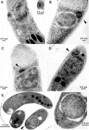
Fig. 1. Ultrastructural morphology of the Cryptosporidium parvum and C. hominis mitochondrion organelle. Transverse (A and B) and longitudinal sections (C and D) show free sporozoites obtained from a C. hominis isolate, while a transverse section from the C. parvum Iowa isolate shows an oocyst with unexcysted sporozoites (E). The sections show a nuclear associated round structure (A–E), surrounded by a double membrane (inset in A), and characterized by electron-dense internal structures (A, C and D).
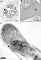
Fig. 2. Immunoelectron microscopy (IMEM) by hsp60 labelling of Cryptosporidium parvum sporozoites. The labelling was achieved by exploiting the heterologous reactivity of an anti-hsp60 polyclonal antibody raised against the Entamoeba histolytica chaperonin 60 (Tovar et al. 1999). C. parvum oocyst and free sporozoite cross-sections (A and C respectively) show a consistent compartmentalized labelling. In the free sporozoite the labelling is concentrated in an electron-dense region just beside the nuclear region in the same area in which the ultrastructure images indicate the double-membrane organelle presence (C). A section of an E. histolytica trophozoite, showing the labelling in a electron-dense structure within the cell, demonstrate the labelling specificity (B).
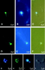
Fig. 3. Immunofluorescence (IMF) by hsp60 labelling and combined DAPI and SYTOX® staining. Using as hsp60 probes, the heterologous Entamoeba histolytica and Toxoplasma gondii polyclonal antibodies (Tovar et al. 1999; Toursel et al. 2000) the labelling indicates a structure close, to the nucleus (A and D) as confirmed by the simultaneous DAPI staining under normal (B and E) and interference light emission (C and F). DAPI staining shows multiple dots for unexcysted sporozoites (G). Multiple dots with DAPI labelling are shown in a free sporozoite (I) with the corresponding interference images (L). The presence of multiple dots in unexcysted sporozoites is also confirmed by using SYTOX® Green staining (H).
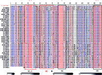
Fig. 4. DbClustal multiple alignment for the Cryptosporidium parvum alternative oxidase (AOX) partial coding sequence cloned from a sporozoite cDNA library and aligned, as translated polypeptide, against other AOX data-base entries. The alignment shows some typical features of the polypeptide: the presence of 3 highly conserved regions of the molecule (H1, H2, H3, boxed blocks), consistent with all AOX retrieved AOX entries, and containing the putative iron-binding motifs EXXH and E (in H1, H3 and H2 domains, marked with red asterisks at the top, Andersson & Nordlund, 1999). Membrane-spanning and surface-exposed regions are designated as M2 and S respectively (red boxed blocks). The symbol [utrif ] indicates the highly conserved residue Y in the transmembrane region M2, proposed to be a component of the potential quinone-binding site (Sluse & Jarmuszkiewicz, 1998). The cylinders under the sequences represent the four-helix bundle regions proposed for the AOX model (Andersson & Nordlund, 1999). Red and blue characters indicate, respectively, residue conservation over 90% and 50%.
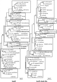
Fig. 5. Phylogenetic analysis of the Cryptosporidium parvum mitochondrial chaperonins 60 (hsp60) and 70 (dnaK, bacterial-like). The clades inferred by the γ-proteobacteria rooted ML tree topology are indicated by boxes and bold numbers indicate the highest significant FITCH bootstrap values for the corresponding supported clades. The scale bars indicate the expected number of substitutions per site.
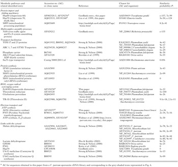
Table 1. Summary of putative mitochondrial pathways and hypothetical links with the cytosol for the mitochondrion-like organelle of Cryptosporidium parvum
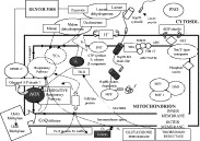
Fig. 6. Conceptual scheme of possible functional links in the mitochondrion-like organelle of Cryptosporidium parvum. Pathway 1. Protein import and folding. Pyridine nucleotide transhydrogenase (PNT) proton pump (also involved in pathway 6), chaperonin hsp70-MI (dnaK), chaperonin hsp60 linked to the cytosolic hsp70 protein activity and mitochondrial processing peptidase (MPP). Pathway 2. Multicomplex assembly. NifS, iron traffic protein and responsible for Fe-S protein assembly. Pathway 3. Transport facilitation. TIM (inner transport system) complex component: TIM 17, 23 proteins, ABC 1, 7, ATM1 transporters, phosphate carrier, ADP shuttle system (AK2) and SecY-type transporter. Pathway 4. Protein synthesis. Translation initiation factor 2 (IFM1), mitochondrial protein phenylalanine-tRNA synthetase (MSF1), mitochondrial protein valyl-tRNA synthetase (MSV). Pathway 5. ROS scavenging system. Superoxide dismutase (FeSOD), glutathione peroxidase, thioredoxin reductase and thioredoxin II (TR-II). Pathway 6. Electron transport and ATP synthesis. Alternative respiratory oxidase (AOX), thioredoxin II, PNT and ATP synthase, F1 subunit β component. Pathway 7. Links with the cytosol. Malate dehydrogenase (MDH), lactate dehydrogenase (LDH), glyceraldehyde-3-P dehydrogenase (GPDHC), pyruvate ferredoxin oxidoreductase (PNO). Grey-shaded components are derived from theoretical translation of C. parvum sporozoite cDNA library amplicons in this study.
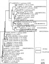
Fig. 7. Phylogenetic analysis of the Cryptosporidium parvum mitochondrial alternative oxidase (AOX) sequence. The clades inferred by the unrooted ML tree are shaded by boxes and identify main subtrees. Dotted and continous lines indicate, as represented, fungi and plantae, respectively. The numbers are clade FITCH bootstrap values. The scale bar indicates the expected number of substitutions per site. The sequences reported in the tree are the database entries included in the multiple alignment of Fig. 4.
 You have
Access
- 73
- Cited by