Patent ductus arteriosus constitutes approximately 5–10 % of all CHDs and warrants closure in all patients with significant left-to-right shunt. Non-surgical closure by device, plugs, or coils is a safe preferred alternative to surgery. Reference Sathanandam, Balduf and Chilakala1,Reference Sathanandam, Agrawal and Chilakala2 One of the most feared complications is the device embolisation. Embolisation into aorta or pulmonary artery could be due to inappropriate device size, abnormal duct anatomy, ductal spasm, device malposition, use of softer devices such as vascular plugs, tension on the cable within the delivery system and ricocheting of the device, and transient rise of systemic or pulmonary artery pressures or from patient movement during procedure. Reference Sen, Jain and Dalvi3,Reference Sathanandam, Gutfinger and Morray4 Retrieval could be surgical or percutaneous using snares and bioptomes. We retrospectively analysed the procedures complicated by device embolisation after non-surgical closure of patent ductus arteriosus along with short and intermediate term outcomes in these patients.
Methods
A retrospective analysis of hospital medical records of all patients who underwent non-surgical duct closure between 2013 and 2021 was approved by institutional review board. Primary coil closures were excluded. Individual patient consent for data collection was waived off as data were anonymised. The patient demographics, clinical profile, echocardiographic and cardiac catheterisation findings, interventional closure methods, and their intermediate term outcomes were analysed.
Echocardiography
Size and shape of the duct and aortic isthmus on maximally expanded systolic frame were assessed in echocardiogram. Colour jet width was not used for measurements due to its dependence on gain settings. Krichenko classification was used for duct morphology. Reference Benson, Burrows, Ml, McLaughlin and Freedom5 The choice of device design and size was entirely based on duct morphology and measurements on echocardiography and not based on angiography. Reference Anil, Sivakumar and Kumar6
Interventional procedure
Informed written consent was obtained for transcatheter closure performed under conscious sedation, routine heparinisation, and intravenous antibiotic prophylaxis. Procedure was done with isolated right femoral venous access followed by antegrade crossing of the duct. Arterial access was often avoided unless patients with severe pulmonary arterial hypertension need a detailed haemodynamic assessment. After pressure measurements, the duct was crossed from pulmonary artery and exchanged to a long venous sheath. Aortogram was done in left lateral view through the side arm of the venous sheath. If duct overlapped with descending thoracic aorta, aortogram was repeated in right anterior oblique projection. Echocardiographic measurements of the narrowest part of the duct were taken as the ductal diameter and were primarily considered for device selection. Aortogram was only used to provide anatomical landmarks of the aortic and pulmonary ends of the duct in relation to the trachea.
Device deployment
In most of the ductal morphologies, conventional duct occluder was chosen 2 mm larger than the duct diameter. In tubular Type C ducts, Amplatzer vascular plug II or IV (Abbott Medical, Plymouth, MN, United States of America) was chosen at least 50% larger than the duct. In very small patients, devices or plugs were sometimes undersized less than the above-mentioned dimensions to avoid aortic or pulmonary artery protrusion. Device stability and lack of its aortic protrusion were confirmed on observing the levophase of a pulmonary angiogram performed through the venous sheath in lateral view before release from the cable. Another angiogram in shallow left anterior oblique projection was used to exclude left pulmonary artery obstruction. Unstable and protruding devices were withdrawn and procedure was reattempted. After device release, patients were monitored in the catheterisation laboratory for device stability and complications using echocardiography.
Definitions
Embolisation was defined as migration of the device out of the duct after release. Malposition was defined as an abnormal unsatisfactory device position within the duct either at risk of embolisation or resulting in aortic or pulmonary artery obstruction. Undersized device was defined as a duct occluder less than 2 mm larger than the narrowest duct diameter and a plug less than 50% larger than the duct diameter. For this retrospective review, two cardiologists performed an independent evaluation of the angiograms and clinical data to determine the possible reason or mechanism of embolisation or malposition.
Percutaneous retrieval
Retrieval of embolised or malpositioned occluder was attempted after ensuring surgical back up and activated clotting time values were maintained over 200 seconds. Snaring of the pin/screw was always initially attempted and when they failed, the device belly was snared. Large braided sheath was advanced into the pulmonary artery and used for retrieval to avoid dragging the device through pulmonary and tricuspid valve whenever feasible. If resheathing was not feasible within the cardiac chambers, it was reattempted again in inferior vena cava. If the device alignment was favourable, the delivery cable was rescrewed into the pin/screw end of the device to facilitate its retrieval. Unsuccessful retrieval warranted surgical referral.
Post-procedural assessment
Procedural success was defined as successful complete duct occlusion without any complication. Residual shunt was defined as incomplete closure with residual flows across the duct that may occur around the device. Monitoring included assessment of vascular access site, residual flows, stability of device, obstruction of pulmonary artery or aorta at three-monthly intervals till one year and yearly thereafter.
Statistical analysis
Analysis used SPSS software version 20.0 (IBM Corp., Armonk, NY, United States of America). Categorical variables were expressed as percentages and Chi square test was used for the categorical variables between the different classes. Continuous variables in normal distribution were presented as mean ± standard deviation and compared by student t test. If the continuous variables were in non-normal distribution, they were expressed in median and range and compared using Mann Whitney test. Multi-variate regression analysis was performed to explore statistical significance between multiple classes. All hypothesis tests of significance were two-tailed and significance was defined as p < 0.05.
Results
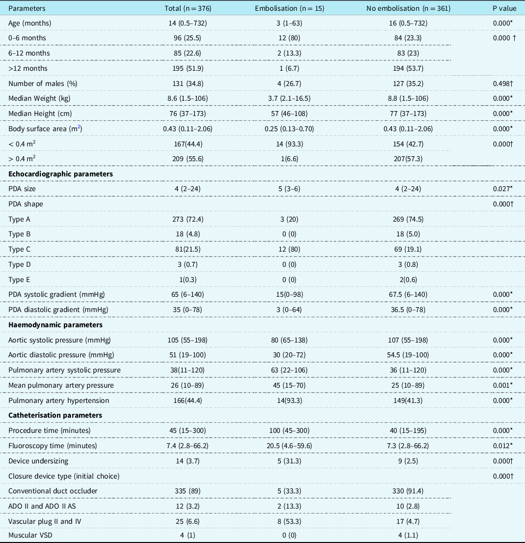
We analysed case records of 376 consecutive cases of transcatheter device closure of ducts in 8 years since 2013, at our institution. The median age of intervention was 14 months (CI: 0.5–732 months). The male to female ratio was 131:245. The mean duration of follow-up was 4.75 ± 2.18 years. Fifteen patients (4%) among 376 procedures had device embolisation or malposition. This study analysed the procedural details of these 15 patients (Table 1). There were no deaths related to the embolisation.
Demographic and clinical profile.

* Mann Whitney U test, data expressed in median and range;
† Chi square test, data expressed in frequency and percentage
Patient characteristics
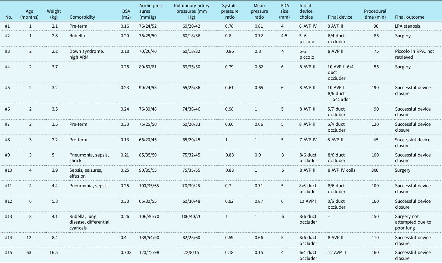
The median age of these 15 patients was 3 months and all except one patient were infants under 1 year of age (Table 2). The median weight was 3.7 (2.1–16.5) kg; all except one patient weighed under 9 kg. The median height being 57 (46–108) cm. Comorbidities in nine patients (60%) included pre-maturity in three patients, rubella syndrome in two patients, Down syndrome with anorectal malformation in one patient, and pneumonia with respiratory failure and sepsis in three patients. The ratio of pulmonary artery to aortic systolic pressure was 0.77 ± 0.21 and more than 0.6 in 13 out of 15 patients. The ratio of pulmonary artery to aortic mean pressure was 0.78 ± 0.22.
Individual patient details.

Specific procedural details
The mean duct size measured on echocardiography was 5 ± 0.9 mm. Eighty percent had a tubular Type C duct and the rest were conical Type A ducts. The embolised devices included Amplatzer vascular plug II in six (40%), conventional duct occluder in six (40%), Amplatzer vascular plug IV in two and Piccolo device in one patient. The procedural time including the additional time for device retrieval was 122 ± 64 minutes.
Embolisation of Amplatzer vascular plug II
There were six embolisations of Amplatzer vascular plug II plugs in patients weighing between 3.2 and 5.8 kg. All patients had a Type C tubular duct. The size of the plug was undersized in three patients, where the plug was less than 50% larger than the duct diameter. In four patients, the embolised plugs could be successfully snared out and the duct was closed with a conventional duct occluder device that was 2 mm larger than the duct. Amplatzer vascular plug II was initially chosen instead of the conical duct occluder with an intention to avoid protrusion of the aortic retention skirt. The remaining two patients had procedural failure and underwent uneventful surgery. The first of the two was a 2-month-old infant weighing 3.7 kg with 6 mm duct, where attempts with 8 mm Amplatzer vascular plug II resulted in embolisation and subsequent attempts with 10 mm Amplatzer vascular plug II as well as 6–4 conventional duct occluder led to aortic protrusion. In the second patient aged 4 month weighing 3.9 kg with 5 mm duct, after retrieval of 8 mm Amplatzer vascular plug II, trials with 8 mm Amplatzer vascular plug IV plug and bioptome-assisted multiple coils failed.
Embolisation of conventional duct occluder
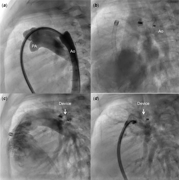
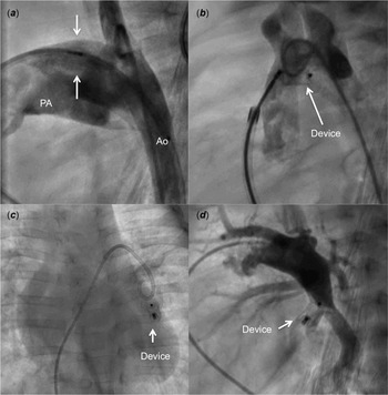
Six patients had embolisation of conical conventional duct occluder after release. The devices were at least 2 mm larger than the duct diameter. In three of the patients, the device embolised to descending aorta. Two of the devices were snared at the screw end and pulled back into the conical duct, and the same device closed the duct successfully (Fig 1). Aortic protrusion after repositioning in the duct warranted a device retrieval in the third patient and replacement with Amplatzer vascular plug II plug. Embolisation occurred into pulmonary artery in three patients, one of which was successfully snared out and replaced with a larger vascular plug. The embolised device could not be retrieved in two patients. One 8-month-old infant, weighing 4.1 kg with rubella syndrome, 6 mm duct, chronic lung disease, severe pulmonary arterial hypertension, and differential cyanosis, had embolisation of 8–6 conventional duct occluder to left pulmonary artery that could not be retrieved in catheterisation laboratory (Fig 2). Despite the fact that this infant had systemic pulmonary artery pressures before the procedure, duct closure was attempted in catheterisation laboratory after detailed parental discussions with plans to continue home oxygen and pulmonary vasodilators after the closure. After device embolisation, heart team decision was to observe the child conservatively without surgery, considering risks involved in cardiopulmonary bypass owing to high pulmonary vascular resistance. Device closure in the second infant aged 1 month weighing 2.8 kg with rubella syndrome and 4.5 mm duct was attempted with 5–6 Piccolo device initially leading to significant residual flows. After duct closure with 6–4 conventional duct occluder, a catheter was advanced into the pulmonary artery to measure the pressures. This led to embolisation of a precariously placed occluder into the mediastinal left pulmonary artery. Considering the small patient size, transcatheter retrieval was not attempted and surgery was successfully performed.
Angiogram from the side arm of a transvenous long Mullins sheath ( a ) placed in the aorta through the duct delineates the anatomy and its relations to tracheal radiolucency. Stable position of a conventional duct occluder after deployment was confirmed on observing the levophase of a sheath side arm angiogram ( b ). After few minutes, device embolises into the aorta ( c ). The screw end was snared and pulled back into the duct ( d ).

After delineating a large tubular duct (arrows) in a patient with rubella and hypertensive duct ( a ), a duct occluder was deployed within the ampulla to avoid aortic protrusion ( b ). Device embolises into the left pulmonary artery ( c ) with screw end facing away from the catheter proving retrieval very difficult ( d ).

Embolisation of Piccolo device
One infant had embolisation of Piccolo device. A 2-month-old infant weighing 2.2 kg and Down syndrome with colostomy for anorectal malformation had a 4 mm tubular duct, near systemic pulmonary artery pressures and large pericardial effusion. After closure with 5–4 Piccolo device, echocardiogram in the post-intervention recovery room showed device embolisation. The device was deeply lodged into a smaller right lower lobe branch and failed attempts at retrieval. The duct was closed with 8 mm Amplatzer vascular plug II plug, pericardial effusion drained, and the infant was conservatively observed for the right lower lobe Piccolo device embolisation. Recovery was uneventful and follow-up evaluation after 1 year showed adequate somatic growth, normalisation of pulmonary artery pressures, and successful duct closure.
Embolisation of Amplatzer vascular plug IV plug
Two infants had embolisation of Amplatzer vascular plug IV plug, which was undersized in one patient. A 4 mm tubular duct with near systemic pulmonary pressures in a pre-term infant aged 1 month weighing 2.1 kg was closed with 6 mm Amplatzer vascular plug IV plug. When it embolised to right pulmonary artery after few minutes, it was retrieved and replaced with 8 mm Amplatzer vascular plug II plug. Despite complete duct closure and normal somatic growth, follow-up echocardiogram and angiogram at 3 months showed complete left pulmonary artery occlusion that could not be recanalised as the pulmonary end of the plug covered its ostium. As the pulmonary artery pressures were normal and distal hilar left pulmonary artery was very hypoplastic on left pulmonary vein wedge angiography, a conservative approach was decided by the heart team. The compromise of the lumen of the left pulmonary artery was not evident immediately after the procedure. Another 3-month-old pre-term infant weighing 2.2 kg had embolisation of 7 mm Amplatzer vascular plug IV that was successfully retrieved and replaced by a 8 mm Amplatzer vascular plug II plug.
Successful device closure group
Successful closure with the initial chosen device based on echocardiography was noted in 361 (96.5%) patients with a median age of 16 months (range 0.5–732 months) and a median weight of 8.8 kg (range 1.5–106 kg). Type A conical duct was the most common in 75% of patients, followed by Type C tubular duct in 19% and Type B window like duct in 5%. The mean size of duct in this group of patients with successful closure was 4.6 ± 1.9 mm. Conical duct occluder devices were the most commonly used in this group in 92% of patients, followed by vascular plugs in 5% and Amplatzer duct occluder II in 3% of patients.
Comparison between groups
The cases with device embolisation had significantly lower age, body weight and height compared to successful cases (Table 1). There was significantly higher rate of embolisation in cases < 6 months of age versus 6–12 months versus > 12 months, when compared to group where it was done successfully. Similarly, embolisation was significantly higher in babies with smaller body surface area of < 0.4 m2 as compared to > 0.4 m2on comparing the two groups. The mean duct size on echocardiography was larger in patients who experienced embolisation compared to others. The Doppler gradients across the duct were significantly less in patients who had embolisation compared to the others. Embolisation was seen significantly more commonly in Type C tubular ducts and was also seen significantly more frequently with vascular plugs as compared to other devices. The embolised group had significantly higher mean pulmonary artery pressures. Patients with severe pulmonary arterial hypertension had a significantly higher rate of embolisation than others. Logistic regression analysis also showed device embolisation was significantly higher in age group of < 6 months compared to 6–12months (p = 0.02), higher in with those with tubular ducts versus conical ducts (p = 0.003), and in duct closure with undersized devices (p = 0.001)(Supplemental Table 1).
Comparison between embolisation to aorta and pulmonary artery
In the 15 cases of device embolisation, 12 devices embolised to pulmonary artery and the remaining 3 embolised to aorta (Supplemental Table 2). There was no significant difference between the two groups, on analysing factors such as age, body surface area, total procedural/ fluoroscopic time, duct size, or morphology. The peak and mean pulmonary artery pressure was higher in cases of device embolisation to aorta, but it too did not reach level of statistical significance. Interestingly, only single disc devices like conical duct occluder embolised to aorta, while double disc devices and vascular plugs embolised to pulmonary arteries. All devices could be successfully retrieved in cases of aortic embolisation, while two devices could not be retrieved from pulmonary vascular bed.
Discussion
This study is one of the largest single centre study aimed at identifying risk factors that lead to device embolisation after transcatheter duct closure. Four percent of procedures were complicated by device embolisation, which was similar to the world literature. Reference Kang, Jivanji and Mehta7–Reference El-said, Bratincsak and Foerster10 A retrospective analysis of 408 consecutive procedures done over 11 years from 13 institutions was the previous major report of the complications following transcatheter duct closure, where the embolisation rates were similar. Reference Backes, Rivera and Bridge8 Device embolisation often occurred immediately, but late diagnosis as late as 6 weeks was also rarely reported in literature. Reference Kang, Jivanji and Mehta7,Reference Backes, Rivera and Bridge8
Most embolisations occurred in infants with small body size in our study. A meta-analysis of 38 studies involving transcatheter duct closure in infants reported 5% incidence of device embolisations, with statistically significant increase when the infants were < 6 kg in weight. Reference Backes, Rivera and Bridge8 A three-fold increase in adverse events and five-fold increase in serious adverse events were observed in patients < 6 kg in another multi-centre registry. Reference Tan, Levi and Moore9,Reference El-said, Bratincsak and Foerster10 Use of Amplatzer duct occluder is off-label in infants < 6 kg, though multiple authors reported its safe and effective use. Reference Sivakumar, Francis and Krishnan11
Embolisation occurred more often to pulmonary artery rather than aorta, similar to our experience. While reporting the procedural serious adverse events, embolisation with successful transcatheter retrieval was classified as a moderate event and that needing surgical retrieval was classified under major event. Reference Kang, Jivanji and Mehta7 Eighty percent of embolised duct occluders had been percutaneously retrieved in published literature, while the others needed surgery. Reference Tan, Levi and Moore9 Three patients in our group among the 15 embolisations needed surgical retrieval.
Embolisation to aorta increases afterload to left ventricle and increases left-to-right ductal shunt, along with risks of bowel gangrene and kidney injury. Embolisation to pulmonary artery reduces cardiac output. Reference Faella and Hijazi12 Percutaneous retrieval of devices carries risk of vascular trauma, tricuspid valve injury, or compartment syndrome. Reference Jang, Son, Lee, Lee and Kim13 There was no mortality in our group of patients. Two devices could not be retrieved as they lodged deep within the small pulmonary lobar branches. A collective heart team decision was made to conservatively observe due to their significant comorbidities and high pulmonary vascular resistance.
Our study observed statistically higher rate of embolisation with the new generation of softer devices such as vascular plugs and Piccolo device, similar to others experience. Reference Kang, Jivanji and Mehta7–Reference Tan, Levi and Moore9 This could be attributed to difficulty in correctly assessing the dimensions of complex ductal anatomy or a learning curve with the recently introduced softer devices and vascular plugs. Device embolisation was significantly higher in those where the device was undersized, which was a known risk factor. Reference El-said, Bratincsak and Foerster10 While four among the eight vascular plugs were undersized in patients who had embolisation, all the 17 plugs used in the other patients who did not have embolisation were selected more than 50% larger than the duct diameter. Unlike older patients, device was not always oversized 2 mm larger than the ductal dimensions in infants weighing under 6 kg to avoid device protrusion into the aorta or pulmonary artery. Risk of device-related obstruction of left pulmonary artery or descending aorta was higher in small infants, though improvement might occur over time with vascular growth. Reference Kang, Jivanji and Mehta7,Reference Tomasulo, Gillespie and Munson14 Softer vascular plugs and Piccolo devices were often chosen to facilitate intraductal deployment, thereby circumventing vessel obstruction. Left pulmonary artery obstruction might be underestimated on echocardiography due to preferential redistribution to the right lung. Reference Kharouf, Heitschmidt and Hijazi15 One among the 15 patients in this group had a successful larger Amplatzer vascular plug II deployment after retrieval of a 6 mm Amplatzer vascular plug IV plug. Left pulmonary artery occlusion observed on late follow-up could not be rectified and it additionally led to marked hypoplasia of the hilar branches.
The reasons behind device embolisation in young infants include inadequate echocardiographic images, device undersized intentionally to avoid protrusion into adjacent vessels, ductal spasm, tension on delivery system, operator related factors such as forward push of the cable, delay in time to release or inadvertent unscrewing, vigorous patient activity, or a complex duct morphology. Reference Sathanandam, Gutfinger and Morray4,Reference Kang, Jivanji and Mehta7 Four patients with pneumonia, lung disease, and significant lung hyperinflation had suboptimal echocardiographic images that could have contributed for the event. Intentional device undersizing was noted in 8 patients where either a chosen duct occluder was similar in size to the duct dimension or the selected vascular plug was less than 150% of the duct diameter. Operator-related factors could not be deciphered in this retrospective analysis. However, there was one instance, where a catheter advanced into the pulmonary artery to record pressures inadvertently dislodged a device. Patient-related factor such as physical movements could be an additional factor as 13 out of the 15 procedures were done on conscious monitored sedation under ketamine.
Limitations
The analysis carries the limitations of a retrospective, single centre study. Reliance on echocardiography for ductal measurements and avoiding conventional aortogram through arterial access could have played a role in inadequate assessment of duct dimensions. Reference Anil, Sivakumar and Kumar6 Newer generation softer devices such as vascular plugs and Piccolo devices could be associated with a learning curve and could have contributed to embolisation. Other limitations include unequal representation of different devices and skewed distribution of ductal morphology.
Conclusions
Even though transcatheter duct closure is preferred instead of surgery in most patients, device embolisation is a complication in 4% of procedures. While 80% of embolisations are amenable for transcatheter retrieval, few may warrant surgical retrieval and duct closure. Larger ducts with high pulmonary artery pressures in younger and smaller infants are more often associated with device embolisation. Tubular ducts are more prone for embolisation compared to usual conical ducts. Softer occluders such as vascular plugs and Piccolo devices are often associated with device embolisations. Earnest efforts to reduce embolisations should include proper echocardiographic sizing, tailoring the appropriate occluder depending on the duct size and morphology, proper technique, minimising patient movements, and additional care in patients with high pulmonary artery pressures.
Supplementary material
For supplementary material accompanying this paper visit https://doi.org/10.1017/S1047951122003973
Acknowledgements
None.
Financial support
This research received no specific grant from any funding agency, commercial, or not-for-profit sectors.
Conflicts of interest
None.
Ethical standards
The authors assert that all procedures contributing to this work comply with the ethical standards of Indian Council of Medical Research and with the Helsinki declaration of 1975, as revised in 2008 and have been approved by the Institutional committee of Madras Medical Mission, Chennai, India.