There are few data concerning tricuspid valve replacement in infancy. We report on a novel technique of valve replacement using an autologous pericardial patch.
Case report
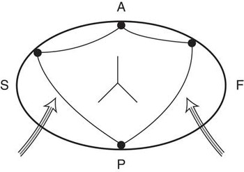
A 10-month-old infant, weighing 7.4 kilograms, was admitted with persistent fever of several days’ duration. Routine investigations included echocardiography, which demonstrated multiple vegetations on the tricuspid valve. Blood cultures were positive for staphylococcus aureus, the possible source being infected eczematous skin lesions. At surgical inspection, large vegetations were shown to be attached both to the septal and anterior leaflets of the valve and extending into the perimembranous septum. All three leaflets of the tricuspid valve were extremely friable, and could not be preserved. Following valve excision and removal of all macroscopic vegetations, a 17-millimetre St. Jude mechanical aortic valve prosthesis was implanted in an inverted manner in a supra-annular position within the right atrium (maximum native annular diameter of 13 millimetres). This, however, resulted in compression of the right coronary artery by the sewing ring, and was removed. Next, a freestyle valve (14 millimetres in diameter) was sewed to the valve annulus, but resulted in inflow obstruction as the valve leaflets would not open satisfactorily in diastole. Finally, a triangular piece of autologous pericardium was harvested. The base of the triangle was sutured onto the anterior part of the annulus, and the apex to the remnants of the septal leaflet, leaving two small openings (see schematic; Fig 1). In addition, an inverted Y-shaped incision was made centrally in the patch, to create a central valve orifice. Intraoperative inspection demonstrated that the valve appeared to function adequately, without significant stenosis or regurgitation. The patient was weaned off bypass with a mean central venous pressure of 15 millimetres of mercury, in sinus rhythm. The post-operative course was uneventful, apart from a transient second degree atrioventricular block in the immediate post-operative period, which resolved spontaneously within 7 days. All cardiac medications could be weaned off during follow-up. His physical and neurological development is normal to date, with no recurrence of endocarditis.
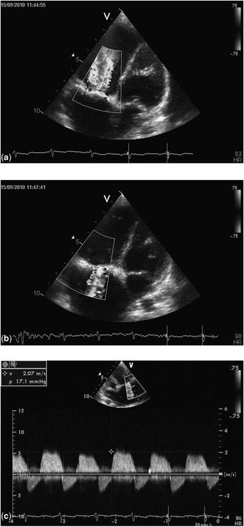
Over 3 years of follow-up (current weight 17 kilograms), the patient has remained without symptoms. He has borderline elevation of hepatic enzyme levels and mild hepatomegaly. Serial echocardiographic assessment reveals a mean gradient across the tricuspid valve of 8–10 millimetres of mercury (peak gradient 17 millimetres of mercury). There appears to be a functional valve (Figs 1 and 2) with moderate central insufficiency. The tricuspid valve annulus diameter measures between 20 and 22 millimetres in diameter, and the patient is currently under consideration for elective valve replacement using a mechanical prosthesis.
Schematic of the reconstructed tricuspid valve. The suture points are indicated anteriorly and posteriorly, as in the central Y-shaped incision. The arrows point to the gaps between the suture points, which allow tricuspid inflow. The septal (S), anterior (A), and posterior (P) aspects of the tricuspid valve annulus are as seen from the surgeon's viewpoint.

Haemodynamic profiles: (a) colour flow mapping demonstrating diastolic inflow, (b) systolic tricuspid valve regurgitation, (c) continuous wave Doppler flow profile across the tricuspid valve, showing a peak inflow gradient of between 17 millimetres of mercury (mean gradient 8 to 10 millimetres of mercury), and normal (physiological) right ventricular systolic pressure.

Comment
There are few reports of successful tricuspid valve replacement in infancy, with protracted follow-up. Pasque et al reported on a series of 11 children undergoing valve replacement, and all their youngest patients died.Reference Pasque, Williams, Coles, Trusler and Freedom1 The majority of their patients had structural malformations affecting the valve. Alternative approaches to the management of tricuspid valve endocarditis include valve repair, which may be possible if a sufficient amount of valvar tissue is spared from destruction and the infectious process no longer active, or complete valve excision.Reference Luciani, Casali, Viscardi, Marcora, Prioli and Mazzucco2, Reference du Plessis, Helbing and Bogers3 Valve excision in infancy is however poorly tolerated, and to date there are reports of one survivor.Reference du Plessis, Helbing and Bogers3 Given these limited data and generally bad outcome, it appears reasonable to take steps to create a functioning valve, even if only for a limited period, until the annular diameter has increased sufficiently to allow safe insertion of a standard sized prosthetic valve. Despite the supra-annular positioning of a valve larger than the native annular diameter being technically feasible, it can on occasion still produce coronary artery compression, as was seen in our patient. The solution adopted in our patient facilitated easy weaning off cardiopulmonary bypass with acceptable central venous pressures. The symptom-free follow-up also attests to the efficacy of the procedure. The patient's current tricuspid annular diameter allows safe implantation of a wide variety of commercially available prosthetic valves.