

Introduction
The Uravan Mineral Belt of the Colorado Plateau, which spans the Colorado–Utah border, has been a rich source of uranium and vanadium ores. The mines in this belt have also yielded many new secondary U and V minerals, the first of which was carnotite, K2(UO2)2(VO4)2·3H2O, described from the Rajah mine in the northern portion of the belt by Friedel and Cumenge (Reference Friedel and Cumenge1899). Although mining in what is now called the Slick Rock district in the southern portion of the belt dates to around 1900, apparently the Burro mine in that district was not active until the mid-1950s. The first new mineral to be described from the Burro mine was metamunirite, NaVO3 (Evans, Reference Evans1991), and recently burroite, Ca2(NH4)2(V10O28)·15H2O, was described (Kampf et al., Reference Kampf, Nash, Marty and Hughes2017). Herein, we describe ammoniomathesiusite, the third new mineral from the Burro mine.
Ammoniomathesiusite is named as the ammonium analogue of mathesiusite, K5(UO2)4(SO4)4(VO5)·4H2O (Plášil et al., Reference Plášil, Veselovský, Škoda, Novák, Sejkora, Čejka, Škácha and Kasatkin2014), with NH+4 in place of K+. The new mineral and name were approved by the Commission on New Minerals, Nomenclature and Classification of the International Mineralogical Association (IMA2017-077). The holotype and three cotypes are deposited in the collections of the Natural History Museum of Los Angeles County, 900 Exposition Boulevard, Los Angeles, CA 90007, USA, catalogue numbers 67248 (holotype), 67249, 67250 and 69251.
Occurrence
Ammoniomathesiusite was collected underground at the Burro mine, Slick Rock district, San Miguel County, Colorado, USA (38°2'42''N, 108°53'23''W). The Burro mine is near the southern end of the Uravan Mineral Belt, in which uranium and vanadium minerals occur together in bedded or roll-front deposits in the sandstone of the Salt Wash member of the Jurassic Morrison Formation (Carter and Gualtieri, Reference Carter and Gualtieri1965; Shawe, Reference Shawe2011). The uranium and vanadium ore mineralisation was deposited where solutions rich in U and V encountered pockets of strongly reducing solutions that had developed around accumulations of carbonaceous plant material, still in evidence as carbonised plant remains and notable logs. Mining operations have exposed both unoxidised and oxidised U and V phases. Under ambient temperatures and generally oxidising near-surface conditions, water reacts with pyrite and chalcopyrite to form aqueous solutions with relatively low pH, which then react with the earlier-formed montroseite–corvusite assemblages, resulting in diverse suites of secondary minerals. The  ${\rm NH}_{\rm 4}^ + $ presumably derives from organic matter in the deposit.
Ammoniomathesiusite is rare and occurs on asphaltum/quartz matrix in association with ammoniozippeite (Kampf et al., Reference Kampf, Plášil, Olds, Nash and Marty2018), gypsum, jarosite and natrozippeite. Other secondary minerals verified by us to occur in the mine include: andersonite, ansermetite, barnesite, brochantite, burroite (Kampf et al., Reference Kampf, Nash, Marty and Hughes2017), calciodelrioite, calcite, chalcomenite, grantsite, gunterite, hewettite, huemulite, hughesite, hydrocerussite, kokinosite, lasalite, lindgrenite, magnesiopascoite, martyite, metamunirite, metarossite, metaschoepite, munirite, navajoite, orthoserpierite, pascoite, rossite, schindlerite, schröckingerite, serpierite, sherwoodite, strelkinite, tyuyamunite, uranopilite, volborthite, wernerbaurite, zippeite and numerous other potentially new minerals, currently under study.
Physical and optical properties
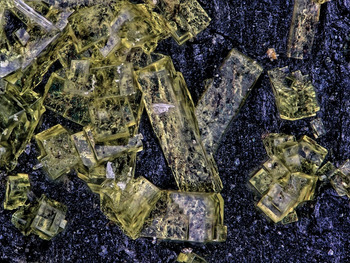
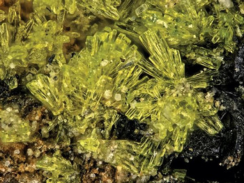
Ammoniomathesiusite crystals are {110} prisms, up to ~0.3 mm long, with square cross-sections and flat {001} terminations, sometimes modified by {111} pyramids (Fig. 1). Broad prisms are typically isolated or intergrown in random orientations (Fig. 2); narrow prisms, often tapering slightly towards their terminations, occur in sprays or bow-tie-like intergrowths (Fig. 3). No twinning was observed.
Crystal drawing of ammoniomathesiusite crystal; clinographic projection.

Ammoniomathesiusite prisms on asphaltum. The field of view is 0.84 mm across.

Sprays of ammoniomathesiusite prisms on asphaltum. The field of view is 0.84 mm across.

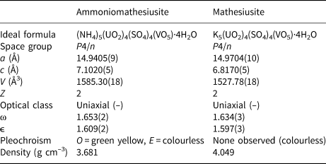
Crystals are yellow to greenish yellow and transparent with vitreous lustre. The streak is very pale yellow. The mineral fluoresces bright yellow–green under a 405 nm laser. The Mohs hardness is 2½, based upon scratch tests. Crystals are brittle with stepped fracture and two cleavages: excellent on {110} and good on {001}. At room temperature, the mineral decomposes in H2O. The density could not be measured because the mineral decomposes in Clerici solution. The calculated density based on the empirical formula is 3.672 g/cm3. Optically, ammoniomathesiusite is uniaxial (–), with ω = 1.653(2) and ε = 1.609(2), measured in white light. The mineral is distinctly pleochroic: O = green yellow, E colourless; O > E.
Raman spectroscopy
Raman spectroscopy was conducted on a Horiba XploRA PLUS spectrometer. Pronounced fluorescence was observed using a 532 nm diode laser; consequently, a 785 nm diode laser was utilised. The power density of the laser beam at the sample was 9.6 mW. The spectrum was recorded from 2000 to 100 cm−1, but was featureless between 2000 and 1200 cm−1. The spectrum from 1400 to 100 cm−1, is shown in Fig. 4.
Raman spectrum of ammoniomathesiusite recorded using a 785 nm diode laser.

The broad band of low intensity at ~1200 cm–1 is probably an overtone or combination band; a band at nearly the same frequency was observed in the spectrum of mathesiusite (Plášil et al., Reference Plášil, Veselovský, Škoda, Novák, Sejkora, Čejka, Škácha and Kasatkin2014), however, with an incorrect assignment. Very weak Raman bands at 1110 cm–1 and at 1090 cm–1 and a broader band at 1057 cm–1 with a shoulder at 1065 cm–1 are attributed to split triply degenerate ν3 antisymmetric stretching vibrations of the SO4 tetrahedron. A sharp, slightly asymmetric two-component band of medium intensity at 1010 cm–1 is assigned to the ν1 symmetric stretching vibration of the SO4 tetrahedron. A sharp band at 977 cm–1 is attributed to the symmetric ν1 (V–O) stretching mode (cf. Plášil et al., Reference Plášil, Veselovský, Škoda, Novák, Sejkora, Čejka, Škácha and Kasatkin2014; Frost et al., Reference Frost, Erickson, Weier and Carmody2005). A weak two-component overlapping band at 904 and 894 cm–1 is attributed to the ν3 antisymmetric stretching vibration of the uranyl ion, U ${\rm O}_{\rm 2}^{{\rm 2 +}} $. A very strong band at 834 cm–1 is assigned the ν1 symmetric U–O stretching vibration of U
 ${\rm O}_{\rm 2}^{{\rm 2 +}} $. The inferred U–O bond lengths (after Bartlett and Cooney, Reference Bartlett and Cooney1989) for the uranyl ion of ~1.78 Å (from ν1), 1.79 Å (from ν3; 894) and 1.78 Å (from ν3; 904) are in line with those derived from our X-ray structure study (see below). A very weak band at 764 cm–1 can be attributed to the libration mode of the H2O or to the stretching V–Oeq vibration. A broader band at 688 cm–1, along with the weak band at 650 cm–1, can be related to V–Oeq vibrations (cf. Plášil et al., Reference Plášil, Veselovský, Škoda, Novák, Sejkora, Čejka, Škácha and Kasatkin2014; Knyazev, Reference Knyazev2000; Chernorukov et al., Reference Chernorukov, Suleymanov, Knyazev, Vyshinski and Klimov2000). The two-component band with maxima at 625 and 615 cm–1 is attributed to the ν4 (δ) triply degenerated antisymmetric stretching vibrations of the SO4 tetrahedron. Broad bands at 590 and 550 cm–1 are probably related to the V–Oeq vibrations as well. Raman bands at 482 and at 459 cm–1 are related to the split ν2 (δ) doubly degenerate bending vibrations of the SO4 tetrahedron. A very weak band at 373 cm–1 is related to the νRotational of
 ${\rm NH}_{\rm 4}^{\rm +} $ (Heyns et al., Reference Heyns, Venter and Range1987). The split ν2 (δ) UO22+ doubly degenerate bending vibrations are represented by the broader component band at 244 cm–1. The rest of the bands are related to the unclassified lattice modes.
Chemical composition
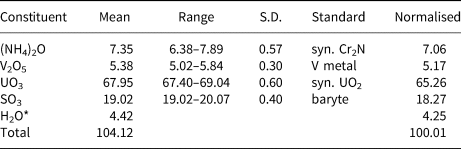
Chemical analyses (seven points on six crystals) were performed at the University of Utah, USA on a Cameca SX-50 electron microprobe with four wavelength dispersive spectrometers and using Probe for EPMA software. Analytical conditions were 15 kV accelerating voltage, 10 nA beam current and a beam diameter of 5 µm. Counting times were 30 s on peak and 30 s on background for each element. Raw X-ray intensities were corrected for matrix effects with a ϕρ(z) algorithm (Pouchou and Pichoir, Reference Pouchou, Pichoir, Heinrich and Newbury1991). Time-dependent intensity corrections were applied to N, U, V and S. Wave-scans across Mg, Al, Na and K peak positions showed these elements to be absent.
Because insufficient material is available for a direct determination of H2O, it is calculated based upon the structure determination. The crystals did not take a good polish and there was minor beam damage. The loss of weakly held H2O under vacuum and/or during analyses would account for the high electron microprobe analytical totals when calculated H2O is included. The somewhat lower analysed (NH4)2O content compared to that indicated by the structure refinement (4.75 vs. 4.92 N atoms per formula unit) could be due to a small amount of H3O+ in place of  ${\rm NH}_{\rm 4}^{\rm + }$ in the structure; however, the presence of H3O+ would imply highly acidic conditions. Considering that H3O+ has not been confirmed in any other phases in the Burro mine mineral assemblages, it seems more likely that the somewhat low analysed (NH4)2O content is due to loss under vacuum (along with the H2O noted above) prior to analyses. The results are given in Table 1.
Chemical composition (in wt.%) for ammoniomathesiusite.

* Based on structure with O = 33; S.D. – standard deviation.
The empirical formula (calculated on the basis of 33 O atoms per formula unit) is [(NH4)4.75(U1.00O2)4(S1.00O4)4(V1.00O5)·4(H2.07O) [Excess H is included for charge balance]. The ideal formula is (NH4)5(UO2)4(SO4)4(VO5)·4H2O, which requires (NH4)2O 7.41, V2O5 5.17, UO3 65.10, SO3 18.22 and H2O 4.10, total 100.00 wt.%. The Gladstone-Dale compatibility index 1 – (K P/K C) for the empirical formula is 0.009, in the superior range (Mandarino, Reference Mandarino2007), using k(UO3) = 0.118, as provided by Mandarino (Reference Mandarino1976).
X-ray crystallography and structure refinement
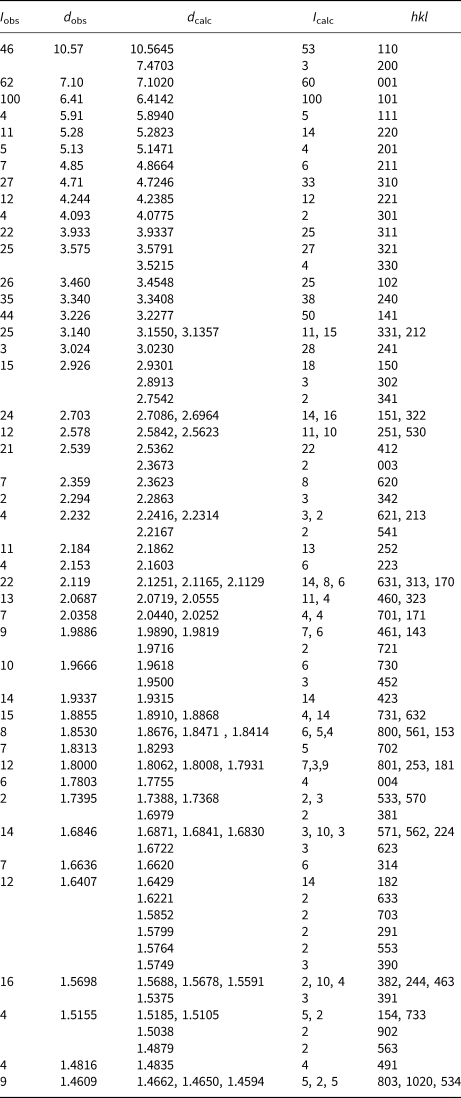
Powder X-ray studies were done using a Rigaku R-Axis Rapid II curved imaging plate microdiffractometer, with monochromatised MoKα radiation (λ = 0.71075 Å). A Gandolfi-like motion on the ϕ and ω axes was used to randomise the samples. Observed d values and intensities were derived by profile fitting using JADE 2010 software (Materials Data, Inc.). The powder data presented in Table 2 show good agreement with the pattern calculated from the structure determination. Unit-cell parameters refined from the powder data using JADE 2010 with whole pattern fitting are a = 14.9519(14), c = 7.1083(8) Å and V = 1589.1(3) Å3.
Powder X-ray data (d in Å) for ammoniomathesiusite. Only calculated lines with I ≥ 2 are included.

The single-crystal structure data were collected at room temperature using the same diffractometer and radiation noted above. The data were processed using the Rigaku CrystalClear software package and an empirical (multi-scan) absorption correction was applied using the ABSCOR program (Higashi, Reference Higashi2001) in the CrystalClear software suite. The structure was solved by direct methods using SIR2011 (Burla et al., Reference Burla, Caliandro, Camalli, Carrozzini, Cascarano, Giacovazzo, Mallamo, Mazzone, Polidori and Spagna2012). SHELXL-2016 (Sheldrick, Reference Sheldrick2015) was used for the refinement of the structure.
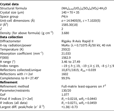
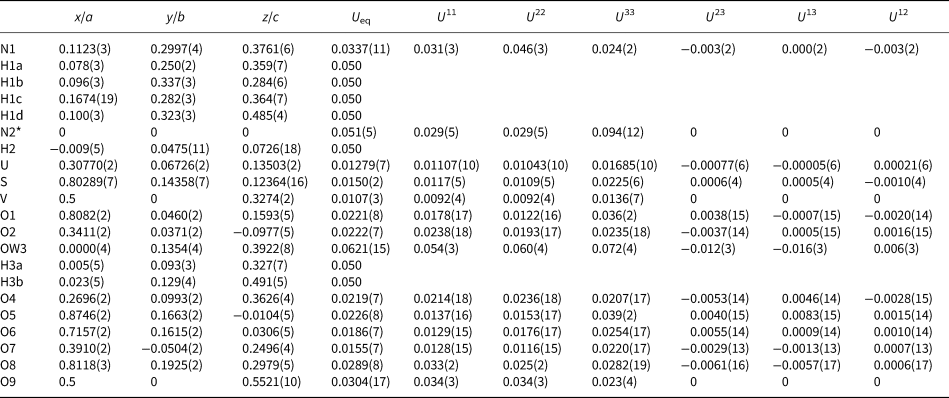
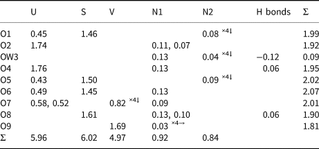
Difference-Fourier syntheses located all non-hydrogen atoms not located in the original structure solution, and subsequent cycles located all H sites. The structure was found to be identical to that of mathesiusite (Plášil et al., Reference Plášil, Veselovský, Škoda, Novák, Sejkora, Čejka, Škácha and Kasatkin2014) and all atom sites were transformed to correspond to that structure, with two NH4 sites in place of the two K sites in mathesiusite. The H atom sites associated with OW3 were refined with soft restraints of 0.82(3) Å on the O–H distances and 1.30(3) Å on the H–H distances, and those for the H sites associated with N1 and N2, with soft restraints of 0.90(3) Å on the O–H distances and 1.45(3) Å on the H–H distances. The U eq of each H was set to 0.05. All non-hydrogen sites refined to full occupancy, except the N2 site, which refined to an occupancy of 0.92(4). Data collection and refinement details are given in Table 3, atom coordinates and displacement parameters in Table 4, selected bond distances in Table 5 and a bond-valence analysis in Table 6. A comparison of the ideal formulas, cell parameters, optical properties and calculated densities (ideal) for ammoniomathesiusite and mathesiusite is provided in Table 7.
Data collection and structure refinement details for ammoniomathesiusite.

R int = Σ|F o2–F o2(mean)|/Σ[F o2]. GoF = S = {Σ[w(F o2–F c2)2]/(n–p)}1/2. R 1 = Σ||F o|–|F c||/Σ|F o|. wR 2 = {Σ[w(F o2–F c2)2]/Σ[w(F o2)2]}1/2; w = 1/[σ2(F o2)+(aP)2+bP] where a is 0.0147, b is 5.5409 and P is [2F c2+Max(F o2,0)]/3.
Atom coordinates and displacement parameters (Å2) for ammoniomathesiusite.

* N2 site refined occupancy = 0.92(4).
Selected bond distances (Å) for ammoniomathesiusite.

* Note that there are no obvious hydrogen bonds through H3a and there are two apparent hydrogen bonds through H3b.
Bond-valence analysis for ammoniomathesiusite. Values are expressed in valence units.*

*  ${\rm NH}_{\rm 4}^ + $–O bond valence parameters from Garcia-Rodriguez et al. (Reference García-Rodríguez, Rute-Pérez, Piñero and González-Silgo2000). U6+–O, V5+–O and S6+–O bond-valence parameters are from Gagné and Hawthorne (Reference Gagné and Hawthorne2015). Hydrogen-bond strengths associated with OW3 are based on O–O bond lengths from Ferraris and Ivaldi (Reference Ferraris and Ivaldi1988).
Comparison of ideal formulas, cell parameters, optical properties and calculated densities (ideal) for ammoniomathesiusite and mathesiusite (Plášil et al., Reference Plášil, Veselovský, Škoda, Novák, Sejkora, Čejka, Škácha and Kasatkin2014).

The crystallographic information files have been deposited with the Principal Editor of Mineralogical Magazine and are available as Supplementary material (see below).
Description and discussion of the structure
The U site in the structure of ammoniomathesiusite is surrounded by seven O atoms forming a squat UO7 pentagonal bipyramid. This is the most typical coordination for U6+, particularly in uranyl sulfates, where the two short apical bonds of the bipyramid constitute the UO2 uranyl group (Burns, Reference Burns2005). Sulfur is coordinated by four O atoms at the distances typical for tetrahedral coordination, ~1.47 Å. Vanadium is in square pyramidal coordination, bonded strongly to one O atom at the distance of 1.596 Å (vanadyl bond; cf. Schindler et al., Reference Schindler, Hawthorne and Baur2000) and four O atoms at the distances of 1.876 Å. This (4 + 1) coordination is one of the characteristic environments for the V5+ cation (Schindler et al., Reference Schindler, Hawthorne and Baur2000). The N1 atom (NH41 group) is [9]-coordinated with an average bond length ~3.10 Å, while the N2 atom (NH42 group) is [12]-coordinated with an average N–O bond length of ~3.24 Å (Table 5). Both NH4 groups are linked to the only H2O molecule (OW3) in the structure.
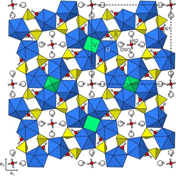
The structure (Figs 5 and 6) contains heteropolyhedral sheets based on [(UO2)4(SO4)4(VO5)]5– clusters. These clusters arise from linkages between corner-sharing quartets of uranyl pentagonal bipyramids, which define a square-shaped void at the centre of which is the base of the VO5 square pyramid. Adjacent corner-shared uranyl pentagonal bipyramids are also linked through SO4 tetrahedra, with which they share corners. Each SO4 shares a third vertex with a uranyl pentagonal bipyramid in another cluster to form the sheets. The  ${\rm NH}_{\rm 4}^{\rm +} $ cations are located between the sheets, together with the H2O group. The corrugated sheets are stacked perpendicular to c. These heteropolyhedral sheets are similar to those in the structures of synthetic uranyl chromates (Unruh et al., Reference Unruh, Quicksall, Pressprich, Stoffer, Qiu, Nuzhdin, Wub, Vyushkova and Burns2012) and molybdates (Obbade et al., Reference Obbade, Yagoubi, Dion, Saadi. and Abraham2003).
The structure of ammoniomathesiusite viewed down c. The unit-cell outline is shown by dashed lines.

The structure of ammoniomathesiusite viewed down a2. The unit-cell outline is shown by dashed lines.

Supplementary material
To view supplementary material for this article, please visit https://doi.org/10.1180/mgm.2018.112
Acknowledgements
Structures Editor Peter Leverett and an anonymous reviewer are thanked for their constructive comments on the manuscript. A portion of this study was funded by the John Jago Trelawney Endowment to the Mineral Sciences Department of the Natural History Museum of Los Angeles County. We would like to thank Don Coram, the owner of the Burro mine, for allowing us access to the property, and to Okie Howell and Jess Fulbright for providing logistical support during our visits.