Introduction
The World Health Organization Classification of Head and Neck Tumours (2017) recognises multiple phenotypes of laryngeal tumour.Reference El-Naggar, Chan, Grandis, Takata and Slootweg1 The vast majority of laryngeal cancer cases are conventional squamous cell carcinoma (SCC). Therefore, clinical guidelines for the management of laryngeal cancer are derived from studies relating to the management of SCC.
A significant minority of patients with laryngeal malignancy (10 per cent) present with primary tumours that harbour different phenotypes. Because of their relative rarity, less is known about the biological behaviour and radiosensitivity of these tumours.
The larynx is the most common site for neuroendocrine carcinomas in the head and neck. Neuroendocrine carcinomas encompass the largest group of non-squamous carcinomas originating in this site.Reference Ferlito, Silver, Bradford and Rinaldo2 Neuroendocrine carcinomas have been reported with increasing frequency in the larynx in recent decades. They are uncommon lesions of considerable scientific interest and clinical importance.
The putative cells of origin are non-neoplastic cells of the diffuse neuroendocrine system that are normal residents of the mucosal epithelium.Reference Chung, Lee, Shim, Kim, Nam and Kim3 They have been identified in the basal and middle layer of the respiratory epithelium of the larynx, particularly in the ventricle and subglottis. Other possible cells of origin are the pluripotent stem cells of the surface or glandular epithelium.
Neuroendocrine carcinomas of the larynx account for less than 1 per cent of laryngeal tumours.Reference Ferlito, Silver, Bradford and Rinaldo2 All types of epithelial-derived laryngeal neuroendocrine neoplasms, including carcinoid, atypical carcinoid, small cell neuroendocrine carcinoma and large cell neuroendocrine carcinoma, are considered malignant. These lesions share specific morphological, immunohistochemical, ultrastructural and molecular characteristics.Reference Ferlito, Lewis and Rinaldo4 The neuroendocrine nature of neuroendocrine carcinomas can be elucidated by various techniques, such as: histochemistry, showing argyrophilic cytoplasmic granules; immunohistochemistry, showing expression of a variety of neuroendocrine markers (e.g. synaptophysin, chromogranin, Leu-7, neuron-specific enolase, clusters of differentiation 56 and 57, and neurofilament protein); and ultrastructural examination, showing dense core granules.Reference Ferlito, Silver, Bradford and Rinaldo2
Hundreds of laryngeal neuroendocrine carcinomas, diagnosed using strict criteria, have been reported in the literature.Reference Ferlito, Lewis and Rinaldo4 An accurate classification of laryngeal epithelial neuroendocrine carcinoma is essential, because these tumours have widely varying prognoses, clinical management approaches and therapeutic responses.
While many classifications have been proposed, laryngeal neuroendocrine neoplasms can be divided into epithelial (carcinoid, atypical carcinoid, small cell neuroendocrine carcinoma and large cell neuroendocrine carcinoma) and neural (paraganglioma) types. Lewis et al. suggested including the terms low grade (well differentiated) for carcinoid tumours, intermediate grade (moderately differentiated) for atypical carcinoid tumours, and high grade (poorly differentiated) for small and large cell neuroendocrine carcinomas.Reference Lewis, Ferlito, Gnepp, Rinaldo, Devaney and Silver5
This review focused on epithelial-derived neuroendocrine neoplasia, of which the atypical carcinoid is the most frequent type.Reference Ferlito, Lewis and Rinaldo4 It aimed to critically analyse clinical presentation and management data pertaining to neuroendocrine carcinomas of the larynx.
Materials and methods
A PubMed search was performed using the term ‘neuroendocrine carcinoma’. English-language articles on neuroendocrine carcinoma of the larynx were reviewed in detail.
Overview of review findings
While many historical classifications have been proposed, in contemporary practice these tumours are sub-classified into four subtypes: carcinoid, atypical carcinoid, small cell neuroendocrine carcinoma and large cell neuroendocrine carcinoma. These tumour types are described in more detail below.
Discussion
Carcinoid tumour
Laryngeal carcinoid is an extremely rare tumour. It is a well-differentiated malignant lesion that possesses a well-demonstrated capacity for both regional and distant metastases.Reference Ferlito, Devaney, Hunt and Rinaldo6
Carcinoid tumours have a distinct male predominance, most commonly occurring in middle-age.Reference Devaney, Ferlito and Rinaldo7 These tumours have been reported both in smoking and non-smoking patients.Reference Ferlito and Rinaldo8
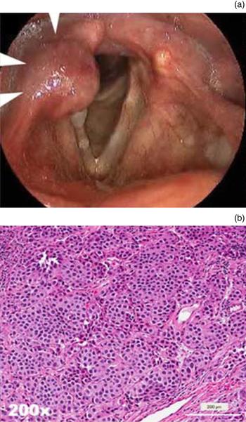
The site of origin is most commonly the supraglottic larynx, especially the arytenoid or the aryepiglottic fold (Figure 1).Reference Ferlito and Rinaldo8 The presenting symptoms are similar to other laryngeal tumours, namely hoarseness, dysphagia, sore throat or a feeling of a lump. Symptoms may be present from 4 weeks to 10 years.Reference Ferlito and Rinaldo8 The tumour rarely presents with nodal metastases, but may metastasise over the course of extended follow up.Reference Soga9 One case of associated carcinoid syndrome has been reported, after the tumour metastasised to the liver.Reference Ferlito, Rinaldo, Bishop, Hunt, Vander Poorten and Williams10
Carcinoid tumour. (a) A submucosal mass is seen involving the right aryepiglottic fold on stroboscopy (arrows). (b) The tumour cells are uniform, growing in nests without mitoses, necrosis or atypia (H&E; ×200).

Macroscopically, the tumour presents as a submucosal or polypoid mass. Histologically, typical carcinoid tumours consist of small- to medium-sized nests and trabeculae of small- to medium-sized cells, with moderate cytoplasm, and round, regular nuclei with speckled chromatin with a salt and pepper appearance. These cells are fairly regular, without substantial pleomorphism and without significant cytological atypia. The rate of mitotic activity is very low (< 2 per 10 high power fields). There is no necrosis and sparse to absent perineural and vascular invasion.
Carcinoid tumours typically express neuroendocrine markers on immunohistochemistry, including chromogranin and synaptophysin.Reference Dieler and Dämmrich11 The tumours are also positive for cytokeratin stains, and, similar to laryngeal atypical carcinoid tumours, carcinoid tumours are often immunoreactive for calcitonin.Reference Nozières, Chardon, Goichot, Borson-Chazot, Hervieu and Chikh12
As initial biopsies are often small and crushed, tumour grading of carcinoid versus atypical carcinoid tumours may require resection for definitive grading. Although atypical carcinoid tumours are more common, the quality of biopsy and histological features should be verified prior to assuming a well-differentiated carcinoid tumour.
Primary conservation surgery, particularly supraglottic laryngectomy, is the preferred treatment.Reference Ferlito, Silver, Bradford and Rinaldo2, Reference Ferlito and Rinaldo8 Transoral carbon dioxide (CO2) laser surgery can be a good alternative in selected cases because of the potential for improved functional results and lower morbidity.Reference Cevizci, Karakullukçu, van den Brekel and Balm13 Given the very low rate of regional metastasis, elective neck dissection is not indicated. Bilateral neck dissection (levels II and III) should be reserved for clinically or pathologically proven cervical metastases.Reference Ferlito, Lewis and Rinaldo4, Reference Ferlito, Devaney and Rinaldo14 Chemotherapy and radiotherapy have not been found to be of any benefit in treatment.Reference Ferlito, Barnes, Rinaldo, Gnepp and Milroy15
Carcinoid tumours are associated with the best prognosis of the laryngeal neuroendocrine carcinomas. Although many patients do not die of disease, carcinoid tumours are not indolent.Reference Ferlito, Silver, Bradford and Rinaldo2 Soga et al. observed that 33 per cent of patients with carcinoid of the larynx developed metastases.Reference Soga, Ferlito and Rinaldo16 The five-year survival rate reported in one large series was 49 per cent.Reference Soga, Ferlito and Rinaldo16 However, this low survival rate may be due to the erroneous inclusion of cases of atypical carcinoid tumour as typical carcinoid tumours.Reference Ferlito, Devaney and Rinaldo14
Atypical carcinoid tumour
Atypical carcinoid tumour is the most frequent laryngeal neuroendocrine carcinoma.Reference Ferlito, Barnes, Rinaldo, Gnepp and Milroy15 It is a moderately differentiated tumour, with aggressive biological behaviour, and frequent regional and distant metastases. In contrast to other disease sites, atypical carcinoid of the larynx is said to be associated with particularly high rates of metastatic disease.Reference Soga, Ferlito and Rinaldo16
It occurs most frequently in men. The mean age at diagnosis is 61 years (range, 33–83 years), with a peak incidence in the sixth and seventh decades of life. Most patients are heavy smokers.Reference Ferlito and Rinaldo8
The tumour usually involves the supraglottic larynx (over 90 per cent of reported tumours), with most tumours being situated in the aryepiglottic fold, arytenoid or laryngeal surface of the epiglottis. Occasionally, tumours occur in the glottis and subglottis.Reference Ferlito and Rinaldo8 Presenting symptoms include hoarseness, dysphagia, sore throat, haemoptysis and otalgia. On occasion, patients may be asymptomatic. Atypical carcinoid metastasises to lymph nodes in approximately 50 per cent of casesReference Woodruff and Senie17 and metastasises distantly in as many as 40–45 per cent.Reference Soga, Ferlito and Rinaldo16 One peculiar aspect of the natural history of these tumours is the predilection to give rise to disseminated skin or subcutaneous metastases.Reference Woodruff and Senie17 Carcinoid syndrome associated with this tumour has been reported,Reference Ferlito, Rinaldo, Bishop, Hunt, Vander Poorten and Williams10, Reference Ferlito and Rinaldo18 and elevated serum levels of calcitonin may be detected.Reference Chung, Lee, Shim, Kim, Nam and Kim3
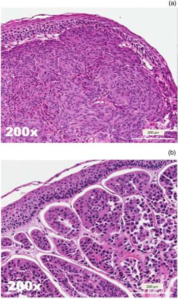
Tumours usually present as nodular, pedunculated or polypoid submucosal masses (Figure 2), with or without ulceration of the overlying mucosa.Reference Chung, Lee, Shim, Kim, Nam and Kim3 Histologically, atypical carcinoid tumours show a similar morphology and growth pattern to typical carcinoid tumours, but with a moderate degree of atypia, with nuclei exhibiting moderate pleomorphism and prominent nucleoli, and a slight increased mitotic activity (more than 2 but less than 10 mitoses per 10 high power fields). If there is abundant mitotic activity (more than 10 mitoses per 10 high power fields), a diagnosis of large cell neuroendocrine carcinoma must be considered. Mucin can be present,Reference McCluggage, Cameron, Arthur and Toner19 and there may be patchy necrosis. Atypical carcinoid tumours are positive for chromogranin and synaptophysin, and frequently calcitonin.Reference Chung, Lee, Shim, Kim, Nam and Kim3, Reference Woodruff and Senie17, Reference Abiko, Ogawa, Hattori, Kusano, Nishimura and Ohuchi20 Occasionally, sustentacular cells can be positive for the S100 stain.Reference Alujević, Jurić, Separović and Kruslin21 Rarely, patients have also been described as showing elevated serum levels of calcitonin.Reference Chung, Lee, Shim, Kim, Nam and Kim3
Atypical carcinoid tumour. (a) A submucosal mass of spindled cells with mild nuclear variability (H&E; ×200). (b) Another example with more oncocytic cytoplasm, variable nuclear hyperchromasia and increased anaplasia with occasional mitoses (H&E; ×200).

The differential diagnoses must include typical carcinoid tumour (histological criteria), paraganglioma (negative for cytokeratin), medullary thyroid carcinoma (positive for thyroid transcription factor 1), and small and large cell neuroendocrine carcinomas (high-grade neoplastic lesions).
The mainstay of treatment for atypical carcinoid tumours is surgical resection. Supraglottic laryngectomy, by transoral CO2 laser, is often the procedure of choice.Reference Chang, Lee, Yeh, Dai and Hao22 In patients considered clinically free of regional disease, elective neck dissection is indicated in view of the high incidence of both early cervical metastasis and subsequent involvement of cervical lymph nodes. However, surgery may be restricted to bilateral dissection of sublevel IIA and level III if the tumour does not extend beyond the supraglottis.Reference Ferlito, Lewis and Rinaldo4 For patients with regional disease, therapeutic neck dissection is indicated. Post-operative (chemo)radiotherapy is indicated in the presence of confirmed cervical lymph node metastases.Reference Ferlito, Silver, Bradford and Rinaldo2 Radical chemoradiotherapy has not been considered as an effective primary treatment option.Reference Ferlito, Barnes, Rinaldo, Gnepp and Milroy15
Death usually results from metastatic disease rather than local recurrence. The size of the primary tumour is important prognostically; patients with tumours greater than 1 cm demonstrate twice the mortality rate to those with small volume disease (44 per cent vs 23 per cent).Reference Woodruff and Senie17 The 5-year survival rate is approximately 50 per cent,Reference Ferlito, Silver, Bradford and Rinaldo2, Reference Soga9, Reference Woodruff and Senie17 with a 10-year survival rate of 30 per cent.Reference Woodruff and Senie17 For patients with atypical carcinoid tumour, extended follow up is advised, as late recurrences are common.Reference van der Laan, van der Laan, Plaat, Wedman, Van Hemel and Halmos23
Small cell neuroendocrine carcinoma
Small cell neuroendocrine carcinoma is quite rare, representing about 0.5 per cent of all laryngeal cancers.Reference Ferlito and Rinaldo24 The larynx is one of the most common sites for extrapulmonary small cell neuroendocrine carcinoma.Reference Ferlito25 It is a biologically aggressive malignancy,Reference Gnepp26 with an increased tendency to present with nodal metastases and a very high rate of distant metastasis. Therefore, most patients die of their disease within two to three years.Reference Ferlito and Rinaldo24, Reference Ferlito and Rinaldo27 Small cell neuroendocrine carcinoma can be regarded as a systemic disease,Reference Gripp, Risse, Leverstein, Snow and Meijer28 and the absence of regional or distant metastasis on staging scans does not preclude the existence of micrometastasis.Reference Ferlito and Rinaldo24
The majority of patients are male and aged over 50 years. This subtype of neuroendocrine carcinoma develops in an older age group than seen in carcinoid or atypical carcinoid tumours. Most patients are heavy smokers.
The disease can involve any laryngeal subsite, but the supraglottis is again the most common.Reference Iqbal, Paleri, Moor, Dobrowsky, Kelly and Kovarik29 Presenting symptoms and clinical signs vary according to the region involved and the extent of the disease, with hoarseness and a neck mass being the most common presenting complaints.Reference Iqbal, Paleri, Moor, Dobrowsky, Kelly and Kovarik29 About half of patients present with cervical lymph node metastasis, and over 90 per cent eventually develop systemic metastatic disease. Different paraneoplastic syndromes, including Schwartz–Bartter, Cushing's and Eaton–Lambert syndromes, have been reported in association with small cell neuroendocrine carcinoma, and all such patients died.Reference Ferlito, Rinaldo, Bishop, Hunt, Vander Poorten and Williams10
A correct diagnosis and comprehensive staging are essential for choosing the most suitable therapeutic approach. Staging investigations should include computed tomography and/or positron emission tomography to assess the burden of disease.
The tumour presents as a submucosal lesion, often with surface ulceration. Histological analysis (Figure 3) reveals that small cell neuroendocrine carcinoma encompasses oat cell, intermediate cell and combined variants. The oat cell variant has small, irregular and very hyperchromatic cells, with sparse cytoplasm and frequently abundant crush artifact. Nuclei are small and darkly staining, with finely granular, salt-and-pepper chromatin and inconspicuous nucleoli.Reference Lewis, Ferlito, Gnepp, Rinaldo, Devaney and Silver5 The nuclei can display what is referred to as nuclear moulding, where one nucleus conforms to the shape of adjacent cells. The size of small cell neuroendocrine carcinoma cells is around the size of three lymphocytes, or less.Reference Lewis, Ferlito, Gnepp, Rinaldo, Devaney and Silver5
Small cell neuroendocrine carcinoma. (a) A low power micrograph shows an infiltrating tumour with extensive necrosis (lower right side of image, arrows) (H&E; ×40). (b) The tumour cells overlap without clear cytoplasmic borders. Nuclei are hyperchromic with numerous mitoses, and apoptotic bodies (H&E; ×200).

The tumours show immunoreactivity for typical neuroendocrine markers, such as chromogranin, synaptophysin, neuron-specific enolase, clusters of differentiation 56 and 57, and neuropeptides. The cells will also be positive for cytokeratins, epithelial membrane antigen, carcinoembryonic antigen, calcitonin, somatostatin, adrenocorticotropic hormone, bombesin and serotonin. In addition, small cell neuroendocrine carcinoma may be positive for thyroid transcription factor 1.Reference Ferlito, Silver, Bradford and Rinaldo2
The intermediate cell variant has slightly more abundant cytoplasm, and the combined (or composite) type is a tumour with an obvious component of small cell neuroendocrine carcinoma mixed with SCC or adenocarcinoma.Reference Ferlito, Silver, Bradford and Rinaldo2 These composite tumours are rare in the larynx.Reference Jaiswal and Hoang30
The differential diagnoses for small cell neuroendocrine carcinoma includes other small round blue cell tumours that can arise primarily in the larynx, such as melanoma (positive for melan-A, tyrosinase, HMB45, SOX10), rhabdomyosarcoma (positive for desmin), extraskeletal Ewing's sarcoma or peripheral neuroectodermal tumour (positive for cluster of differentiation 99, though this is not specific), Merkel cell carcinoma (positive for keratin 20), and metastatic lesions such as metastatic pulmonary small cell neuroendocrine carcinoma.
A therapeutic protocol using a combination of local irradiation and chemotherapy, similar to that used for pulmonary small cell carcinoma, is indicated. Radical surgical procedures (total laryngectomy and radical neck dissection) have failed in the majority of cases reported.Reference Ferlito, Silver, Bradford and Rinaldo2, Reference Ferlito and Rinaldo24, Reference Ferlito and Rinaldo27, Reference Ferlito, Pesavento, Recher, Caruso, Dal Fior and Montaguti31, Reference Ferlito, Shaha and Rinaldo32 The commonly used chemotherapeutic agents are cisplatin, carboplatin, cyclophosphamide, doxorubicin, vincristine, methotrexate and lomustine.Reference Jiang, Yang, Guan, Mi and Wang33 Galanis et al. reported a higher response rate to platinum-based chemotherapy (72 per cent, with a median duration of 8.5 months) compared with a doxorubicin-based regimen (57 per cent, with a median duration of 4.5 months) for extensive or recurrent extrapulmonary small cell carcinomas.Reference Galanis, Frytak and Lloyd34 However, in some highly selected cases, minimally invasive approaches, such as transoral CO2 laser surgery, may be used to remove the primary tumour.Reference Iqbal, Paleri, Moor, Dobrowsky, Kelly and Kovarik29 Prophylactic cranial irradiation is advocated by some authors because of the high incidence of intracranial metastasis; however, a recent review by Coca-Pelaz et al. concluded that it does not appear to have a role in the treatment of these patients.Reference Coca-Pelaz, Devaney, Rodrigo, Halmos, Strojan and Mendenhall35
The prognosis of this tumour is dismal and the clinical course tends to be rapidly fatal.Reference Ferlito and Rinaldo27, Reference Ferlito, Shaha and Rinaldo32 The two- and five-year survival rates have been reported as 16 per cent and 5 per cent, respectively, similar to those for small cell lung cancer, and these outcomes do not correlate with the tumour size.Reference Ferlito, Silver, Bradford and Rinaldo2, Reference Ferlito and Rinaldo24, Reference Ferlito and Rinaldo27 Gnepp reported a mean survival time of less than 10 months (range, 1–26 months), with 73 per cent of patients dying of widespread metastasis.Reference Gnepp26 Soga reported a five-year overall survival rate of 8 per cent.Reference Soga9
Large cell neuroendocrine carcinoma
Large cell neuroendocrine carcinoma is a high-grade malignant neuroendocrine tumour that was first defined in the lung by Travis et al. in 1991.Reference Travis, Linnoila, Tsokos, Hitchcock, Cutler and Nieman36 It is rare in the larynx. This tumour has generated considerable discussion.Reference Thompson, Stelow, Mills, Westra and Bishop37 In the most recent edition of the World Health Organization Classification of Tumours, large cell neuroendocrine carcinoma is described as a subtype of poorly differentiated neuroendocrine carcinoma.Reference El-Naggar, Chan, Grandis, Takata and Slootweg1 The tumour has a unique morphology and behaviour, and should be classified as a specific tumour diagnosis.Reference Devaney, Ferlito and Rinaldo7 There are reports of these tumours arising in different head and neck subsites, with the most common site being the larynx.Reference Ferlito, Strojan, Lewis, Perez-Ordoñez and Rinaldo38
Large cell neuroendocrine carcinoma is an extremely aggressive tumour. It is poorly differentiated, and can share biological and clinical characteristics with small cell neuroendocrine carcinoma and atypical carcinoid tumour.Reference Ferlito, Strojan, Lewis, Perez-Ordoñez and Rinaldo38, Reference Alos, Hakim, Larque, de la Oliva, Rodriguez-Carunchio and Caballero39 However, the prognosis is similar to that for small cell neuroendocrine carcinoma; it appears to be exceedingly poor, with most patients developing lymph node metastases and distant metastatic disease, with subsequent demise within two years.Reference Lewis, Ferlito, Gnepp, Rinaldo, Devaney and Silver5, Reference Ferlito, Strojan, Lewis, Perez-Ordoñez and Rinaldo38
Given the limitations of the current classifications, estimation of the true epidemiology of large cell neuroendocrine carcinoma is unreliable.Reference Ferlito, Strojan, Lewis, Perez-Ordoñez and Rinaldo38 In a meta-analysis of neuroendocrine carcinoma, the median age of patients who presented with large cell neuroendocrine carcinoma was comparable to those with small cell neuroendocrine carcinoma. Although a male preponderance still exists, the proportion of female patients was somewhat larger in comparison with small cell neuroendocrine carcinoma (29.6 vs 18.6 per cent). Patients are usually heavy smokers. Some recent studies found a potential relationship between large cell neuroendocrine carcinoma and human papillomavirus (HPV), as up to 30 per cent of cases may be HPV-positive.Reference Thompson, Stelow, Mills, Westra and Bishop37, Reference Alos, Hakim, Larque, de la Oliva, Rodriguez-Carunchio and Caballero39 Unfortunately, the presence of HPV does not confer an altered prognosis in these tumours, which remains poor.
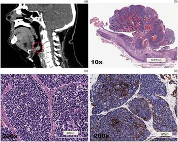
Patients with large cell neuroendocrine carcinoma usually present with a painless mass located in the supraglottic region (Figure 4). Cervical nodal metastases are very common and should raise strong suspicion clinically. Association with paraneoplastic syndromes has not been reported for large cell neuroendocrine carcinoma.Reference Ferlito, Rinaldo, Bishop, Hunt, Vander Poorten and Williams10
Large cell neuroendocrine carcinoma. (a) Sagittal computed tomography image shows a pedunculated mass of the epiglottis (within dotted circle). (b) Microscopic overview of the resection specimen, with the pedunculated tumour arising from the epiglottic surface with underlying cartilage (H&E; ×10). (c) Neoplastic cells are hyperchromatic, overlapping with enlarged nuclei, apoptotic bodies, mitoses and focal necrosis (H&E; ×200). (d) Tumour cells show expression for chromogranin (immunohistochemical evaluation; ×200).

The diagnosis of large cell neuroendocrine carcinoma is based on high-grade features, and on the presence of both neuroendocrine morphology and immunohistochemical evidence of neuroendocrine differentiation. The criteria for diagnosis of large cell neuroendocrine carcinoma for lung tumours, according to Travis et al., include: (1) the presence of neuroendocrine differentiation features, including organoid nesting, trabecular growth, rosettes and peripheral palisading; (2) the presence of enlarged tumour cells, with vesicular chromatin, small to prominent nucleoli, and moderate to abundant cytoplasm; (3) increased mitotic activity (greater than 10 mitoses per 10 high power fields (2 mm2)); and (4) confirmation of neuroendocrine differentiation using immunohistochemical staining for chromogranin and/or synaptophysin.Reference Travis, Linnoila, Tsokos, Hitchcock, Cutler and Nieman36 Markers including neuron-specific enolase, and/or neural cell adhesion molecule, and clusters of differentiation 56 and 57 are not specific for neuroendocrine differentiation, and interpretation should be made in conjunction with morphology and other markers.
Macroscopically, the tumour presents as a submucosal unencapsulated mass. More specifically, from a histopathological point of view, large cell neuroendocrine carcinoma has large polygonal cells with generous eosinophilic cytoplasm. There is no absolute cell size cut-off between large cell neuroendocrine carcinoma and small cell neuroendocrine carcinoma, although cell size in the latter is usually smaller than the diameter of three resting lymphocytes. Nuclear characteristics, however, are key to this distinction. Large cell neuroendocrine carcinoma usually has vesicular or coarse salt-and-pepper chromatin and prominent nucleoli. This is in contrast to small cell neuroendocrine carcinoma, where nucleoli are inconspicuous or absent, and chromatin is finely granular. The growth pattern shows characteristic neuroendocrine features, with sheets and nests of tumour cells, and organoid and trabecular architecture. The tumours frequently display necrosis and lymphovascular invasion.
The distinguishing feature to separate large cell neuroendocrine carcinoma from atypical carcinoid tumour, which can have quite similar histological features, is mitotic activity. Mitotic activity is more than 10 per 10 high power fields in large cell neuroendocrine carcinoma (although most have higher rates, usually more than 50 per 10 high power fields), whereas it is less than or equal to 10 in atypical carcinoid tumour.Reference Lewis, Ferlito, Gnepp, Rinaldo, Devaney and Silver5, Reference Travis, Linnoila, Tsokos, Hitchcock, Cutler and Nieman36
Although not necessarily discriminating, large cell neuroendocrine carcinoma typically has much more cytological atypia and more necrosis than atypical carcinoid tumour. On immunohistochemistry, large cell neuroendocrine carcinoma is positive for chromogranin, synaptophysin, cluster of differentiation 56, neuron-specific enolase and calcitonin.Reference Ferlito, Strojan, Lewis, Perez-Ordoñez and Rinaldo38, Reference Kusafuka, Ferlito, Lewis, Woolgar, Rinaldo and Slootweg40
The treatment of large cell neuroendocrine carcinoma has not been standardised. Chemoradiotherapy is accepted as the primary treatment modality.Reference Ferlito, Strojan, Lewis, Perez-Ordoñez and Rinaldo38 It appears that in all but the most early stage tumours, some forms of systemic therapy acting against possible micrometastases should be considered as part of the treatment regimen.Reference Ferlito, Strojan, Lewis, Perez-Ordoñez and Rinaldo38 In tumours with a high propensity for distant dissemination, such as large cell neuroendocrine carcinoma, the administration of systemic therapy seems to be important. For patients with systemic disease, palliative chemotherapy or best supportive care remain appropriate options.Reference Ferlito, Strojan, Lewis, Perez-Ordoñez and Rinaldo38 Combined cisplatin-based chemotherapy, similar to that used for small cell neuroendocrine carcinoma, seems to be most effective.
Despite aggressive therapy, survival remains poor. The five-year disease-specific survival rate is 15 per cent for large cell neuroendocrine carcinoma, the worst of all four histological neuroendocrine tumour subtypes.
Conclusion
Neuroendocrine carcinoma of the larynx is rare; however, it is important for clinicians and pathologists to be fully aware both of the diagnosis and the existence of different disease subtypes. This is because such tumours exhibit a wide range of biological behaviour, ranging from the extremely aggressive nature of small and large cell neuroendocrine carcinomas, which usually have a fatal prognosis, to the less aggressive clinical course of carcinoid tumours. The distinction is critical given the impact on the therapeutic approach. The findings highlight the need for both a high index of clinical suspicion and adequate representative pre-treatment biopsies. In small and large cell neuroendocrine carcinomas, a combination of irradiation and chemotherapy is indicated, while the management of carcinoid and atypical carcinoid tumours is surgical.
Competing interests
None declared.