Post-operative junctional ectopic tachycardia is a severe complication after paediatric cardiac surgery and is reported to occur in 3–10% of cases.Reference Zampi, Hirsch and Gurney 1 Risk factors for a junctional ectopic tachycardia are long extracorporeal bypass time, hypomagnesaemia, and an operation site near the ventricular septum.Reference Andreasen, Johnsen and Ravn 2 – Reference Rekawek, Kansy and Miszczak-Knecht 3 The narrow complex tachycardia typically starts with atrioventricular dissociation a few hours after surgery and is soon followed by a significant increase in the heart rate. In combination with restrictive diastolic ventricular function after cardiopulmonary bypass, this often results in low cardiac output syndrome starting a futile cycle that requires immediate intervention. Currently, this includes antiarrhythmic drugs such as amiodarone, deep sedation, and cooling the core temperature to 34–35°C.Reference Kovacikova, Hakacova, Dovos, Skrak and Zahorec 4 Attempts to restore synchronised atrial contraction by A00-pacing frequently fail because of defective atrioventricular conduction. In this situation, R-wave synchronised atrial pacing may be an effective alternative strategy. However, intact temporary epimyocardial pacing wires are a prerequisite for atrial pacing. We present a case of successful R-wave synchronised atrial pacing in a case with non-functioning atrial pacing wires via a transoesophageal pacing lead.
Case report
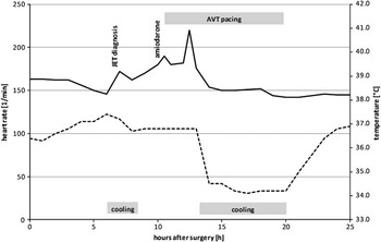
A 3-month-old female baby was admitted to our paediatric intensive care unit after uneventful surgical closure of a perimembranous ventricular septal defect. At the time of admission, we documented sinus rhythm with a heart rate of 160 beats/min. The patient was extubated 5 h after surgery. During the first 6 post-operative hours, the heart rate remained constant and then rapidly increased to more than 175 beats/min (Fig 1).
Clinical course of a 3-month-old with junctional ectopic tachycardia (JET). The figure shows the progression of the heart rate (solid line), body temperature (dashed line), and the therapeutic measures.

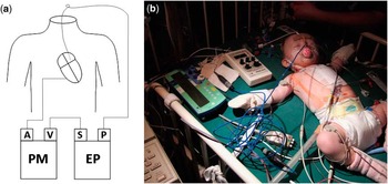
The electrocardiogram revealed the typical signs of junctional ectopic tachycardia with narrow QRS complexes and no detectable P-waves. R-wave synchronised atrial pacing was unsuccessful because of dislocated atrial epimyocardial pacing wires stimulating the ventricle with broad QRS complexes. The patient was cooled to a core temperature of 36.0°C, which resulted in a short period of declining heart rate, but soon it began to rise again. Amiodarone was administered with three intravenous boli of 1 mg/kg, followed by a continuous intravenous infusion of 600 mg/m2 body surface area per day. With the heart rate continuously rising, we established R-wave synchronised atrial pacing using a transoesophageal pacing lead (Fig 2).
R-wave synchronised atrial pacing using a transoesophageal pacemaker. ( a ) Schematic drawing and ( b ) original setting of the oesophageal pacemaker connected in series to an external pacemaker. Sequential pacing is performed by connecting the ventricular pacing wire to the atrial input of the external pacemaker to sense the QRS complexes. The oesophageal pacemaker is triggered by the stimulus from the external pacemaker's atrial output channel. A = atrial input channel; EP = esophageal pacemaker; P = pacing channel; PM = external pacemaker; S = sensing channel; V = ventricular input channel.

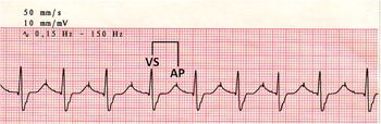
The transoesophageal pacemaker (PACE 50 E, Osypka, Germany) was synchronised with a capable external pacemaker (PACE 203H JJ, Osypka) via the special function cable of the transoesophageal pacemaker in a master–slave manner. The pacing mode of the PACE 203H JJ was set to VAT by choosing the VDD mode and turning the ventricular sensing to endless high. The post-ventricular atrial refractory period was set to 100 ms, the maximum tracking rate to 200 beats/min, and the atrioventricular delay to 220 ms. The ventricular epimyocardial wires were connected to the atrial input channel of the pacemaker, thereby changing the VAT mode into an effective AVT mode and the atrioventricular delay into an effective ventriculoatrial interval. The output current of the external pacemaker's ventricular channel was set to 3 V. Then the ventricular channel of the PACE 203H JJ was connected to the sensing channel of the oesophageal pacemaker using a special function cable (Osypka, Germany) and a custom-made connecting cable. The transoesophageal pacemaker was adjusted to external synchronisation, the impulse duration was set to 5 ms, and the impulse amplitude to 10 mA. After identifying the correct position of the oesophageal pacing lead through a continuously recording electrocardiography machine (Pagewriter 200, Hewlett Packard, Palo Alto, California, United States of America), R-wave synchronised transoesophageal atrial pacing was started (Fig 3).
Electrocardiogram showing effective R-wave synchronised atrial pacing. AP = atrial pacing; VS = ventricular sensing.

After establishing synchronised atrial pacing, the heart rate immediately declined. Paroxysmal breakthrough tachycardia 1 h later required cooling of the patient to a core temperature of 34.5°C and deep sedation. Thereafter, the heart rate continuously decreased, and within <2 h rhythmic control was achieved. The patient was rewarmed, sedation was stopped, and the continuous infusion of amiodarone was completed. The baby recovered well and was discharged from the paediatric intensive care unit on the 3rd post-operative day.
Discussion
R-wave synchronised atrial pacing is an inventive and effective therapeutic tool of post-operative junctional ectopic tachycardia. The method was first described by Till and Rowland in 1991.Reference Till and Rowland 5 In 2003, Janousek et alReference Janousek, Vojtovic and Gebauer 6 published the successful use of a modified commercially available pacemaker in nine patients with junctional ectopic tachycardia. Owing to its good results, the technique has become a standardised pacing strategy in the treatment of post-operative junctional ectopic tachycardia.Reference Haas, Plumpton, Justo, Jalali and Pohlner 7 Janousek et al also described the successful administration of R-wave synchronised atrial pacing in patients with dysfunctional ventricular pacing wires. In these cases, ventricular impulses can be sensed through surface electrocardiogram leads connected to the atrial channel of the pacemaker.
Transoesophageal pacing in patients with junctional ectopic tachycardia has been described by two authors: Guccione et alReference Guccione, Di Carlo and Papa 8 used non-synchronised transoesophageal atrial pacing at a higher rate. Janousek et alReference Janousek, Vojtovic and Chaloupecky 9 used the pacing pulse of a transoesophageal pacemaker in cases with poor signal of the atrial pacing wire. A temporary DDD pacemaker sensed the high voltage signal of the transoesophageal pacemaker from the otherwise non-functional atrial pacing wires and thereby achieved synchronised pacing of the ventricles via the epicardiac pacing wires. The method allows atrioventricular sequential pacing and goes back to Roth and Huertas 1995.Reference Roth and Huertas 10
However, R-wave synchronised transoesophageal pacing in case of dysfunctional atrial pacing wires via two connected pacemakers has not yet been described in the literature. Similarly, it would be possible to perform R-wave synchronised atrial pacing without any functional pacing lead, simply by sensing the R-wave via surface leads and pacing via a transoesophageal pacemaker.
Acknowledgement
The authors gratefully thank the parents of the patient for their kind support and their informed consent to the transoesophageal pacing and publishing the photograph.