Background
Alzheimer's disease (AD) is a burden that affects many aspects of society, including economics, health, and society in general (Aguero-Torres et al., Reference Aguero-Torres, Fratiglioni, Guo, Viitanen, von Strauss and Winblad1998, Reference Aguero-Torres, Fratiglioni, Guo, Viitanen and Winblad1999, Reference Aguero-Torres, von Strauss, Viitanen, Winblad and Fratiglioni2001). In addition to cognitive deficits, up to 80% of patients with AD exhibit neuropsychiatric symptoms over the course of the disease (Bedford et al., Reference Bedford, Melzer and Guralnik2001). In addition to cognitive impairment, the neuropsychiatric symptoms of AD lead to increased disability, promote institutionalization, hasten the death of patients, and escalate the pre-existing suffering of their families (Teri, Reference Teri1997; Murman and Colenda, Reference Murman and Colenda2005). Of all neuropsychiatric symptoms, apathy is one of the most common (Mega et al., Reference Mega, Cummings, Fiorello and Gornbein1996).
Apathy is generally defined as a lack of interest and motivation, and is characterized by diminished initiation, poor persistence, low social engagement, and emotional indifference (Marin, Reference Marin1990). It is manifested in the behavioral, cognitive, and emotional aspects of goal-directed behavior (Marin, Reference Marin1991), and can be categorized into three subdomains: emotional, behavioral, and cognitive domains (Marin et al., Reference Marin, Biedrzycki and Firinciogullari1991). Apathy is aggravated as the disease progresses, and is associated with reduced function, early institutionalization, and an increased mortality risk (Mega et al., Reference Mega, Cummings, Fiorello and Gornbein1996; Banerjee et al., Reference Banerjee, Murray, Foley, Atkins, Schneider and Mann2003; Colombo et al., Reference Colombo, Vitali, Cairati, Vaccaro, Andreoni and Guaita2007; Vilalta-Franch et al., Reference Vilalta-Franch, Calvo-Perxas, Garre-Olmo, Turro-Garriga and Lopez-Pousa2013). It also causes distress to caregivers and increases their care burden (Landes et al., Reference Landes, Sperry, Strauss and DS2001; Matsumoto et al., Reference Matsumoto2007).
It is believed that biological factors could be significantly involved in the development of apathy in patients with AD. However, this has not yet been adequately explored in the literature. Neuroimaging studies using single-photon emission computerized tomography (SPECT), positron emission tomography (PET), and magnetic resonance imaging (MRI) have provided evidence to demonstrate that patients who receive a diagnosis of AD and exhibit apathy have a higher incidence of structural or functional brain abnormalities, predominantly in the frontal area (Benoit et al., Reference Benoit1999; Migneco O et al., Reference Migneco, Benoit and PM2001; Lanctôt et al., Reference Lanctôt2007; Marshall et al., Reference Marshall, Monserratt, Harwood, Mandelkern, Cummings and Sultzer2007b). Therefore, abnormalities in the frontal lobe may be a substrate of apathy. In addition to structural and functional imaging studies, studies that employ imaging techniques to detect neurochemical changes in the brain may provide further information. However, to date, no data of neurochemical changes in the brain related to apathy in AD patients have been obtained. Magnetic resonance spectroscopy (MRS) is a non-invasive method through which levels of neurochemical metabolites can be measured. Compared with traditional MRI, MRS has demonstrated superior sensitivity in detecting neurochemical alterations in the brain. It can be employed to detect subtle changes, even when the clinical symptoms have not yet appeared (Chan et al., Reference Chan, Shungu, Douglas-Akinwande, Lange and Rowland1999). The main neurochemical metabolites that can be detected using MRS are N-acetyl-aspartate (NAA), creatine (Cr), choline (Cho), and myo-inositol (MI). Each metabolite represents a different physiological or pathological marker in the brain. NAA is a marker of neuronal heath, and neuronal injury or death may decrease the NAA concentration in the brain (Urenjak et al., Reference Urenjak, Williams, Gadian and Noble1993; Moffett et al., Reference Moffett, Ross, Arun, Madhavarao and Namboodiri2007). Higher choline levels indicate a higher cell density or an accelerated turnover rate of the cell wall (Ross et al., Reference Ross, Kreis and Ernst1992). MI is a marker of astrocytes and intracellular osmotic integrity (Ross, Reference Ross1991). Cr is a substrate and product of creatine kinase, which plays a role in cell energy (Kato et al., Reference Kato, Inubushi and Kato1998).
In summary, past studies have suggested that structural or functional changes in the frontal area, especially in the orbitofrontal and anterior cingulate regions, are associated with apathy in patients with AD; however, no study has utilized MRS to evaluate neurochemical changes in relation to apathy. In addition, few neuroimaging studies have focused on the different subdomains of apathy. Thus, in this study, we investigated the frontal neurochemical alterations related to apathy among patients with AD. The hypothesis of this study is that certain alternations in neurochemical metabolites are associated with apathy and the subdomains of apathy in patients with AD.
Methods
Subjects
In this study, we conducted a neuropsychiatric study of dementia (Yeh et al., Reference Yeh2014). Study participants were recruited from the outpatient clinics of a teaching hospital. Patients with dementia due to AD were recruited. AD was diagnosed according to the Diagnostic and Statistical Manual of Mental Disorders, Fourth Edition, Text Revision (DSM-IV-TR; American Psychiatric Association, 2000). The exclusion criteria included other neurological disorders which may affect the presentation of apathy and cognitive function such as stroke, traumatic head injury, Parkinson's disease, and epilepsy. The study was approved by the Institutional Review Board of Kaohsiung Medical University. All patients provided written informed consent. If the participants had a compromised capacity to consent, their legally authorized relatives provided written consent on their behalf.
Assessments
The severity of apathy was measured using the Apathy Evaluation Scale (AES) (Marin et al., Reference Marin, Biedrzycki and Firinciogullari1991), which is composed of 18 items, with each rated in terms of severity on a scale of 1–4 (1 = not at all, 2 = slightly, 3 = somewhat, and 4 = a lot). The total AES score ranged from 18 to 72, with a higher score indicating more severe apathy. This method of measurement consisted of three subdomains of apathy: behavioral apathy, cognitive apathy, and emotional apathy. We calculated the total AES score and the scores of the three subscales. As depression and apathy may have common symptoms such as anhedonia and reduced activity (Landes et al., Reference Landes, Sperry, Strauss and DS2001), depression may be a confounding factor in analyzing the association between neuroimaging findings and apathy. Depression was therefore measured using the Cornell Scale for Depression in Dementia (CSDD) (Alexopoulos, Reference Alexopoulos2002), and the severity of dementia was assessed using the Clinical Dementia Rating Scale (CDR) (Morris, Reference Morris1993).
Image acquisition and analysis
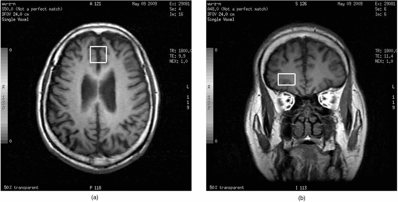
3T 1H MRS was performed in this study (GE Medical Systems, Milwaukee, WI, USA). Conventional MR images were obtained for an anatomic registration of the spectroscopic data, including transverse fast spin echo T2-weighted images, axial fluid-attenuated inversion recovery (FLAIR) spin echo T1-weighted images and T2-weighted images. Point-resolved spectroscopy (PRESS) was employed to obtain single-voxel spectra. Three preceding chemical-shift-selective saturation pulses were used for water suppression. The observed water signal optimized the field homogeneity over the selected voxel of interest. The parameters were TE = 30 ms, TR = 1500 ms, 256 acquisitions, 4 dummy scans, spectral width = 2500 Hz; 2048 data points. Phase correction of the metabolite spectra was achieved using an additional 16 acquisitions without water suppression in sequence. The voxel sizes were 2 × 2 × 2 cm3 over the anterior cingulate region and 2 × 2 × 1 cm3 over the right orbitofrontal region. The locations of these two voxels are presented in Figure 1.
Representation of the location of the voxel of interest in a T2-weighted 1H magnetic resonance spectroscopy image: (a) anterior cingulate region (b) right orbitofrontal region.

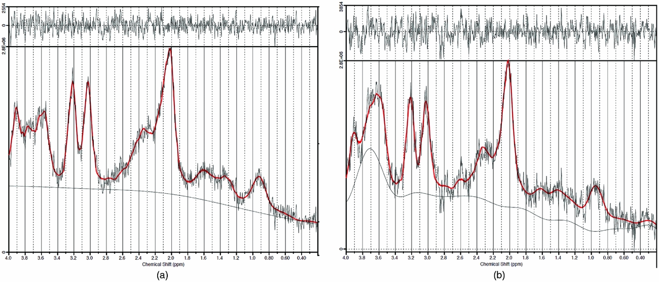
We employed the Linear Combination of Model Spectra, which is a user-independent frequency domain spectral fitting program (Figure 2). To estimate uncertainties, we utilized the Cramer–Rao lower bounds as the main guideline for judging the spectra of metabolite concentrations. Any spectrum with a full width at half-maximum of > 0.070 ppm was defined as poor spectra and was excluded from the analyses. We calculated the ratios of NAA/Cr, choline/Cr, and mI/Cr as markers of neurochemical alteration.
Representative examples of the chemical shift of 1H magnetic resonance spectra in the (a) anterior cingulate region; (b) right orbitofrontal region.

Statistics
A linear regression model was employed to analyze the associations between apathy and neurochemical metabolites, with adjustments for CSDD score, CDR score, age, sex, and educational level. Statistical significance was defined as a p value of less than 0.05.
Results
Forty-four patients were recruited, but eight of them displayed poor MRS spectra. Therefore, 36 patients were included in the analyses. Among 36 patients, fifteen subjects (41.7%) were male. Thirty subjects (83.3%) had mild dementia with CDR = 1. The average age of the participants was 76.0 (SD = 8.5) years old. The mean score of the Mini-Mental State Examination (MMSE) was 20.0 (SD = 5.7). The mean score of the AES was 41.5 (SD = 12.1), and the mean score of the CSDD was 8.4 (SD = 5.6). The NAA/Cr, choline/Cr, and mI/Cr ratios of the MRS were 1.12 (SD = 0.13), 0.31 (SD = 0.03), and 0.85 (SD = 0.14) respectively in the anterior cingulate region; and 1.06 (SD = 0.21), 0.32 (SD = 0.08), and 0.98 (SD = 0.45), respectively in the right orbitofrontal region.
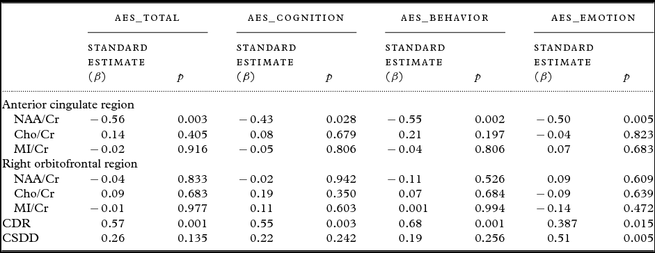
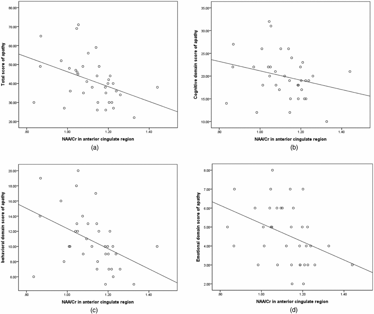
We analyzed the associations of apathy scores with all the MRS data from both voxels, dementia severity and depression severity, adjusting for age, sex, and educational level. More severe dementia, as indicated by a higher CDR score, was associated with more severe apathy, both in terms of the total AES score and the subdomain scores. A higher CSDD score was associated with a higher score in the emotional domain of apathy (β = 0.51, p = 0.005). A higher total AES score was associated with a lower NAA/Cr ratio in the anterior cingulate region (β = −0.56, p = 0.003). Meanwhile, a lower NAA/Cr ratio in the anterior cingulate region was associated with the scores of all AES subdomains (β = −0.43, p = 0.028 in the cognitive domain; β = −0.55, p = 0.002 in the behavior domain; β = −0.50, p = 0.005 in the emotion domain). However, no significant associations were observed in the right orbitofrontal region (Table 1). Since there is likely to be a strong correlation between the NAA/Cr in the two regions, we performed a regression model of the two voxels separately. In the anterior cingulate region, a lower NAA/Cr ratio was associated with a higher total AES score (β = −0.55, p = 0.001) and all AES subscores (β = −0.42, p = 0.020 in the cognitive domain; β = −0.55, p = 0.001 in the behavior domain; β = −0.48, p = 0.003 in the emotion domain). In the right orbitofrontal region, the NAA/Cr ratio had no significant associations with the total AES score (β = −0.15, p = 0.473) or AES subscores (β = −0.10, p = 0.634 in the cognitive domain; β = −0.22, p = 0.289 in the behavior domain; β = −0.02, p = 0.936 in the emotion domain). The results of the separated regression model were similar to those of the regression model that combined all variables in one analysis. In relation to the NAA/Cr ratio in the anterior cingulate region, scatterplots of total apathy scores are provided in Figure 3(a), and those for the subdomains of apathy scores are illustrated in Figures 3(b–d).
Linear regression analysis of variables predicting the total score and subdomain scores of the apathy evaluation scale

*Controlling for age, sex, and educational level.
AES: Apathy Evaluation Scale; NAA: N-acetyl-aspartate; Cho: Choline; MI: myo-inositol; Cr: creatine; CDR: Clinical Dementia Rating scale; CSDD: Cornell Scale for Depression in Dementia.
(a) Scatterplot of the total score of apathy and NAA/Cr ratio in the anterior cingulate region. (b) Scatterplot of the cognitive domain score of apathy and NAA/Cr ratio in the anterior cingulate region. (c) Scatterplot of the behavioral domain score of apathy and NAA/Cr ratio in the anterior cingulate region. (d) Scatterplot of the emotional domain score of apathy and NAA/Cr ratio in the anterior cingulate region.

Discussion
In this study, we demonstrated that a relatively high total AES score was associated with a relatively low NAA/Cr ratio in the anterior cingulate region. In addition, the cognitive, behavioral, and emotional subdomains of apathy were associated with a low NAA/Cr ratio in the anterior cingulate region, but were not associated with neurochemical alterations in the right orbitofrontal region.
Our study indicated that the anterior cingulate cortex may play a central role in causing apathy. This finding was consistent with previous studies. Several studies using SPECT, ((18)F) fluorodeoxyglucose-PET or amyloid PET have indicated that apathy is associated with brain alterations in the anterior cingulate region (Migneco et al., Reference Migneco, Benoit and PM2001; Lanctot et al., Reference Lanctot2007; Marshall et al., Reference Marshall, Monserratt, Harwood, Mandelkern, Cummings and Sultzer2007a; Mori et al., Reference Mori2014). In addition to the correlation between brain structure and apathy, we further examined neurochemical alterations in the anterior cingulate region. Our study demonstrated that a low NAA/Cr ratio is associated with a high degree of apathy. NAA is considered a marker of neuron health, and its level corresponds with neuronal integrity (Cheng et al., Reference Cheng, Newell, Mallory, Hyman and Gonzalez2002). As the level of Cr remains unchanged in patients with AD, researchers have often used Cr as an internal reference to adjust the variability in measurement (Valenzuela and Sachdev, Reference Valenzuela and Sachdev2001; Kantarci, Reference Kantarci2007). Thus, in our study, a low NAA/Cr ratio may indicate a decreased NAA level. Therefore, our findings suggest that apathy is associated with lower neuronal integrity in the anterior cingulate region.
Few studies have investigated the association between brain imaging findings and the different domains of apathy. Only one study using SPECT demonstrated that the behavior, cognitive, and emotional domains of apathy were negatively correlated with perfusion activity in the right middle orbitofrontal gyrus, right anterior cingulate cortex, and left superior dorsolateral prefrontal cortex, respectively (Benoit et al., Reference Benoit, Clairet, Koulibaly, Darcourt and Robert2004). As that study did not control for possible confounders, type I errors may have occurred. With adjustments for confounders, such as depression severity, CDR, age, sex, and educational level, our study evinced that various domains of apathy were associated with a lower NAA/Cr ratio in the anterior cingulate region, but not in the right orbitofrontal region, which implies that the anterior cingulate region may play a central role in causing all domains of apathy. The anterior cingulate cortex is crucial for initiation and for motivation, which drives the commitment of effort to goal-directed activities. Damage to the anterior cingulate cortex is therefore likely to lead to not only cognitive, but also behavioral and emotional inactivity (Devinsky et al., Reference Devinsky, Morrell and Vogt1995; Allman et al., Reference Allman, Hakeem, Erwin, Nimchinsky and Hof2001).
To the best of the authors’ knowledge, this is the first study to use 1H MRS to investigate the associations between neurochemical levels and apathy in patients with AD. 1H MRS, which differs from other neuroimaging techniques such as PET, SPECT, and MRI, can be used to investigate the alternations of neurochemicals in the brain, and provides an alternative view of the pathology of apathy in AD. The key strengths of our study are that we used the AES to evaluate apathy, which achieves good validity and reliability in patients with AD, and that it evaluated three different subdomains of apathy (Marin et al., Reference Marin, Biedrzycki and Firinciogullari1991).
However, there were some limitations in our study. First, the sample size used in this study was small, and the participants were recruited from one teaching hospital. Therefore, the generalizability is limited. In addition, we were unable to judge how the observed NAA/Cr ratio compared with normal levels without a healthy control group. Further research using a control group may provide a more meaningful result. It should also be noted that medications may alter the neurochemical levels as seen on MRS images – as most of our participants were administered psychotropic drugs, we were unable to compare the MRS findings of patients under drug treatment with those of patients who were not receiving medication. Further studies to investigate the effects of drugs are required.
Conclusions
Our study demonstrated that apathy, including its cognitive, behavioral, and emotional subdomains, was associated with a lower NAA/Cr ratio in the anterior cingulate region, but not in the right orbitofrontal region, which indicates that the anterior cingulate region may play a certain role in all aspects of apathy in patients with AD.
Conflict of interest
None.
Description of the author's roles
Y-C Yeh designed and carried out the study, analyzed the data, and wrote the manuscript. C-W Li and Y-T Kuo carried out the study and analyzed the data. M-F Huang and T-L Liu formulated the research question, designed the study, and assisted with writing the paper. T-S Jaw carried out the study and analyzed the data. Y-H Yang was responsible for the statistical design of the study and assisted with the statistical analysis. K-C Kuo formulated the research question and designed the study. C-S Chen formulated the research question, designed the study, supervised the data analysis, and assisted with writing the paper.
Acknowledgments
The study was supported by a grant from the Kaohsiung Medical University Research Foundation (KMU-M098021) and Kaohsiung Medical University Hospital, Kaohsiung Medical University, Taiwan (KMUH 102-2R56).