Implications
The group of protein known as pregnancy-associated glycoproteins, or PAGs, are abundantly produced by the placenta of many agriculturally important livestock. However, their role during pregnancy is a mystery. To further understand these complex proteins, experiments were performed to ‘inactivate’ PAGs in maternal blood by coating them with antibodies. Interestingly, the ability to coat PAGs with antibodies was successful, but there was no obvious negative impact on pregnancy. One interpretation of these results is that the systemic role of PAGs may be secondary to their local role at the interface between the placenta and the maternal uterus.
Introduction
The pregnancy-associated glycoproteins (PAGs) are a large family of glycoproteins produced by placental trophoblasts of even-toed ungulates (animals comprising the Artiodactyla order). In the Ruminantia sub-order, the PAGs are represented by two distinct groupings comprising ancient and modern PAGs – based on the relative times at which they arose during Artiodactyl evolution (Garbayo et al., Reference Garbayo, Green, Mannekin, Beckers, Kiesling, Ealy and Roberts2000; Green et al., Reference Green, Xie, Quan, Bao, Gan, Mathialagan, Beckers and Roberts2000; Hughes et al., Reference Hughes, Green, Garbayo and Roberts2000). The genes for the former are transcribed in both the trophoblast populations of the ruminant placenta (the mononucleate and the binucleate (BNCs) trophoblasts) (Garbayo et al., Reference Garbayo, Green, Mannekin, Beckers, Kiesling, Ealy and Roberts2000; Green et al., Reference Green, Xie, Quan, Bao, Gan, Mathialagan, Beckers and Roberts2000). In contrast, the modern PAG genes represent a much larger group and they are transcribed exclusively by BNCs. Many BNC products are able to gain access to maternal tissues (Duello et al., Reference Duello, Byatt and Bremel1986; Liang et al., Reference Liang, Limesand and Anthony1999). Indeed, the modern/BNC-specific PAGs can be found circulating in maternal blood soon after apposition and attachment of the conceptus to the uterine epithelium (Sasser et al., Reference Sasser, Ruder, Ivani, Butler and Hamilton1986; Zoli et al., Reference Zoli, Guilbault, Delahaut, Ortiz and Beckers1992; Green et al., Reference Green, Parks, Avalle, Telugu, McLain, Peterson, McMillan, Mathialagan, Xie, Hook and Roberts2005). This feature of the modern/BNC PAGs has been exploited as the basis for pregnancy diagnosis in cattle and related ruminants. Not surprisingly, the modern PAGs have received the bulk of attention from animal scientists and have been the subject of several biochemical, molecular and functional studies (Del Vecchio et al., Reference Del Vecchio, Sutherland and Sasser1996; Xie et al., Reference Xie, Green, Bao, Beckers, Valdez, Hakami and Roberts1997; Green et al., Reference Green, Xie, Quan, Bao, Gan, Mathialagan, Beckers and Roberts2000; Hughes et al., Reference Hughes, Green, Piontkivska and Roberts2003; Klisch and Leiser, Reference Klisch and Leiser2003; Weems et al., Reference Weems, Kim, Humphreys, Tsuda and Weems2003; Wooding et al., Reference Wooding, Roberts and Green2005; Klisch et al., Reference Klisch, Jeanrond, Pang, Pich, Schuler, Dantzer, Kowalewski and Dell2008). However, despite such efforts, the functional role of the PAGs remains a mystery. At least a part of the ambiguity arises from the fact that different research groups have come up with rather distinct results, even though most of the experiments have been performed with similar preparations of purified PAG-1 (Pregnancy-specific protein B (PSPB)) – the prototypical member of the BNC-specific PAGs. Some of the proposed functions for PAGs are summarized here.
PAGs have been proposed to have both direct and indirect luteotrophic activity, acting to maintain progesterone production once the pregnancy has become established. When administered to cultured bovine luteal cells, PAG-1/PSPB promoted release of the luteotrophic compound, prostaglandin E2 (PGE2) (Del Vecchio et al., Reference Del Vecchio, Sutherland and Sasser1995a and Reference Del Vecchio, Sutherland and Sasser1996; Weems et al., Reference Weems, Lammoglia, Vera-Avila, Randel, Sasser and Weems1998a) and an increase in progesterone (Del Vecchio et al., Reference Del Vecchio, Sutherland and Sasser1996; Weems et al., Reference Weems, Lammoglia, Vera-Avila, Randel, King, Sasser and Weems1998b). However, other studies reported no increase in progesterone (Del Vecchio et al., Reference Del Vecchio, Sutherland and Sasser1995a and Reference Del Vecchio, Sutherland and Sasser1995b). PAG-1 may also elevate the production of PGE2 by cultured endometrium from both cattle and sheep (Del Vecchio et al., Reference Del Vecchio, Sasser and Randel1990; Weems et al., Reference Weems, Kim, Humphreys, Tsuda and Weems2003). A working model based on these data visualizes PAG-1/PSPB being up-regulated by placental estrogen; PAG-1 then increases the production of PGE2, which is believed to augment the synthesis of progesterone (Weems et al., Reference Weems, Bridges, LeaMaster, Sasser, Vincent and Weems1999, Reference Weems, Bridges, LeaMaster, Sasser, Ching and Weems2001 and Reference Weems, Bridges, Sasser, Ching, LeaMaster, Vincent and Weems2002).
Another role has been proposed in which the PAGs act in an immunomodulatory capacity. In cattle, the phagocytotic and oxidative burst activities of circulating polymorphonuclear neutrophil leukocytes are lowest around the time of parturition and in the first 3 weeks after calving (Kehrli et al., Reference Kehrli, Nonnecke and Roth1989; Saad et al., Reference Saad, Concha and Astrom1989). Dosogne et al. (Reference Dosogne, Burvenich, Freeman, Kehrli, Detilleux, Sulon, Beckers and Hoeben1999) and Hoeben et al. (Reference Hoeben, Monfardini, Opsomer, Burvenich, Dosogne, De Kruif and Beckers2000), both noted that bovine PAG concentrations peak immediately prior to this decline in leukocyte activity. They hypothesized that there could be an association between the high PAG concentrations at parturition and lowered leukocyte killer activities. There are, in fact, some indications that PAG-1 (PSPB) can be immunosuppressive (Hoeben et al., Reference Hoeben, Burvenich, Massart-Leen, Lenjou, Nijs, Van Bockstaele and Beckers1999). For example, bovine PAG-1 can reduce the proliferative activity of bovine hematopoietic cells in vitro (Hoeben et al., Reference Hoeben, Burvenich, Massart-Leen, Lenjou, Nijs, Van Bockstaele and Beckers1999). The PAGs can also associate with uterine serpins – proteins able to inhibit natural killer cell activity and the proliferation of peripheral blood lymphocytes in vitro (Mathialagan and Hansen, Reference Mathialagan and Hansen1996; Peltier et al., Reference Peltier, Liu and Hansen2000). Finally, PAG-1 (PSPB) has been shown to induce the release of the alpha chemokine, granulocyte chemotactic protein-2, from cultured bovine endometrial explants and an endometrial cell line (BEND cells) (Austin et al., Reference Austin, King, Vierk, Sasser and Hansen1999).
Clearly, the role of PAGs is uncertain and complicated by the fact that there are so many of them (Garbayo et al., Reference Garbayo, Green, Mannekin, Beckers, Kiesling, Ealy and Roberts2000; Green et al., Reference Green, Xie, Quan, Bao, Gan, Mathialagan, Beckers and Roberts2000; Brandt et al., Reference Brandt, Parks, Killian, Ealy and Green2007). A further difficulty in understanding how they operate arises from the fact that nearly all earlier functional studies have been performed in vitro rather than on intact animals. In this report, we attempted to gain some preliminary insight into PAG function by auto-immunizing ewes against a mixture of purified native ovine PAGs. The underlying assumption was that either eliminating or suppressing the ability of PAG to act outside the placenta would either compromise the pregnancy as it proceeded, or not allow it to become established in the first place. Similar autoimmune approaches have been used by others to study potential contraceptive vaccines in humans (Stevens, Reference Stevens1979; Talwar et al., Reference Talwar, Singh, Pal, Chatterjee, Sahai, Dhall, Kaur, Das, Suri and Buckshee1994), and to test the functions of specific circulating hormones and placental proteins in other species (Medan et al., Reference Medan, Wang, Watanabe, Suzuki and Taya2004).
Methods
Preparation of proteins for immunizations
A preparation consisting of a mixture of ovine PAG was obtained essentially as described earlier for the isolation of bovine PAG (Green et al., Reference Green, Parks, Avalle, Telugu, McLain, Peterson, McMillan, Mathialagan, Xie, Hook and Roberts2005; Wooding et al., Reference Wooding, Roberts and Green2005; Green and Roberts, Reference Green and Roberts2006). Briefly, ovine cotyledons were collected from late stage pregnant ewes (∼Day 100; n = 3) by manual separation of the placentomes into cotyledon and caruncular components. Developmental stage was estimated by measurement of crown–rump lengths (Hammond, Reference Hammond1927; Noakes et al., Reference Noakes, Parkinson and England2001). The isolated cotyledons were homogenized in 20 mM Tris (pH 8.0), 50 mM NaCl, 2 mM ethylenediamine tetraacetic acid (EDTA), 0.2 mM phenylmethylsulfonylfluoride (PMSF), 0.02% (w/v) NaN3. Insoluble material was removed by centrifugation at 5000 × g for 30 min. The cleared supernatant was placed in 50 000 MWCO dialysis tubing and dialyzed against >100 volumes of 20 mM Tris (pH 7.0), 150 mM NaCl, 1 mM EDTA, 0.02% (w/v) sodium azide, 20 mM PMSF, 1 mM 2-mercaptoethanol. The presence of immunoreactive PAG was confirmed by immunoblotting with a rabbit anti-ovine PAG-1 antiserum (#80) raised against recombinant ovine PAG-1 that had been produced in bacteria (Wooding et al., Reference Wooding, Roberts and Green2005). Since PAGs belong to the aspartic peptidase family of enzymes, the aspartic peptidase inhibitor, pepstatin, was used to selectively bind PAGs present in cotyledons. The cleared cotyledonary extract was applied to a column containing 50 ml of beaded pepstatin-A agarose (4% cross-linked; Sigma, St Louis, MO, USA) equilibrated in the same buffer. After loading, the matrix was washed with 40 column volumes of the loading buffer, followed by 10 column volumes of 20 mM Tris (pH 7.0), 1 M NaCl, 1% Triton X-100, 1 mM EDTA, 0.02% (w/v) sodium azide, 20 mM PMSF, 1 mM 2-mercaptoethanol. Proteins remaining bound to the column, were eluted by increasing the pH of the buffer in a stepwise fashion (pH 8.0, 9.5 and 10.5). Elution fractions were collected and analyzed by dot blot for the presence of PAG immunoreactivity. The fractions that contained PAG were pooled and their protein concentrations were determined by Bradford assay (Bradford, Reference Bradford1976).
The material that flowed-through from the pH 7 pepstatin column was dialyzed, as described above, against 20 mM sodium citrate (pH 5.0), 150 mM NaCl, 1 mM EDTA, 0.02% (w/v) sodium azide, 20 mM PMSF, 1 mM 2-mercaptoethanol. After centrifugation, the supernatant was applied to the pepstatin column equilibrated in the same pH 5.0 buffer. The column was washed (40 column volumes of loading buffer followed by 10 column volumes of 20 mM sodium citrate (pH 5.0), 1 M NaCl, 1% Triton X-100, 1 mM EDTA, 0.02% (w/v) sodium azide, 20 mM PMSF, 1 mM 2-mercaptoethanol). Bound PAGs were eluted and analyzed for the presence of PAG. Since both the neutral and acidic-binding preparations had similar purity and immunoreactivity, aliquots from each were pooled for immunizing ewes.
To gain some insight into which PAGs were represented in the preparations, the eluted proteins were subjected to SDS-PAGE and the main immunoreactive bands were subjected to reduction, alkylation of cysteines with iodoacetamide and digested with trypsin for 16 h (Shevchenko et al., Reference Shevchenko, Wilm, Vorm and Mann1996; Belghazi et al., Reference Belghazi, Bathany, Hountondji, Grandier-Vazeille, Manon and Schmitter2001). The positive ion mass spectra of the resulting peptides were obtained in the presence of alpha-cyano-4-hydroxycinnamic acid matrix. The most abundant ions were further subjected to sequencing by MS/MS (tandem mass spectrometry). The ion spectra were obtained on a 4700 MALDI TOF/TOF mass spectrometer (Applied Biosystems, Foster City, CA, USA) (Shevchenko et al., Reference Shevchenko, Wilm and Mann1997; Link et al., Reference Link, Eng, Schieltz, Carmack, Mize, Morris, Garvik and Yates1999; Baldwin et al., Reference Baldwin, Medzihradszky, Lock, Fisher, Settineri and Burlingame2001; Belghazi et al., Reference Belghazi, Bathany, Hountondji, Grandier-Vazeille, Manon and Schmitter2001). The monoisotopic masses of the acquired spectra were used for searching against a nonredundant translated mammalian sequence database (NCBInr) by using the Mascot (www.matrixscience.com) or Protein Prospector MS FIT programs (http://prospector.ucsf.edu/) (Clauser et al., Reference Clauser, Baker and Burlingame1999; Perkins et al., Reference Perkins, Pappin, Creasy and Cottrell1999). No significant matches were obtained with just the peptide mass fingerprint data, so searches were repeated with both the MS and MS/MS spectra. The predicted peptide sequences matched several known ovine, bovine and caprine PAGs.
Immunization of sheep
PAGs in the eluted fractions were dialyzed against >1000 volumes of 10 mM sodium phosphate (pH 7.0), 150 mM NaCl and cross-linked to keyhole limpet hemocyanin (KLH) by using glutaraldehyde (Harlow and Lane, Reference Harlow and Lane1988). The PAG was included at a 30-fold molar excess relative to the amount of KLH and the final glutaraldehyde concentration was at a 20-fold molar excess relative to the PAG. Cross-linking was allowed to proceed overnight at room temperature and the reaction was terminated by adding glycine (pH 7.2) to a final concentration of 20 mM (Harlow and Lane, Reference Harlow and Lane1988).
The animals used in this study were mostly Romanov-crossbred ewes that were maintained at the University of Missouri Sheep Farm. The ewes were immunized subcutaneously with either 100 μg of KLH (n = 9 animals) or 300 μg of ovine PAG, in the form of a PAG–KLH conjugate (n = 22 animals). The protein solutions were emulsified with an equal volume of Freund’s complete adjuvant (Sigma) immediately prior to the injections. Subsequent ‘booster’ injections were performed with Freund’s incomplete adjuvant. The animals were re-exposed to the immunogens at approximately 8-week intervals for a total of five immunizations. The final injection was administered 2 weeks prior to breeding.
The ewes were synchronized for estrus by two intramuscular injections of a prostaglandin analog (Lutalyse; Pharmacia & Upjohn Co., Kalamazoo, MI, USA) administered 9 days apart. The ewes were bred to two rams of proven fertility. Four animals that cycled back on Day 16 after breeding were re-bred to the same rams and their samples and lambing data were also included in the study. Blood samples for serum were collected from the jugular vein, from each animal, at the time of standing heat (Day 0) and on Days 10, 14 to 28 and then weekly, throughout the remainder of pregnancy. The number and weight of the lambs born to each ewe were collected within 3 h of birth. The procedures used in this study were carried out under protocols approved by the University of Missouri Animal Care and Use Committee.
PAG ELISA
To determine if PAG autoimmunization had any influence on circulating PAG concentrations during pregnancy, an ovine PAG ELISA was established by using methods that have been described earlier, in detail, for bovine PAGs (Green et al., Reference Green, Parks, Avalle, Telugu, McLain, Peterson, McMillan, Mathialagan, Xie, Hook and Roberts2005; Green and Roberts, Reference Green and Roberts2006). Briefly, ovine PAGs were purified by pepstatin-affinity chromatography, as described above, and used for the production of polyclonal antibodies in rabbits and as a standard in the ELISA (Green et al., Reference Green, Parks, Avalle, Telugu, McLain, Peterson, McMillan, Mathialagan, Xie, Hook and Roberts2005; Green and Roberts, Reference Green and Roberts2006). The resulting antibodies were purified by using protein A-Sepharose (Pierce Biotechnology Inc., Rockford, IL, USA) and the immunoglobulin was quantified by Bradford assay with rabbit IgG (Sigma) as the standard (Bradford, Reference Bradford1976; Harlow and Lane, Reference Harlow and Lane1988).
The ELISA was an indirect ‘sandwich’ design. Purified rabbit anti-ovine-PAG antibodies (4 μg) were coated in each well of a 96-well ELISA plate. The wells were blocked in 2% BSA and 1% non-fat dry milk. Serially diluted ovine-PAG standard (diluents: non-pregnant ewe serum) or unknown serum samples were added to the wells and the plate was incubated at 4°C overnight. Unbound protein was removed by washing in a 96-well plate washer (BioTek, Winooski, VT, USA) and the wells were then incubated with 3 μg of biotinylated rabbit anti-PAG IgG for 30 min at room temperature. The plate was washed again and neutravidin–AP (alkaline phosphatase) conjugate (Pierce Biotechnology) was added and allowed to incubate at room temperature for 30 min. After washing, 0.1 ml of 1 mg/ml p-nitrophenyl phosphate (PNPP) (Sigma) was added to each well. After 15 to 30 min, the absorbance at 405 nm was measured by using an EL808 plate reader (BioTek). A PAG standard dilution series was included on every ELISA plate.
Assay validation
Two serum samples representing distinct PAG concentrations were assayed seven times in quadruplicate to calculate intra- and inter-assay variation. Parallelism was assessed by serially diluting late pregnant ewe sera, and measuring PAG reactivity in the sera relative to the PAG standards.
Data analysis
Standard curves were generated by using purified native ovine PAG isolated by pepstatin-affinity chromatography. The PAG standards were adjusted to provide a range from 0.039 to 40 ng. A standard curve was included on every ELISA plate and was generated by non-linear regression of a LOG (ng PAG std) v. absorbance plot, using the Graphpad Prism software (version 3.02 for Windows; San Diego, CA, USA; www.graphpad.com).
Differences in lamb number and weights, as well as mean PAG concentrations between groups were tested by ANOVA, using the data processing functions of the GraphPad Prism Software.
Measurement of anti-PAG titers present in the immunized ewes
To determine the extent to which the immunized animals developed antibodies to the immunogens, 1 μg of KLH or purified PAGs was coated on 96-well ELISA plates. The plates were blocked as described above and serial dilutions of sera from each ewe were incubated in the wells overnight. The plates were washed and incubated with anti-sheep–AP conjugate (Jackson ImmunoResearch, West Grove, PA, USA) and hydrolysis of PNPP was measured at 405 nm (Green and Roberts, Reference Green and Roberts2006). The respective titers representing ewe sera reactivity toward the plated immunogens were calculated.
Results
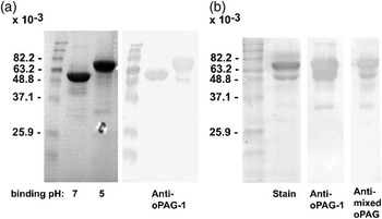
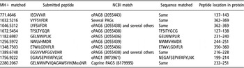
The aspartic peptidase inhibitor, pepstatin A, was used to affinity purify PAGs from D100 cotyledonary extracts (Stewart et al., Reference Stewart, Piggott, May and Westley1994; Wright et al., Reference Wright, Levy, Patel and Alhadeff1997; Green and Roberts, Reference Green and Roberts2006). The isolated PAGs were represented in two major bands (relative Mr varied from approximately 50 000 to 65 000) when analyzed by one-dimensional SDS-PAGE (Figure 1). Some differences in electrophoretic mobility were observed based on the binding conditions (i.e. pH) employed during PAG isolation (Figure 1a). It was notable that when only a single binding condition was used (pH 5.0), both main PAG protein bands were represented in the isolated material (Figure 1b). Despite such mobility differences, however, there was considerable overlap in the PAGs represented in the bands. Tryptic digests and peptide sequencing of the most abundant peaks revealed several sequences from the major PAG bands that most closely matched ovine PAG-5 (AAB53226) and PAG-6 (AAB53227), with additional matches with ovine PAG-8 (AAB53229) and weaker matches with other bovine and caprine PAGs (Table 1). The preparations shown in Figure 1a were part of those used to immune the ewes. The material shown in Figure 1b was used for antibody production in rabbits and as a native PAG standard in the ELISA. The generated antiserum reacted with all the original components in the antigen preparation (Figure 1b).
Rapid isolation of native pregnancy-associated glycoprotein (PAG) by pepstatin affinity chromatography: (a) ovine PAG isolated by binding to pepstatin at pH 7 and 5 (labels are shown below the lanes) and used for immunization of ewes. (b) Results from a pepstatin–agarose batch purification of ovine PAGs at pH 5 for use in anti-PAG antibody production in rabbits and as PAG standard in the ELISA. The panel on the left is a stained gel showing the isolated proteins. The middle and right panels are western blots performed on duplicate gels with antibodies raised against recombinant ovine PAG-1 and the mixture of native ovine-PAGs, respectively. Normal rabbit serum did not produce any staining (data not shown).

Selected ovine pregnancy-associated glycoproteins (PAG) tryptic peptide sequences obtained by MS/MS (tandem mass spectroscopy)

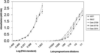
In order to assess the effectiveness of the autoimmunization protocol, it was necessary to have an assay capable of quantifying PAGs in pregnant ewes. Therefore, part of this work involved the establishment of an ELISA for the detection of circulating ovine PAGs. The assay could efficiently detect PAG standard diluted in non-pregnant ewe serum, down to 1 ng/ml. A plot of serial dilutions of the PAG standard revealed a sigmoidal-type curve typical of most antibody–antigen interactions (Figure 2). Parallelism between PAGs present in pregnant ewe sera and the purified PAG standard, indicated that the PAG detected in maternal sera is very similar to that used as standard (Figure 2). The intra- and inter-assay repeatability of the ELISA, used to measure PAG concentrations in pregnant ovine serum, was determined from ewes representing distinct concentrations of PAG. The intra-assay CV averaged 8.8% and the inter-assay CV averaged 13.5%.
On the left are typical standard curves for purified ovine pregnancy-associated glycoprotein (PAG) in the ELISA. Serially diluted PAG standard was incubated overnight at 4 to 8°C, to allow antibody–antigens interactions to achieve equilibrium. A dilution series of pregnant ewe sera, to assess parallelism in the ELISA between purified PAGs and those in circulation, is illustrated on the right.

Active immunization of ewes against PAGs produced a range of immune responses in the animals. Of the 22 animals that were auto-immunized before breeding, seven developed no or little reactivity toward PAGs while 15 produced high titers toward native PAGs. Those seven animals that did not respond to the immunization either did not react at all or the reactivity was indistinguishable from background upon dilution of the sera 1 : 2000. Those animals that did produce auto-antibodies to native PAGs exhibited robust reactivity even at a dilution of >1 : 16 000. All of the immunized animals (PAG–KLH and KLH alone) exhibited strong reactivity toward KLH in the standard ELISA. The KLH-immunized animals exhibited no reactivity toward PAGs in the titering ELISA.
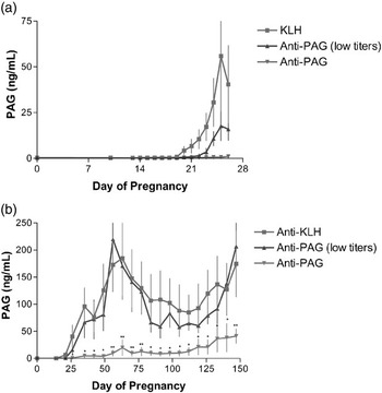
Figure 3a shows the profiles of PAG concentrations in the serum of the 29 pregnant ewes collected at Day 0, 10 and 13 to 26 after breeding. Weekly sampling throughout pregnancy is shown in Figure 3b. PAG immunoreactivity in the KLH-immunized (control) animals rose rapidly between Days 20 and 26, reaching an average concentration of 40.4 ng/ml by Day 26 (Figure 3a). When viewed over the course of the entire pregnancy, the PAG profile was biphasic – with one peak at week 9 (185 ng/ml) and the other peak at term (175 ng/ml) (Figure 3b).
Group comparisons – profiles of pregnancy-associated glycoprotein (PAG) concentrations in pregnant ewe serum: (a) PAG concentrations in ewe sera on Days 0, 10 and 13 to 26 after breeding. The PAG concentration of the anti-PAG (low) animals differed from the keyhole limpet hemocyanin (KLH) animals only at Day 25 (P < 0.01). The concentration in the anti-PAG animals differed from the KLH animals at Day 24 (P < 0.01) and Days 25 and 26 (P < 0.001). (b) PAG concentrations from ewe sera collected at weekly intervals throughout pregnancy. The PAG concentration in the anti-PAG animals significantly differed from both the KLH and ‘low’ anti-PAG animals by the 4th week and remained lower throughout pregnancy. Each data point represents the mean PAG concentration for each group ± s.e. *P < 0.01; **P < 0.001.

The concentrations between the low-titer anti-PAG animals and the KLH controls did differ significantly early in pregnancy. However, the PAG profiles eventually came to mirror one another by the 4th week of pregnancy (Figure 3). For those animals in the high-titer anti-PAG group, the average concentration of circulating PAGs deviated from the control KLH group by Day 24 post-breeding and, remained much lower than the KLH and low-titer animals throughout the remainder of the pregnancy (Figure 3b). The maximum PAG concentration measured in the high-titer animals was 41 ng/ml during the final week of pregnancy (Figure 3b).
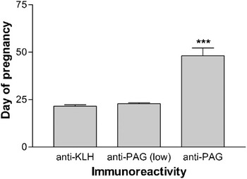
The average time at which circulating PAGs exceeded 1 ng/ml was another method used to make comparisons between groups. PAGs were detectable in the anti-KLH and anti-PAG (low-titer) animals by Days 21.6 ± 2.2 and 22.9 ± 1.3, respectively. The high-titer animals differed from the other groups; in these animals, circulating PAGs did not become detectable until Day 48.1 ± 15.6 (Figure 4) – well after the time in which a mature placenta has become established in these animals.
Group comparisons – the average day of pregnancy when the concentration of pregnancy-associated glycoprotein (PAG) in maternal sera exceeded 1 ng/ml. There was no difference between the ewes immunized with keyhole limpet hemocyanin (KLH) and those anti-PAG animals that failed to react strongly with PAG. The PAG in maternal sera of immunoreactive anti-PAG animals did not become detectable until Day 48. Each bar represents the mean ± s.d. ***P < 0.001.

The overall pregnancy rates for the ewes in this study were well above 90%. Only two of the animals failed to become pregnant or take a pregnancy to term (Table 2). Despite that approximately ⅔ of the PAG-immunized animals developed high titers to the antigens, this had no effect upon pregnancy rates, the number of lambs born per ewe, or in the birth weight of the lambs (Table 2). There was no difference in the average length of gestation between the groups (data not shown).
Pregnancy rates, average lambing rate and average lamb birth-weights between the immunization groups

KLH = keyhole limpet hemocyanin; PAG = pregnancy-associated glycoproteins.
aAnimals were placed into the low-reactive group if they failed to recognize PAGs in a direct ELISA or if the reactivity was lost upon dilution >1 : 2000.
bAnimals were placed into the high-reactive group if they could recognize PAGs in a direct ELISA at a dilution ⩾1 : 16 000.
cThere were no statistically significant differences between the values in each column.
Discussion
The PAG-1 protein has been implicated as both a luteotrophin and as an immunomodulator during pregnancy in ruminants (Del Vecchio et al., Reference Del Vecchio, Sutherland and Sasser1995a; Weems et al., Reference Weems, Lammoglia, Vera-Avila, Randel, King, Sasser and Weems1998b; Hoeben et al., Reference Hoeben, Monfardini, Opsomer, Burvenich, Dosogne, De Kruif and Beckers2000). It remains unknown if these disparate functional roles are due to the ability of PAG-1 to act in many ways (i.e., it is a multi-functional protein) or, if the protein preparations used in those studies consisted of multiple PAGs, each with distinct functions. Most of the studies describing PAG action have been completed in vitro, and the relevance of such studies to animal physiology, as a whole, is a legitimate question. With these issues in mind, the goal of this work was relatively simple and straightforward: to determine if circulating PAGs are critical for maintaining pregnancy in ewes. These experiments employed autoimmunization of ewes to suppress PAGs immunologically as a way to gain some insight into PAG function. The hypothesis being tested was that, if PAGs circulating in maternal blood were playing a major role during pregnancy, then a decrease in PAG concentrations would be reflected in increased pregnancy losses or in the birth of compromised offspring.
In the control ewes immunized against KLH, the biphasic serum profiles of PAGs were quite similar to those obtained by other laboratories (Ruder et al., Reference Ruder, Stellflug, Dahmen and Sasser1988; Ranilla et al., Reference Ranilla, Sulon, Carro, Mantecon and Beckers1994). The PAG-immunized animals that failed to develop strong reactivity toward PAGs (the ‘low-titer’ group) exhibited profiles that were essentially identical to the KLH-immunized animals. In contrast, the ability to detect circulating PAGs in the high-titer ewes was delayed and the concentration of PAGs in pregnant sera was markedly decreased (Figures 3 and 4). However, despite that approximately two-thirds of the immunized animals developed good reactivity toward PAGs, no significant effect was observed in the subsequent pregnancy (Table 2).
The inability to detect PAGs in sera from immunized animals presumably was due either to auto-antibodies interfering with the ability of the anti-PAG antibodies in the ELISA to bind to PAGs in sera, or to the selective removal of PAGs from the maternal circulation via clearance mechanisms (Simister et al., Reference Simister, Jacobowitz Israel, Ahouse and Story1997; Vaughn and Bjorkman, Reference Vaughn and Bjorkman1998; Gorgani and Theofilopoulos, Reference Gorgani and Theofilopoulos2007). At present, it is not possible to determine which mechanism was most responsible for the decrease in circulating PAG concentrations. However, one might predict that the rabbit anti-PAG antibodies employed in the ELISA probably had a greater affinity for PAGs than would the auto-anti-PAG immunoglobulins. The rabbit antibodies would have likely displaced the auto-antibodies during the overnight incubation of the sera in the ELISA plates. Obviously such points are merely conjecture, however. But regardless of the mechanism, it is likely that either the coating of circulating PAGs with auto-antibodies, or the selective removal of antibody–PAG complexes, would be predicted to interfere with PAG function in the maternal system. Clearly, the decrease in circulating PAGs did not disrupt pregnancy – suggesting that circulating PAGs are present in amounts much higher than are required for normal function, or that a systemic role of circulating PAGs may not be absolutely required for the maintenance of pregnancy.
On the other hand, paracrine-like activities of the PAGs might well remain operational in such PAG-immunized ewes. There was no way to measure the amount of immunoreactive PAGs present at extra-vascular sites near or at the maternal–placental interface, where a substantial amount of PAG immunoreactivity is known to accumulate (Wooding et al., Reference Wooding, Roberts and Green2005). What was clear was that cell-based immune-directed attack on the fetal placenta, whether through complement or activated T-cells, was clearly not pervasive in these animals since no signs of increased pregnancy failure, late term miscarriage, or birth of small lambs were observed. Therefore, it appears that local actions of the PAGs remained relatively unaffected, although further studies are needed to test this contention.
In summary, this work sought to study the function of PAGs in pregnant animals by the immunological removal or blocking of circulating PAGs in pregnant ewes. The major outcome from these studies was that auto-immunization of ewes against PAGs was successful in decreasing circulating PAGs, but did not produce an obvious impact on the pregnancy. Although these results do not exclude a systemic role for the PAGs, one can extrapolate from these data that future work on PAG function might, more productively, be focused on events taking place in the microenvironment at the placenta–uterine interface.
Acknowledgements
This work was supported by NIH Grant HD21896 and the Animal Reproductive Biology Group of the University of Missouri Food for the 21st Century Program. The authors thank Jon Bader for his help with animal handling and management. They also thank Mary P. Avalle for assistance with blood sampling. The authors acknowledge The Proteomics Center at the University of Missouri-Columbia for their assistance with the mass spectrometry analyses.