Introduction
Dementia with Lewy bodies (DLB) is the second most common cause of degenerative dementia after Alzheimer's disease (AD) and also one of the major and most challenging differential diagnoses that clinicians face at the memory clinic. A number of studies conducted in the 1990s emphasized the importance of hallucinations and delusions to diagnose DLB and distinguish it from AD (Weiner et al., Reference Weiner1996; Ballard et al., Reference Ballard1999; Hirono et al., Reference Hirono1999; Del Ser et al., Reference Del Ser, McKetih, Anand, Cicin-Sain, Ferrara and Spiegel2000; Ballard et al., Reference Ballard, O'Brien, Swann, Thompson, Neill and McKeith2001; McKeith, Reference McKeith2002). In 2005, this prior work contributed to an update of the previous consensus criteria for the diagnosis of DLB (McKeith et al., Reference McKeith1996) so that the current set of criteria consider “recurrent visual hallucinations that are typically well formed and detailed” as a core diagnostic feature of DLB, whereas hallucinations in other modalities and systematized delusions are considered supportive features “commonly present but not proven to have diagnostic specificity” (McKeith et al., Reference McKeith2005).
However, two important aspects of the neuropsychiatry of DLB remain controversial. First, to date no neuropsychiatric questionnaire has been developed specifically for the diagnosis of DLB, and it is not fully documented whether those aimed at characterizing the neuropsychiatric manifestations (NPM) of AD are useful in this differential diagnosis. Second, while a correlation between the prevalence of hallucinations and delusions and the severity of cognitive decline have been demonstrated for AD (Lyketsos et al., Reference Lyketsos, Steinberg, Tschanz, Norton, Steffens and Breitner2000) and Parkinson's disease with dementia (Aarsland et al., Reference Aarsland2007), in DLB this relationship between NPM and progression is not completely understood. These psychotic symptoms seem to correlate poorly with the progression of DLB, presumably because they appear early after the onset of cognitive deficits and then plateau (Ballard et al., Reference Ballard1999; Del Ser et al., Reference Del Ser, McKetih, Anand, Cicin-Sain, Ferrara and Spiegel2000; Rockwell et al., Reference Rockwell, Choure, Galasko, Olichney and Jeste2000; Engelborghs et al., Reference Engelborghs2005; Stavitsky et al., Reference Stavitsky2006), but these prior studies may have incurred a selection bias due to the circularity of using visual hallucinations to diagnose DLB.
Therefore our aim was two-fold: (1) To investigate the usefulness of the Neuropsychiatric Inventory (NPI; Cummings et al., Reference Cummings, Mega, Gray, Rosenberg-Thompson, Carusi and Gornbein1994) and the Columbia University Scale for Psychopathology in Alzheimer's Disease (CUSPAD; Devanand et al., Reference Devanand1992) to discriminate between probable DLB and probable AD in a memory clinic, and (2) to explore the relationship between hallucinations and delusions, and the stage of progression of cognitive and functional impairments in DLB.
Methods
Participants
Participants were patients diagnosed with DLB (n = 80) and AD (n = 85) at the Memory Disorders Unit of the Hospital Virgen del Rocío in Seville (Spain), a university tertiary referral hospital in Southern Spain. The local Institutional Review Board approved the study protocol, and the patients or their next-of-kin gave informed written consent to participate in the study. Inclusion criteria required that AD patients met the National Institute of Neurological and Communicative Disorders and Stroke and Alzheimer's Disease and Related Disorders Association (NINCDS–ADRDA) criteria for probable AD (McKhann et al., Reference McKhann1984), and DLB patients met the Third Consortium criteria for the diagnosis of probable DLB (McKeith et al., Reference McKeith2005). Specifically, besides the central feature of dementia, all DLB patients had at least one core feature other than visual hallucinations (fluctuating cognition or spontaneous parkinsonism) and one or more suggestive features, including a positive dopamine transporter (DAT) SPECT scan (123I-FP-CIT DATscan, GE Healthcare). Thus, the study protocol was designed so that visual hallucinations were recorded but not used in the diagnostic process of DLB. Parkinsonism was considered present when at least two parkinsonian signs among rigidity, tremor, bradykinesia, and gait instability were present in the neurological examination. Among the core features listed above, 27 (33.7%) patients were noted to have cognitive fluctuations using the Mayo Fluctuation Scale (Ferman et al., Reference Ferman2004), 40 (50.0%) patients reported visual hallucinations, and 74 (92.5%) patients exhibited parkinsonism at the neurological examination. In our memory unit, DAT scan is routinely performed as part of the diagnostic work-up in all patients with possible and probable DLB. One of the authors (DGS), who was blinded to the clinical diagnosis, visually assessed and confirmed the striatal decrease in the radiotracer uptake typically seen in DLB patients (McKeith et al., Reference McKeith2007). Patients with probable AD did not undergo DAT scan but the proportion of positive DAT scan in this group is very low (6.9%, McKeith et al., Reference McKeith2007). Regarding other DLB suggestive features, no prior episode of severe neuroleptic sensitivity was noted, and 24 (30%) patients reported sleep disturbances consistent with rapid eye movement (REM) sleep behavior disorder (i.e. answered “yes” to the question “Have you ever been told, or suspected yourself that you seem to ‘act out your dreams’ while asleep, for example, punching, flailing your arms in the air, making running movements, etc.?”). To prevent bias due to the NPM of vascular dementia, patients with significant ischemic burden (scoring 1 or more in the Fazekas scale (Fazekas et al., Reference Fazekas, Chawluk, Alavi, Hurtig and Zimmerman1987)) in their routine brain CT/MRI scan were excluded from the study.
Data collection
Data collection included age, gender, treatment with neuroleptic and dopaminergic drugs and with acetyl-cholinesterase inhibitors (AChEi), duration of clinical disease from symptom onset, and time from clinical onset to first hallucination and delusion.
Neuropsychological evaluation
Patients and their main caregiver underwent a comprehensive neuropsychiatric evaluation that included the ten-item NPI and CUSPAD. The ten-item NPI is a questionnaire that evaluates the presence, frequency, and severity of ten different NPMs (delusions, hallucinations, agitation, depression, anxiety, euphoria, apathy, disinhibition, irritability, and aberrant motor behavior; Cummings et al., Reference Cummings, Mega, Gray, Rosenberg-Thompson, Carusi and Gornbein1994). The CUSPAD is a structured questionnaire that describes with great detail the subtypes of delusions and hallucinations that can be found in AD patients (Devanand et al., Reference Devanand1992). The results of both NPI and CUSPAD here refer to the NPM present within the month prior to the evaluation. In this study, the subtypes of delusional misidentifications inquired as part of the CUSPAD were those described by Ballard et al. (Reference Ballard1995; Reference Ballard, Lowery, Harrison, McKeith, Perry, McKeith and Perry1996) and Burns et al. (Reference Burns, Jacoby and Levy1990).
The level of global cognitive deterioration was quantitatively estimated with the Mini-Mental State Examination (MMSE; Folstein et al., Reference Folstein, Folstein and McHugh1975), whereas the functional impact of cognitive decline on basic and instrumental activities of daily living was assessed with the Spanish version of the Interview for Deterioration in Daily Living Activities in Dementia (IDDD; Teunisse et al., Reference Teunisse, Derix and Crevel1991) (Spanish version validated by Böhm et al., Reference Böhm, Peña-Casanova, Aguilar, Hernandez, Sol and Blesa1998). The IDDD has a cut-off score of >36 in the Spanish population, with a higher IDDD score indicating a more severe functional impairment.
Statistical analyses
The frequencies of each NPM included in the NPI and the CUSPAD subtypes of delusions and hallucinations were compared between the DLB and the AD groups using χ 2 with Fisher's exact test. Following methods published elsewhere (Tiraboschi et al., Reference Tiraboschi, Salmon, Hansen, Hofstetter, Thal and Corey-Bloom2006), we calculated the sensitivity and specificity to DLB versus AD of each NPM, as well as the positive likelihood ratio (LR+) and the odds ratio (OR) of the association between each NPM and DLB versus AD. If any cell of the contingency tables had a zero, then 0.5 was added to all cells to be able to calculate OR. Sensitivity is the proportion of patients with a diagnosis of probable DLB who report a given NPM. Specificity is the proportion of patients with a diagnosis of probable AD who do not report a given NPM. OR represents how many times is a given NPM more likely to be present in DLB than in AD and LR+ represents how many times is DLB more likely to happen than AD if a given NPM is present. Unlike positive and negative predictive values, LR+ is independent of prevalence (LR+ = sensitivity/(1 – specificity).
The duration of clinical disease and the latency to the first delusion and first hallucination were compared between both groups using the Mann–Whitney U test. The frequency of hallucinations and delusions was correlated with the measures of clinical severity using Pearson's χ 2 test for trend. For these analyses, the MMSE scores were categorized as mild (MMSE > 20), moderate (MMSE = 10–20), and severe (MMSE < 10) cognitive impairment, following the guidelines of the UK National Institute for Clinical Excellence (NICE; 2011) and in agreement with Ballard et al. (Reference Ballard1999). Likewise, the level of functional impairment was categorized according to the distribution of IDDD scores as mild (IDDD < 42), moderate (IDDD = 42–55), and severe (IDDD > 55) (see the Supplemental Content 1 for further details about these subgroups). Lastly, survival analyses were performed with the appearance of the first hallucination or delusion as the end-point and with the Kaplan–Meier curves representing the survival time free of the symptom (latency). Patients free of hallucinations/delusions were identified at the time of clinical evaluation. Log-rank tests were run to compare survival curves from AD and DLB patients. The significance level was set at a two-tailed p < 0.05. All analyses were performed with SPSS version 16.0 (IBM) or GraphPad Prism version 5.0 for Mac (GraphPad Software Inc., La Jolla, CA). (See supplementary material attached to the electronic version of this paper at www.journals.cambridge.org/jid_IPG.)
Results
Demographic characteristics
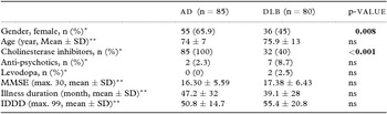
Table 1 summarizes the demographic characteristics of DLB and AD. No differences were found between groups in age, duration of clinical disease (from symptom onset), severity of cognitive decline (MMSE), and functional impairment (IDDD), indicating that any significant difference observed in the frequency or phenomenology of NPM cannot be just explained by a different stage of the disease. Two AD patients and seven DLB patients were taking risperidone, and two DLB patients (but no AD patient) were taking levodopa at the time of evaluation (p > 0.05). All 85 AD patients and 32 DLB patients were on AChEi at the time of evaluation (100% AD vs. 40% DLB, p < 0.0001). However, the frequency of hallucinations did not significantly differ between treated and non-treated DLB patients (62.5% with AChEi vs. 43.75% without AChEi, p = 0.1154), and the frequency of delusions was actually significantly higher among the DLB patients treated (62.5% with AChEi vs. 37.5% without AChEi, p = 0.0398), suggesting that it is unlikely that both symptoms are masked by the AChEi treatment.
Demographic and clinical characteristics of the study groups

Notes: Values expressed as mean ± SD or percentage as indicated. Statistically significant differences are in bold.
*Pearson's χ 2 with Fisher's exact test; **Mann–Whitney U test.
MMSE = Mini-Mental State Examination; IDDD = Interview for Deterioration in Daily Living Activities in Dementia.
Frequency of hallucinations and delusions
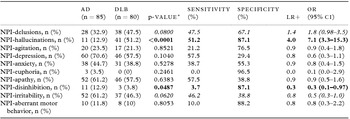
We used the ten-item NPI (Cummings et al., Reference Cummings, Mega, Gray, Rosenberg-Thompson, Carusi and Gornbein1994) as a screening tool for the presence of NPM in both diseases within the last month. Importantly, despite both groups having similar disease severity, the results of NPI revealed that disinhibition is significantly more common in AD than in DLB, whereas hallucinations are more frequent in DLB than in AD, and delusions tend to be more prevalent also in DLB. Although the sensitivity of these NPI items to diagnose DLB or AD was relatively low, these differences yielded a very high specificity of hallucinations for DLB and that of disinhibition for AD (Table 2).
Neuropsychiatric Inventory (NPI)

Notes: *Pearson's χ 2 with Fisher's exact test.
Statistically significant differences are in bold, and trends toward statistical significance are in italics.
AD = Alzheimer's disease; DLB = dementia with Lewy bodies; NPI = Neuropsychiatric Inventory; LR+ = positive likelihood ratio; OR = odds ratio; CI = confidence interval.
The recurrence of hallucinations and delusions was assessed using the two categories included in the NPI, that is, ≤1/week versus >1/week or daily. While no significant differences were observed regarding delusions, hallucinations were significantly more persistent in DLB than in AD patients (p = 0.04).
Phenomenology of hallucinations and delusions
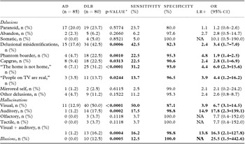
To further describe the characteristics of delusions and hallucinations, we applied CUSPAD (Devanand et al., Reference Devanand1992) to both AD and DLB patients. Although CUSPAD was originally designed to characterize the NPM associated with AD, we took advantage of the detailed categorization of hallucinations and delusions to investigate the existence of distinct patterns specific to AD or DLB. The CUSPAD results revealed that both visual and auditory hallucinations are significantly more frequent in DLB than in AD. Although auditory hallucinations were very specific to DLB, they were almost always accompanied by visual hallucinations and the occurrence of isolated auditory hallucinations was extremely rare. The most frequent auditory hallucinations were voices of people speaking (35.3%). Some patients reported hearing noises behind them and finding a person (a visual hallucination) when they turned back around. Other auditory hallucinations involved well-defined sounds (23.5%) such as a doorbell ringing, people's footsteps, and unintelligible whisperings and voices. Of note, the occurrence of illusions, defined as perceptive misinterpretation of real visual stimuli, was observed exclusively in DLB patients (Table 3).
The Columbia University Scale for the Psychopathology of Alzheimer Disease (CUSPAD)

Notes: *Pearson's χ 2 with Fisher's exact test.
Statistically significant differences are in bold.
AD = Alzheimer's disease; DLB = dementia with Lewy bodies; LR+ = positive likelihood ratio; NA = not applicable; OR = odds ratio; CI = confidence interval.
In addition, the CUSPAD results indicated that delusional misidentifications are highly specific of DLB, whereas paranoid delusions do not help to discriminate between the two diseases, and other types of delusions, such as abandon and somatic delusions, are similarly rare in both dementias. Among the delusional misidentifications, the phantom boarder delusion, the Capgras delusion, the delusion that “people on TV are in the room,” and the delusion that “their home is not their home” were significantly more common in DLB than in AD, whereas the mirrored self-misidentification was similarly rare in both dementias (Table 3).
Relationship of NPM with the clinical severity of DLB and AD
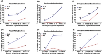
To investigate the relationship between hallucinations and delusions and the progression of dementia in both AD and DLB, we correlated their presence with the stages of cognition and functional decline defined by the MMSE and IDDD score intervals (see Methods), and with the duration of clinical illness from symptom onset. Figures 1 and 2 summarize the results of these analyses (see also Supplemental Content 1 for detailed analysis of relationship between hallucinations/delusional misidentifications and cognitive and functional impairment).
Relationship between neuropsychiatric manifestations and cognitive and functional decline in DLB and AD. Frequencies of visual hallucinations (a and d), auditory hallucinations (b and e), and delusional misidentifications (c and f) are depicted for DLB and AD using MMSE (a–c) and IDDD (d–f) intervals to categorize the severity of cognitive and functional decline. *p < 0.05, **p < 0.01, ***p < 0.0001, Pearson's χ 2 with Fisher's exact test between DLB and AD within the same MMSE or IDDD interval.

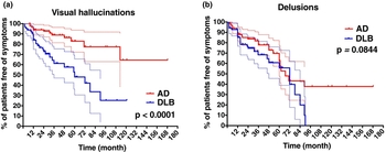
Survival free of visual hallucinations and delusions in DLB and AD. The survival free of visual hallucinations (a) and delusions (b) is represented with the Kaplan–Meier curves for DLB and AD patients based on the date of appearance of the first hallucination or systematized delusion with respect to the clinical onset of the disease. Ticks along the curves represent censored patients, that is, those who did not have visual hallucinations or delusions until the moment of the clinical evaluation. p-values correspond to the Log-rank test comparing AD and DLB curves.

Visual hallucinations
Visual hallucinations were significantly more frequent in DLB than in AD patients from the early stages of the disease. Indeed, the prevalence of visual hallucinations paralleled the progression of cognitive and functional impairment in DLB, whereas they only appeared at severe stages of the disease in AD (Figure 1a). The latency to the first visual hallucination was significantly shorter in DLB than in AD patients (17.0 ± 24.0 months vs. 36.0 ± 32.0 months, p = 0.023), confirming that DLB patients have visual hallucinations much earlier than AD patients during the course of their diseases. Indeed, 15 DLB patients (18.75%) reported visual hallucinations at presentation, whereas no AD patient did. Analysis of survival free of hallucinations excluding these 15 DLB patients confirmed that DLB patients develop visual hallucinations significantly earlier than AD patients (Figure 2a).
Auditory hallucinations
As mentioned above, auditory hallucinations in DLB patients were typically associated with visual hallucinations. In agreement with this observation and supporting the idea that visual hallucinations precede auditory hallucinations, Figure 1 shows that auditory hallucinations tend to appear in moderate and severe stages of DLB. In sharp contrast, auditory hallucinations are very infrequent at any clinical stage of AD.
Delusional misidentifications
Delusional misidentifications paralleled the progression of cognitive and functional impairment in both DLB and AD. These correlations revealed that they were significantly more frequent at moderate and severe stages of DLB as compared with AD, but not at earlier stages. In keeping with this observation, the latency from symptom onset to the first delusion was not significantly different between DLB and AD (23.6 ± 29.0 months vs. 31.3 ± 26.0 months, p > 0.05). Analysis of survival free of delusions only yielded a trend toward a significant difference between the DLB and the AD groups (Figure 2b).
Discussion
Our results indicate that NPI and CUSPAD are useful tools to discriminate between DLB and AD in the clinical setting. While originally designed to evaluate the NPM of AD, the CUSPAD revealed that hallucinations and delusions in DLB and AD differ not only quantitatively but also qualitatively. Besides being more prevalent in DLB than in AD, both hallucinations and delusions also have distinct patterns of manifestation and progression. Importantly, the differences observed cannot be explained by differences in age, clinical disease duration, or dementia severity, as these characteristics were equivalent in both groups of patients.
Visual hallucinations
Visual hallucinations were not only more frequent and more persistent in DLB but they appeared at an earlier stage of dementia and tracked better with the progression of disease as compared with AD. The higher prevalence and persistence of visual hallucinations in DLB compared with AD is in agreement with previous studies (Ballard et al., Reference Ballard1995; Reference Ballard, Lowery, Harrison, McKeith, Perry, McKeith and Perry1996; Reference Ballard, O'Brien, Swann, Thompson, Neill and McKeith2001; Del Ser et al., Reference Del Ser, McKetih, Anand, Cicin-Sain, Ferrara and Spiegel2000; Rockwell et al., Reference Rockwell, Choure, Galasko, Olichney and Jeste2000; Engelborghs et al., Reference Engelborghs2005; Auning et al., Reference Auning2011). The earlier onset of visual hallucinations with respect to AD is also consistent with prior studies (Ballard et al., Reference Ballard, O'Brien, Swann, Thompson, Neill and McKeith2001; Auning et al., Reference Auning2011). However, compared with our study, these other studies reported an overall higher prevalence of visual hallucinations in their DLB sample (i.e. 72% (Ballard et al., Reference Ballard1999), and 77.5% (Del Ser et al., Reference Del Ser, McKetih, Anand, Cicin-Sain, Ferrara and Spiegel2000)). In addition, other authors have reported no or a poor correlation between the frequency of visual hallucinations and the progression of cognitive impairment in DLB (Ballard et al., Reference Ballard1999; Del Ser et al., Reference Del Ser, McKetih, Anand, Cicin-Sain, Ferrara and Spiegel2000; Rockwell et al., Reference Rockwell, Choure, Galasko, Olichney and Jeste2000; Engelborghs et al., Reference Engelborghs2005; Stavitsky et al., Reference Stavitsky2006). These differences could be explained by their use of the core feature of visual hallucinations as diagnostic criteria.
Hallucinations in other modalities
Although auditory hallucinations were substantially less frequent than visual hallucinations, they were very highly specific to DLB. Likewise visual hallucinations, we observed a lower prevalence of auditory hallucinations compared with that reported by other authors (19–39%; Ballard et al., Reference Ballard1999; Reference Ballard, O'Brien, Swann, Thompson, Neill and McKeith2001). Of note, while auditory hallucinations have been previously described to occur independently of visual hallucinations (Ballard et al., Reference Ballard, O'Brien, Swann, Thompson, Neill and McKeith2001), in our DLB sample auditory hallucinations seldom presented alone, but were usually accompanied and preceded by visual hallucinations. Indeed, they typically develop at moderate and late stages of the disease, thus justifying their consideration as a supportive rather than a suggestive or core feature (McKeith et al., Reference McKeith2005). Along the same line, hallucinations in other sensory modalities (olfactory and tactile), while absent in AD patients, were reported even less frequently than auditory hallucinations by our DLB patients (8% each) in agreement with other authors (Weiner et al., Reference Weiner1996).
Illusions
Illusions were also infrequent but highly specific of DLB. Interestingly, an earlier study also using the CUSPAD obtained a prevalence of illusions as 32% in DLB, which was significantly higher than that in the AD group (Stavitsky et al., Reference Stavitsky2006). Hence, although more studies are needed, illusions might be useful as a supportive feature for the diagnosis of DLB.
Delusions
Likewise visual hallucinations, delusions also tended to be more frequent in DLB than in AD, but the most striking difference regarding delusions between both dementias was qualitative: while paranoid delusions occurred at a similar frequency in both groups of patients, delusional misidentifications were far more common in DLB than in AD patients, and therefore more specific of DLB. The delusional misidentifications more commonly associated with DLB than with AD included phantom boarder delusion, the Capgras delusion, the delusion that “their home is not their home,” and the delusion that “people on TV are in the room.” These findings largely agree with the descriptions of previous studies (Ballard et al., Reference Ballard1999; Hirono et al., Reference Hirono1999; Del Ser et al., Reference Del Ser, McKetih, Anand, Cicin-Sain, Ferrara and Spiegel2000; Rockwell et al., Reference Rockwell, Choure, Galasko, Olichney and Jeste2000) and add evidence in favor of the usefulness of the subtype of delusional misidentifications as a supportive feature for the diagnosis of DLB and its differentiation from AD. However, unlike visual hallucinations, delusional misidentifications appear primarily associated with moderate and severe stages of DLB, thus precluding an early differential diagnosis between DLB and AD and arguing against their upgrade to the core diagnostic features of DLB.
Strengths and limitations
The main strengths of our study are the large size of the DLB and AD groups, their consecutive enrollment based on current consensus criteria, the use of a positive dopamine transporter SPECT scan in substitution of visual hallucinations for the diagnosis of probable DLB, and the use of validated detailed questionnaires to systematically evaluate the NPM associated with both dementias.
Several limitations should also be recognized: (1) All the study participants were recruited in a memory clinic of a teaching hospital so that our conclusions might not be generalizable to the general population. Community-based studies and studies based at a movement disorders clinic or a psychiatry clinic may well yield different results regarding the prevalence and characteristics of the psychotic symptoms of DLB. (2) The design of the study was cross-sectional rather than longitudinal and our conclusions might be affected by the bias inherent to this class of studies. We tried to minimize sampling bias by enrolling the participants in a consecutive fashion. Recall bias should not be concerning, as DLB and AD patients had similar severity of cognitive decline. (3) The diagnosis of AD and DLB was based on clinical criteria, and neuropathological confirmation was not available. Considering the high pathological overlap between AD and DLB described in autopsy studies, a certain degree of cross-contamination in both groups is expected. We attempted to minimize this source of noise by strictly applying current clinical diagnostic criteria to enroll only patients with probable AD and probable DLB, and by supporting the diagnosis of probable DLB with a positive dopamine transporter SPECT scan (O'Brien et al., Reference O'Brien2004; McKeith et al., Reference McKeith2007; Walker et al., Reference Walker2007). However, it should also be noted that the requirement of a positive dopamine transporter SPECT scan for the diagnosis of probable DLB might have introduced another selection bias in our sample, such as a higher prevalence of parkinsonism. All patients enrolled in this study have now been followed in our memory clinic for at least two years after the study evaluation without changes in the initial diagnosis.
In summary, we observed that the differential early diagnosis between AD and DLB can benefit from a detailed neuropsychiatric evaluation, and that both NPI and CUSPAD are valuable tools to discriminate between both diseases. In addition, extending previous observations, we found that visual hallucinations are more frequent in DLB than in AD at all stages of dementia and track better with dementia severity in DLB than in AD. By contrast, auditory hallucinations and delusional misidentifications (a specific subtype of delusions), while highly specific of DLB, are more frequent only at moderate and severe stages.
Conflict of interest
None
Description of authors’ roles
Aida Suárez-González and Alberto Serrano-Pozo equally contributed to this work. Aida Suárez-González was involved in research project (formulation of research questions, organization, and execution), statistical analysis (execution, data interpretation, review, and critique), and manuscript (writing of the first draft, review, and critique). Alberto Serrano-Pozo was also involved in research project (organization and execution), statistical analysis (design, execution, data interpretation, review, and critique), and manuscript (writing, review, and critique). Eva M. Arroyo-Anlló was involved in research project (organization) and manuscript (review and critique). Emilio Franco-Macías was involved in research project (execution); and Juan Polo was involved in statistical analysis (design, execution, review, and critique), manuscript (review and critique). David García-Solís was involved in research project (execution); and Eulogio Gil-Néciga was involved in research project (formulation of research questions and execution) and manuscript (writing, review, and critique).
Acknowledgments
The authors thank patients and caregivers involved in the research at the Memory Disorders Unit of the University Hospital Virgen del Rocío in Seville (Spain). Alberto Serrano-Pozo was supported by a fellowship from the Fondo de Investigaciones Sanitarias – Instituto de Salud Carlos III of the Ministerio de Ciencia e Innovación (CM06/00161), Madrid, Spain.