Implications
The results of this study indicate that the dietary supplementation of seaweed extract (SWE) or galacto-oligosaccharides (GOS) improved the intestinal health of pigs after an experimental challenge with Salmonella Typhimurium. Dietary supplementation with SWEs reduced faecal shedding of S. Typhimurium. This could provide a dietary means to reduce faecal shedding of S. Typhimurium in pigs and this could eventually reduce contamination of pork in slaughter houses.
Introduction
S. Typhimurium is a foodborne pathogen that can cause serious illness in humans, and is the predominant serotype found in pigs and humans in Europe (Prendergast et al., Reference Prendergast, Duggan, Gonzales-Barron, Fanning, Butler, Cormican and Duffy2009). Pork and pork products are recognised as sources of S. Typhimurium infection in humans (Prendergast et al., Reference Prendergast, Duggan, Gonzales-Barron, Fanning, Butler, Cormican and Duffy2009). Salmonella infections in pigs are often asymptomatic, making diagnosing on farms very difficult (Rostagno and Callaway, Reference Rostagno and Callaway2012).
Supplementation with in-feed antibiotics have been widely used to reduce pathogen infection in swine, however the European Union ban of these in-feed growth promotors has prompted the search for alternatives (Casewell et al., Reference Casewell, Friis, Marco, McMullin and Phillips2003). The ban on the inclusion of antimicrobial growth promotors in feed is likely to have caused adverse effects on public and animal health due to increased likelihood of exposure to higher level of (foodborne) pathogens such as Salmonella (Rostagno and Callaway, Reference Rostagno and Callaway2012). Recently, SWE and GOS have been explored as novel sources of bioactive compounds that contain immunomodulatory and antimicrobial properties (McDonnell et al., Reference McDonnell, Figat and O’Doherty2010; Searle et al., Reference Searle, Cooley, Jones, Nunez, Crudgington, Weyer, Dugdale, Tzortzis, Collins, Woodward and La Ragione2010; Walsh et al., Reference Walsh, Sweeney, O’Shea, Doyle and O’Doherty2013).
The main bioactive polysaccharides from the seaweed species Laminaria are laminarin and fucoidan. Supplementation with these polysaccharides has resulted in decreased abundance of Enterobacteriaceae and pathogenic strains of Escherichia coli, while improving the number of Lactobacillus spp. in the large intestine of pigs (McDonnell et al., Reference McDonnell, Figat and O’Doherty2010; Walsh et al., Reference Walsh, Sweeney, O’Shea, Doyle and O’Doherty2013; Heim et al., Reference Heim, Sweeney, O’Shea, Doyle and O’Doherty2014). This indicates that SWE supplementation has potential to reduce S. Typhimurium infections and shedding in pigs. Supplementation with SWE has shown to modify the gene expression of several pro- and anti-inflammatory cytokines in both challenged and unchallenged ileal and colonic tissues (Smith et al., Reference Smith, O’Doherty, Reilly, Ryan, Bahar and Sweeney2011; Sweeney et al., Reference Sweeney, Collins, Reilly, Pierce, Ryan and O’Doherty2012; Walsh et al., Reference Walsh, Sweeney, O’Shea, Doyle and O’Doherty2013).
Galacto-oligosaccharides are classed as non-digestible prebiotics that exhibit beneficial effects on the host by stimulating the growth and/or activity of beneficial bacteria such as Bifidobacteria and Lactobacillus (Gibson and Roberfroid, Reference Gibson and Roberfroid1995). Galacto-oligosaccharides possess immunomodulatory properties by either modifying the intestinal microbiota and/or ameliorating the effects of intestinal inflammation (Vulevic et al., Reference Vulevic, Drakoularakou, Yaqoob, Tzortzis and Gibson2008; Tanabe and Hochi, Reference Tanabe and Hochi2010). Previous research has shown that GOS inclusion in an in vitro assay with mucus-secreting three-dimensional human colonic cells can reduce the colonisation and invasion of S. Typhimurium (Searle et al., Reference Searle, Cooley, Jones, Nunez, Crudgington, Weyer, Dugdale, Tzortzis, Collins, Woodward and La Ragione2010).
The objective of the present study was to investigate the effects of dietary supplementation of GOS and SWE on parameters of intestinal health after in vivo S. Typhimurium challenge. It was hypothesised that GOS and SWE would decrease S. Typhimurium numbers and faecal shedding, and reduce the pro-inflammatory response in the intestinal tract after S. Typhimurium challenge. Both GOS and SWE were likely to have the potential to alter the Lactobacillus and Bifidobacterium numbers in colonic and caecal digesta in the presence of S. Typhimurium.
Material and methods
All procedures described were conducted under experimental license from the Irish Department of Health in accordance with the Cruelty to Animals Act 1876 and the European Communities (Amendments of the Cruelty to Animals Act, 1876) Regulations.
Experimental design, animal management and diets
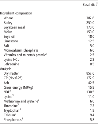
The experiment was designed as a complete randomised block design. In total, 30 female pigs were selected (30.9 kg, SD 3.65 kg) and blocked on basis of initial weight and litter of origin. Within each block, they were randomly assigned to one of three treatments (n=10 pigs/treatment) and housed in individual pens. The pigs were offered the following diets: (T1) basal diet, (T2) basal diet+2.5 g GOS/kg diet (Clasado Ingredients Ltd, Buckinghamshire, UK) and (T3) basal diet+SWE (containing 180 mg laminarin/kg diet+340 mg fucoidan/kg diet). The SWE (NeoShield2®) containing laminarin and fucoidan was derived from Laminaria spp. and sourced from BioAtlantis Ltd (Tralee, Co. Kerry, Ireland). The diet composition is shown in Table 1. The basal diet contained 13.8 MJ/kg of digestible energy and 9.2 g/kg standardized ileal digestible lysine contents. The GOS and SWE were included with the basal diet at manufacture.
Composition and chemical analysis (g/kg, unless otherwise indicated)

1 Treatments: (1) basal diet; (2) basal diet+2.5 g galacto-oligosaccharides/kg feed; (3) basal diet+seaweed extract containing 180 mg laminarin/kg feed and 340 mg fucoidan/kg feed.
2 The premix provided vitamins and minerals (per kg diet) as follows: 4.2 mg of retinol, 0.07 mg of cholecalciferol, 80 mg of α-tocopherol, 120 mg of copper as copper sulphate, 100 mg iron as ferrous sulphate, 100 mg of zinc as zinc oxide, 0.3 mg of selenium as sodium selenite, 25 mg of manganese as manganous oxide, 0.2 mg of iodine as calcium iodate on a calcium sulphate/calcium carbonate carrier, 2 mg of thiamine, 15 μm of cyanocobalamin, 7 mg of pantothenic acid, 2 mg of riboflavin, 7 mg of niacin, 3 mg of adenine and 100 mg of phytase (Natuphos) (Nutec, Co. Kildare, Ireland).
3 Calculated for tabulated nutritional composition (Sauvant et al., 2004).
Following an 11-day dietary adaptation period, all pigs were challenged by oral gavage with 1×10⁸ colony-forming units S. Typhimurium (day 0). Fresh faecal samples were collected pre-challenge to confirm that all pigs were S. Typhimurium negative using a presence/absence test. As well, fresh faeces were collected on days 2, 4 and 7 post-challenge to enumerate S. Typhimurium. Fresh faeces were collected through grab sampling. Initial weight, weight at challenge and weight at euthanasia were recorded. The pigs had ad libitum access to feed and water. Feed was available up to weighing of the pigs and then weighed back in order to calculate average daily feed intake and feed efficiency.
Post-challenge, diarrhoea scores were recorded daily by the same operator, using a subjective score on a one-point scale ranging from 1 to 5. The following scoring system was used: 1=hard, firm faeces; 2=slightly soft faeces; 3=soft, partially formed faeces; 4=loose, semi-liquid faeces; and 5=watery, mucous-like faeces (Sweeney et al., Reference Sweeney, Dillon, Fanning, Egan, O’Shea, Figat, Gutierrez, Mannion, Leonard and O’Doherty2011).
Bacterial challenge
The pigs were challenged on day 0 with Salmonella enterica subspecies enterica serovar typhimurium PT12 (hereafter referred to as S. Typhimurium), a porcine isolate obtained from the culture collection of the Central Veterinary Research Laboratory (Backweston Laboratory Campus, Co. Kildare, Ireland). S. Typhimurium culture was prepared from protect beads (Technical Service Consultants, Lancashire, UK) stored at −20°C in 10 ml tryptic soya broth (TSB) (Oxoid, Hampshire, UK) and incubated at 37°C overnight. Following overnight incubation, an aliquot of this culture was aseptically transferred into TSB and incubated at 37°C for 18 h to achieve a stationary-phase culture. The culture was centrifuged at 3000×g for 10 min. The pellet was re-suspended in phosphate buffer saline (PBS) (Oxoid).The pigs were humanely sacrificed on day 17 post-challenge by lethal injection with pentobarbital sodium (Euthatal Solution, 200 mg/ml; Merial Animal Health Ltd, Essex, UK) at a rate of 0.71 ml/kg BW into the cranial vena cava, and the entire intestinal tract was immediately removed. Digesta from the caecum, colon and rectum was collected into sterile containers (Sarstedt, Co. Wexford, Ireland) and frozen immediately to quantify S. Typhimurium, Bifidobacteria and Lactobacilli. Tissue from the tonsils, ileo-caecal lymph node and mesenteric lymph node was collected into sterile containers and frozen immediately to quantify S. Typhimurium. In addition, tissue samples were taken from the ileum and second loop of the proximal colon for cytokine (interleukin (IL)-1α, IL-1β, IL-6, IL-8, IL-10, IL-17A, IL-18, IL-22, tumour necrosis factor-α and interferon γ (IFN-γ)) and other immune-related (mucin 2B, trefoil factor 3 (TFF3), caspase-1 and regenerating islet-derived protein 3-γ (Reg3-γ)) gene profiling by quantitative real-time PCR (qPCR). Ileum and colon tissues were excised, emptied by dissecting them along the mesentery and rinsed using sterile PBS. Tissue sections of 1 cm2, which had been stripped of overlying smooth muscle, were cut from each tissue and stored in 5 ml RNAlater® solution (Applied Biosystems, Foster City, CA, USA) overnight at 4°C. RNAlater® was then removed before storing the samples at −20°C.
Microbiology: DNA extraction and quantitative PCR
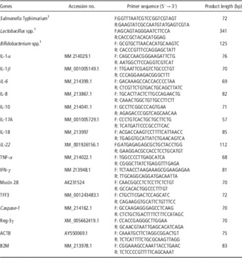
Microbial genomic DNA was extracted from faecal and colonic, caecal and rectal digesta samples using a QIAamp DNA stool kit (Qiagen, West Sussex, UK) in accordance with the manufacturer’s instructions. Microbial genomic DNA was extracted from tonsils, mesenteric and ileo-caecal lymph nodes using a DNeasy Blood & Tissue kit (Qiagen) in accordance with the manufacturer’s instructions. Quantity and quality of DNA was assessed using a NanoDrop ND1000 Spectrophotometer (Thermo Scientific, Wilmington, DE, USA). Standard curves were generated as described by O’Shea et al. (Reference O’Shea, Sweeney, Bahar, Ryan, Thornton and O’Doherty2012). In brief, genomic DNA from all faeces, colonic or caecal digesta samples was pooled and amplified through routine PCR using S. Typhimurium, bifidobacteria and Lactobacilli specific primers (Table 2). The S. Typhimurium primer was designed using Primer Express software (Applied Biosystems) and synthesised by MWG Biotech (Buckinghamshire, UK). The target specific chosen was hyper-invasive locus A, a Type III secretory system regulator previously applied by McCabe et al. (Reference McCabe, Burgess, O’Regan, McGuinnes, Barry, Fanning and Duffy2011) for detection of S. Typhimurium. Validation of the S. Typhimurium primer showed a slope of 3.348 and an efficiency of 98.82%. Serial dilutions of these amplicons served to generate standard curves using qPCR (ABI 7500 Real-Time PCR System; Applied Biosystems), allowing estimations of absolute quantification on the basis of gene copy number (GCN). Quantitative real-time PCR was performed in a final reaction volume of 20 µl containing 3 µl of template DNA, 1 µl of forward (100 pM) and 1 µl of reverse primers (100 pM), 10 µl SYBR Green PCR Master Mix (Applied Biosystems) and 5 µl nuclease-free water. The thermal cycling conditions involved an initial denaturation step at 95°C for 10 min followed by 40 cycles of 95°C for 15 s and 65°C for 1 min. Dissociation analyses of the qPCR product were performed to confirm the specificity of the resulting qPCR products. All samples were prepared in duplicate. A positive control containing template DNA of the S. Typhimurium PT12 that the pigs were challenged with was used, as well as a negative control containing distilled water instead of template DNA to ensure the specificity of the assay. The mean cycle threshold values of duplicates of each sample were used for calculations. The limit of detection (2.24 log GCN) was determined using the method described by Fachmann et al. (Reference Fachmann, Josefsen, Hoorfar, Nielsen and Löfström2015).
Porcine oligonucleotide primers used for quantitative real-time PCR

F=forward; R=reverse; IL=interleukin; TNF-α=tumour necrosis factor α; IFN-γ=interferon γ; TFF3=trefoil factor 3; Reg-3γ=regenerating islet-derived protein-3 γ; ACTB=β-actin; B2M=b2-microglobulin.
1 Reference Salmonella primer: McCabe et al. (Reference McCabe, Burgess, O’Regan, McGuinnes, Barry, Fanning and Duffy2011); Lactobacillus primer: Delroisse et al. (Reference Delroisse, Boulvin, Parmentier, Dauphin, Vandenbol and Portetelle2008); Bifidobacterium primer: Penders et al. (Reference Penders, Vink, Driessen, London, Thijs and Stobberingh2005).
RNA extraction, complementary DNA synthesis and quantitative real-time PCR
Ribonucleic acid was extracted from ~50 mg of tissue samples using the GenElute Mammalian Total RNA Miniprep Kit (Sigma-Aldrich Ireland Ltd, Co. Wicklow, Ireland) according to the manufacturer’s instructions. Total RNA was quantified using 1.5 µl of total RNA on a NanoDrop Spectrophotometer ND1000 . Samples with a 260 : 280 ratio ≥2.0 were considered suitable for complementary DNA (cDNA) synthesis. Total RNA integrity (i.e. quality and quantity) was assessed by analysing 1 µl of total RNA using the Agilent 2100 Bioanalyzer version A.02.12 (Agilent Technologies, Santa Clara, CA, USA) using RNA Nano LabChips® (Caliper Technologies Corporation, Mountain View, CA, USA). Complementary DNA synthesis was performed using 1 µg of total RNA and oligo(dT)20 primers in a final reaction volume of 20 μl using High-Capacity cDNA Reverse Transcription kit (Invitrogen Life Technologies, Inc., Co. Dublin, Ireland) following the manufacturer’s instructions. The final reaction volume of 20 μl was adjusted to 250 μl using nuclease-free water. All primers for the cytokines IL-1α, IL-1β, IL-6, IL-8, IL-10, IL-17A, IL-18, IL-22, tumour necrosis factor-α (TNF-α) and IFN-γ, mucin 2B, TFF3, caspase-1 and Reg3-γ were designed using Primer Express software and synthesised by MWG Biotech. Primer sequences are presented in Table 2. The efficiency of all primer sets was established using a semi-log curve of quantity v. control of twofold serial dilutions of cDNA as reported previously by Smith et al. (Reference Smith, O’Doherty, Reilly, Ryan, Bahar and Sweeney2011). Six previously validated reference genes (Ryan et al., Reference Ryan, Collins, O’Doherty and Sweeney2010) were evaluated for stability in a subset of samples using geNorm (Vandesompele et al., Reference Van Soest, Robertson and Lewis2002), which included: β-actin (ACTB); b2-microglobulin (B2M); glyceraldehyde-3-phosphate dehydrogenase; peptidylprolyl isomerase A; tyrosine 3-monooxygenase/tryptophan 5-monooxygenase activation protein z polypeptide; hydroxymethyl-bilane synthase. b2-Microglobulin and ACTB had the lowest stability M value of all the reference genes tested and were therefore chosen for subsequent normalisation. The stability ‘M’ value generated by the geNorm application was <1.5, which indicated the stability of B2M and ACTB as endogenous controls for these intestinal samples.
Quantitative PCR was subsequently carried out on cDNA using the ABI PRISM 7500 Fast sequence detection system for 96-well plates (Applied Biosystems). All samples were prepared in duplicate using SYBR Green Fast PCR Master Mix, cDNA as template and specific primers for genes selected. For each reaction, 5 μl cDNA, 1.2 μl forward and reverse primer mix (300 nM), 3.8 μl nuclease-free water and 10 μl Fast SYBR Green PCR Master Mix were added and made up to a final volume of 20 μl. The two-step PCR programme was as follows: 95°C for 10 min for one cycle, followed by 95°C for 15 s and 60°C for 1 min for 40 cycles.
Diet analysis
The dietary samples were milled through a hammer mill provided with a 1-mm screen (Christy & Norris Hammer Mill; Christy Turner Ltd, Suffolk, UK). The concentrates were analysed for nitrogen, dry matter (DM), ash, gross energy (GE) and NDF. The DM content was determined after drying overnight at 103°C. Ash content was determined after ignition of a known weight of concentrates in a muffle furnace (Nabertherm, Bremen, Germany) at 500°C for 4 h. Nitrogen content was determined using the LECO FP 528 instrument (Leco Instruments UK Ltd, Cheshire, UK). The NDF content was determined according to the method of Van Soest et al. (Reference Vandesompele, De Preter, Pattyn, Poppe, Van Roy, De Paepe and Speleman1991) using the Ankom 220 Fibre Analyzer (AnkomTM Technology, Macedon, NY, USA). The GE content was determined using a Parr 1201 oxygen bomb calorimeter (Parr, Moline, IL, USA). Total laminarin content of the SWE supplement was determined using a Megazyme kit (Megazyme International Ireland Limited, Bray Co., Wicklow, Ireland). Fucoidan levels were determined using the method of Usov et al. (Reference Usov, Smirnova and Klochkova2001).
Statistical analysis
All data were analysed as a complete randomised block design using the GLM procedure of SAS (SAS Institute Inc., Cary, NC, USA). All data were initially checked for normality using Proc Univariate in SAS. The statistical model included the effects of treatment and block. A contrast statement was used to compare (1) basal treatment v. GOS supplementation and (2) basal treatment v. SWE supplementation. The individual pig served as the experimental unit. Gene copy numbers of Lactobacillus spp. and S. Typhimurium were log-transformed before statistical analysis. The pdiff function of SAS was used to separate means.
For pig diarrhoea score and S. Typhimurium counts in the faeces post-challenge, data were analysed by repeated-measures analysis using the PROC MIXED procedure of SAS. The model included the effects of dietary treatment and time (days after challenge) and the associated two-way interactions. The pdiff function of SAS was used to separate means. All data presented in the tables are expressed as least squares means with their pooled standard errors. The probability level that denotes significance is P<0.05; a numerical trend is P>0.05 and P<0.10, and not significant P>0.10.
Results
Performance
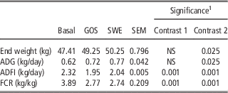
The effects of dietary supplementation with GOS and SWE on growth performance post-challenge are shown in Table 3. The average daily gain and weight at euthanasia were improved in the pigs offered the SWE supplemented diet (P<0.05). Average daily feed intake is reduced in pigs offered a diet supplemented with GOS and SWE. Feed conversion ratio is improved in pigs offered a diet supplemented with GOS and SWE (P<0.05). Faecal score post-challenge was not affected by dietary treatments (data not shown; P>0.10).
Effects of supplementation with GOS or SWE on growth performance of pigs after Salmonella Typhimurium challenge at day 0 until euthanasia at day 17 post-challenge (least squares mean values with pooled standard errors, 10 pigs/treatment)

GOS=oligosaccharides; SWE=seaweed extract; NS=not significant (P>0.10), ADG=average daily gain; ADFI=average daily feed intake; FCR=feed conversion ratio.
1 Contrast 1: basal v. GOS; contrast 2: basal v. SWE.
Microbiology
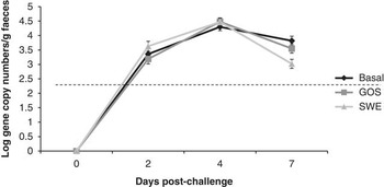
The effects of dietary GOS and SWE supplementation on faecal S. Typhimurium counts are shown in Figure 1. There was a treatment×time interaction observed. There were no differences between faecal S. Typhimurium numbers observed at days 2 and 4. However, dietary SWE supplementation reduced log GCN of S. Typhimurium in faeces at day 7 post-challenge compared with the basal treatment group.
Microbial counts (log gene copy number/g faeces) in faeces over time of pigs after Salmonella Typhimurium challenge at day 0. Values are means, with pooled standard errors represented by vertical bars. Limit of detection of the assay (dashed line) was 2.24 log gene copy numbers/g faeces. There was a significant treatment×time interaction (P<0.001). Basal pigs=receiving basal diet; GOS=galacto-oligosaccharide supplemented pigs; SWE=seaweed supplemented pigs.

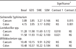
The effects of dietary supplementation with GOS and SWE on selected bacterial species after S. Typhimurium challenge are shown in Table 4. Compared to the basal diet, dietary supplementation with SWE reduced S. Typhimurium log GCN in the caecal and colonic digesta (P<0.05). Dietary supplementation with GOS increased Lactobacillus spp. log GCN in both caecal and colonic digesta compared with the basal diet (P<0.05). The number of S. Typhimurium in the rectum, tonsils, mesenteric and ileo-caecal lymph nodes was below the limit of detection of the assay (data not shown). There was no effect of treatments on Bifidobacterium spp. abundance in caecal and colonic digesta (P>0.10).
Effects of supplementation with GOS or SWE on microbial counts (log gene copy number/g digesta) in digesta of pigs 17 days after a Salmonella Typhimurium challenge (least squares mean values with pooled standard errors, 10 pigs/treatment)

GOS=oligosaccharides; SWE=seaweed extract; NS=not significant (P>0.10).
1 Contrast 1: basal v. GOS; contrast 2: basal v. SWE.
Gene expression profiles
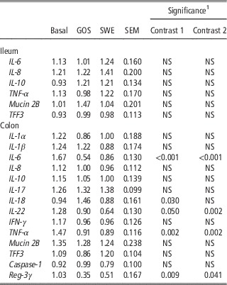
The effects of dietary supplementation with GOS or SWE on the gene expression of a panel of selected immune-related genes after S. Typhimurium challenge are shown in Table 5. There was no effect of dietary treatments on the gene expression of mucin 2B and TFF3 and on the gene expression of the cytokines IL-6, IL-8, IL-10 and TNF-α in ileal tissue (P>0.05).
Effect of dietary treatment with GOS or SWE and intestinal (cytokine) gene expression in ileal and colonic tissue after challenge with Salmonella Typhimurium on day 17 post-challenge (least squares mean values with pooled standard errors)

GOS=oligosaccharides; SWE=seaweed extract; NS=not significant (P>0.10); IL=interleukin; TNF-α=tumour necrosis factor α; TFF3=trefoil factor 3; IFN-γ=interferon γ; REG-3γ=regenerating-derived protein-3 γ.
1 Contrast 1: basal v. GOS; contrast 2: basal v. SWE.
In colonic tissue, both dietary GOS and SWE supplementation resulted in lower messenger RNA (mRNA) expression levels of IL-6 (P<0.001), IL-22 (P<0.01), TNF-α (P<0.01) and Reg3-γ (P<0.05) compared with the basal group. Pigs receiving diets supplemented with GOS had higher mRNA expression levels of IL-18 compared with the basal group (P<0.05).
Discussion
S. Typhimurium is a major food safety concern in humans in the developed world and pork and pork products are recognised as vehicles of infection. Pigs infected with S. Typhimurium not necessarily show symptoms of infection but are carriers of the pathogen (Rostagno and Callaway, Reference Rostagno and Callaway2012). This increases the risk of contamination into the human food chain. The objective of this study was to investigate the effects of dietary GOS and SWE supplementation in vivo on S. Typhimurium numbers and markers of intestinal inflammation in pigs following experimental challenge with S. Typhimurium. It was hypothesised that dietary GOS and SWE supplementation would decrease S. Typhimurium numbers and decrease intestinal inflammation after S. Typhimurium infection. Dietary supplementation with SWE resulted in reduced faecal shedding of S. Typhimurium at day 7 post-challenge and down-regulated pro-inflammatory cytokines expression, whereas dietary GOS supplementation increased Lactobacillus spp. numbers in caecal and colonic digesta samples and down-regulated pro-inflammatory cytokines expression.
Microbial analysis was performed to investigate the effects of the bioactives on selected bacteria. Different methods were used to select the best method for enumeration of the bacteria, showing that the qPCR method applied in the present study had the highest sensitivity (data not shown). The dietary GOS supplementation significantly increased Lactobacillus populations in caecal and colonic digesta samples. Increases in the Lactobacillus population have been associated with reduction in S. Typhimurium numbers (Casey et al., Reference Casey, Casey, Gardiner, Tangney, Stanton, Ross, Hill and Fitzgerald2004; Price et al., Reference Price, Totty, Lee, Utt, Fitzner, Yoon, Ponder and Escobar2010). However, no reduction in the number of S. Typhimurium in caecal and colonic digesta samples or faeces post-challenge was observed in the present study with dietary GOS supplementation.
Previous studies have shown that SWE supplementation containing a combination of laminarin and fucoidan are associated with reductions in Gram-negative bacteria such as Enterobacteriaceae and (enterotoxigenic) E. coli in pigs (Reilly et al., Reference Reilly, O’Doherty, Pierce, Callan, O’Sullivan and Sweeney2008; Walsh et al., Reference Walsh, Sweeney, O’Shea, Doyle and O’Doherty2013; Heim et al., Reference Heim, Sweeney, O’Shea, Doyle and O’Doherty2014). In the current experiment, dietary SWE supplementation reduced the number of S. Typhimurium recovered from caecal and colonic digesta samples by 0.6 and 1.0 log GCN/g digesta, respectively. In faeces on day 7 post-challenge, dietary SWE supplementation resulted in a reduction of 0.8 log GCN/g faeces. It has been previously reported that both β-glucans (laminarin) and fucoidans from different sources such as Saccharomyces cerevisiae and Cladosiphon okamuranus TOKIDA possess antimicrobial activity by preventing attachment of pathogenic bacteria to the intestinal wall (Shibata et al., Reference Shibata, Iimuro, Uchiya, Kawamori, Nagaoka, Ueyama, Hashimoto, Yokokura, Sugimura and Wakabayashi2003; Price et al., Reference Price, Totty, Lee, Utt, Fitzner, Yoon, Ponder and Escobar2010). Sulphated polysaccharides like fucoidan seem to attach to the bacterial membrane, competing against attachment to the mucosal layer (Shibata et al., Reference Shibata, Iimuro, Uchiya, Kawamori, Nagaoka, Ueyama, Hashimoto, Yokokura, Sugimura and Wakabayashi2003). The SWE extract used in the current experiment had high fucoidan contents, this may explain the reduced numbers of S. Typhimurium observed.
The changes in the selected bacteria after offering diets supplemented with SWE or GOS have the potential to affect the intestinal immune system. Knetter et al. (Reference Knetter, Bearson, Huang, Kurkiewicz, Schroyen, Nettleton, Berman, Cohen, Lunney, Ramer-Tait, Wannemuehler and Tuggle2015) stated that pigs with higher S. Typhimurium shedding have an enhanced systemic inflammatory response due to the higher infection rate and higher immune activation. This enhanced inflammatory response is often through an up-regulation of the nuclear factor (NF)-κβ pathway. Stimulation of the NF-κβ pathway is associated with an up-regulation of the pro-inflammatory cytokines IL-6, IL-12, IL-23; the antimicrobial peptide Reg-3γ; IL-8-like chemokines; and the immunosuppressor IL-10 (Vladimer et al., Reference Vladimer, Marty-Roix, Ghosh, Weng and Lien2013). In this study, we observed an up-regulation of IL-6, IL-22, TNF-α and Reg3-γ expression in pigs from the basal treatment group, indicating that the S. Typhimurium challenge increased the activation of the immune system and the NF-κβ pathway. To further examine the effects of SWE and GOS on the immune status of the S. Typhimurium challenged pigs, the expression of a panel of selected genes involved with the immune response was assessed. S. Typhimurium has several ways of activating the (innate) immune response, for example with lipopolysaccharides or the flagella on the outer membrane (Mukherjee and Hooper, Reference Mukherjee and Hooper2015). Lai et al. (Reference Lai, Quarles, López-Yglesias, Zhao, Hajjar and Smith2013) stated that there are at least two flagella recognition pathways: (1) cytosolic flagella are recognised by the Naip5-Naip6/NlrC4/Caspase-1 pathway, resulting in the up-regulation of especially IL-1β and IL-18, and (2) extracellular flagella are recognised by toll-like receptor 5, which in turn activates the NF-κβ pathway. When components of S. Typhimurium activate the NF-κβ pathway, pro-inflammatory cytokines such as IL-6, IL-8 and TNF-α are produced, and tight junctions between epithelial cells are disrupted. This mediates an inflammatory response and increases fluid entering the gut lumen, resulting in diarrhoea and intestinal inflammation (Knetter et al., Reference Knetter, Bearson, Huang, Kurkiewicz, Schroyen, Nettleton, Berman, Cohen, Lunney, Ramer-Tait, Wannemuehler and Tuggle2015).
Dietary supplementation with GOS and SWE resulted in suppression of a panel of selected cytokines that are associated with the NF-κβ pathway, revealing the anti-inflammatory effects of GOS and SWE. The down-regulated expression of the pro-inflammatory cytokines IL-6 and TNF-α was associated with supplementation of diets with either GOS or SWE. A down-regulation of several pro-inflammatory cytokines in blood, as well as an increase in beneficial bacteria has been observed after supplementation with GOS in healthy human adults (Vulevic et al., Reference Vulevic, Drakoularakou, Yaqoob, Tzortzis and Gibson2008). Zheng et al. (Reference Zheng, Yang, Zhou, Zhu, Shu, Wu, Li, Wang and Bo2012) observed a decreased activation of the NF-κβ pathway in newly weaned pigs after supplementation with soyabean oligosaccharides. Galacto-oligosaccharides are thought to not only have anti-inflammatory but also immunomodulatory properties; supplementation with GOS is associated with an improved regulation of the immune balance after infection (Vulevic et al., Reference Vulevic, Drakoularakou, Yaqoob, Tzortzis and Gibson2008). The increased Lactobacillus spp. numbers after dietary GOS supplementation could be related to the altered immune response in the pigs. It is thought that certain Lactobacillus strains can interact with the epithelial cells in the intestinal tract, leading to the observed reduced cytokine gene expression (Vulevic et al., Reference Vulevic, Drakoularakou, Yaqoob, Tzortzis and Gibson2008).
Down-regulation of pro-inflammatory cytokines was observed in other experiments after dietary SWE supplementation (Sweeney et al., Reference Sweeney, Collins, Reilly, Pierce, Ryan and O’Doherty2012; Walsh et al., Reference Walsh, Sweeney, O’Shea, Doyle and O’Doherty2013). The mode of action of these SWE supplements is not yet fully understood. However, previous research has shown that β-glucans such as laminarin enhance activation of intestinal immune cells and improve barrier function through modulation of the intestinal epithelial cells (Vetvicka et al., Reference Vetvicka, Vannucci and Sima2014). It has been previously reported that sulphated polysaccharides such as fucoidan can modify innate immunity through interference with the migration of leucocytes to the site of inflammation and activity of neutrophils, macrophages and natural killer cells (Jiao et al., Reference Jiao, Yu, Zhang and Ewart2011). In this study, pigs dietary supplemented with SWE are shown to have an altered immune response 17 days post-challenge, and have lower S. Typhimurium numbers in faeces at day 7 post-challenge and in the digesta at slaughter (day 17). This indicates that the pigs have dealt with the S. Typhimurium infection quicker and more effectively than the pigs from the basal treatment.
The expression of the antimicrobial protein Reg3-γ was down-regulated in colonic tissue of pigs supplemented with GOS or SWE. This protein is produced after activation of the NF-κβ pathway and is also associated with the expression of the cytokine IL-22, which responds to microbial invasion by producing antimicrobial proteins such as Reg3-γ (Vladimer et al., Reference Vladimer, Marty-Roix, Ghosh, Weng and Lien2013; Behnsen et al., Reference Behnsen, Jellbauer, Wong, Edwards, George, Ouyang and Raffatellu2014). Regenerating islet-derived protein 3-γ is a C-type lectin, binding to peptidoglycans on the bacterial membrane. These proteins are also considered biomarkers of intestinal inflammation in humans and rodents since up-regulation of these proteins is closely related to the activation of inflammasomes, which is often triggered by the recognition of bacterial products (Behnsen et al., Reference Behnsen, Jellbauer, Wong, Edwards, George, Ouyang and Raffatellu2014; Soler et al., Reference Soler, Miller, Nöbauer, Carpentier and Niewold2015). The inflammasome is part of the Naip5-Naip6/NlrC4/Caspase-1 pathway and responsible for the activation of the pro-inflammatory cytokines IL-1β and IL-18 (Lai et al., Reference Lai, Quarles, López-Yglesias, Zhao, Hajjar and Smith2013).
The expression of IL-22 can be linked to the presence of S. Typhimurium in the intestinal tract (Behnsen et al., Reference Behnsen, Jellbauer, Wong, Edwards, George, Ouyang and Raffatellu2014). Both dietary GOS and SWE supplementation reduced expression of IL-22, which is involved in tissue generation and maintenance of the mucosal barrier, especially during microbial challenges. It has previously been shown by Behnsen et al. (Reference Behnsen, Jellbauer, Wong, Edwards, George, Ouyang and Raffatellu2014) that IL-22 plays a unique role in S. Typhimurium infections; it promotes S. Typhimurium numbers through competitive exclusion of E. coli, S. Typhimurium’s closest competitor in the inflamed gut. High IL-22 expression results in high occurrence of antimicrobial proteins, which suppresses E. coli but favours S. Typhimurium and thus enhances colonisation of the mucosal layer by S. Typhimurium (Behnsen et al., Reference Behnsen, Jellbauer, Wong, Edwards, George, Ouyang and Raffatellu2014). The reduced IL-22 expression observed could be related to the reduced numbers of S. Typhimurium found in digesta in the SWE supplemented group. However, the reduced IL-22 gene expression does not coincide with reduced S. Typhimurium numbers in the GOS supplemented treatment group.
A high production of pro-inflammatory cytokines can lead to a rearrangement of nutrients when these are partitioned away from growth to support the immune response, as is observed in the basal group. The reduced inflammatory response observed in pigs offered a GOS or SWE supplemented diet resulted in an improved feed efficiency. With a smaller inflammatory response, fewer nutrients are needed to support the immune response, which increases nutrients available for growth.
In conclusion, the results from the current experiment demonstrate that dietary SWE supplementation reduced S. Typhimurium numbers and shedding in faeces post-challenge and caecal and colonic digesta. In addition, dietary SWE reduced the inflammatory response in colonic tissue. The dietary GOS supplementation did not affect S. Typhimurium numbers in faeces or digesta but reduced the inflammatory response in colonic tissue while increasing the Lactobacillus population in caecal and colonic digesta.
Acknowledgements
The research leading to these results has received funding from the European Union’s Seventh Framework Programme managed by REA Research Executive Agency (http://ec.europa.eu/research/rea; FP7/2007-2013) under grant agreement number 315198. This research was undertaken as part of a project entitled ‘Thrive-Rite: Natural Compounds to enhance Productivity, Quality and Health in Intensive Farming Systems’. Further details are provided on the consortium’s website (www.thriverite.eu) and the EU Commission’s webpage (http://cordis.europa.eu/project/rcn/104395_en.html). None of the collaboration partners had a role in the design and analysis of the present study or in the writing of this article. None of the authors has any conflicts of interest. The authors would like to thank the staff at the Central Veterinary Research Farm, Longtown Farm, Clane, Co. Kildare for the maintenance and husbandry of the pigs.