Introduction
Lymphatic filariasis (LF) has been identified as one of six infectious diseases that have the potential for elimination as a public health problem. Ever since the Global Alliance to Eliminate Lymphatic Filariasis was formed in 2000 at a meeting in Santiago de Compostela, Spain, in support of the World Health Organization's Global Programme to Eliminate Lymphatic Filariasis (GPELF), efforts have been dedicated to eliminating lymphatic filariasis as a public health problem and also reducing suffering in infected populations (GAELF, 2012). The GPELF incorporates disability management to ensure ‘a visible impact’ on those who are already affected by the disease, along with interrupting the transmission of LF by annual mass drug administration (MDA) (Ottesen, Reference Ottesen2000). The GPELF recommends that all those living in at-risk communities should be treated orally, once per year, with an appropriate, two-drug combination (Molyneux & Zagaria, Reference Molyneux and Zagaria2002). Engels, addressing the seventh GPELF mapping the worldwide distribution of lymphatic filariasis, indicated that as of 2011, 73 endemic countries required MDA to 1.39 billion people, even though 538.6 million people were treated in 2010–2011. The South-East Asia Region of nine endemic countries accounted for 63.4%, i.e. 883.7 million, of the population requiring MDA (Engels, Reference Engels2012).
For a successful GPELF, a strong system of evaluation and monitoring of programme outcomes and MDA coverage of transmission interruption is essential. Much of the available information on the geographical distribution of this disease is historical and based on utilizing the microscopic examination of thick blood smears collected at night. However, this method is labour-intensive while being deficient in detecting amicrofilaraemics (Weil, Reference Weil2005). The development of the immunochromatography test (ICT) for filariasis was a ‘breakthrough’ in this field (Weil et al., Reference Weil, Lammie and Weiss1997), as it facilitated filarial diagnostics by enabling antigens from adult Wuchereria bancrofti to be detected in fingerprick samples of blood collected at any time of the day, thereby obviating the need for inconvenient blood sampling during the night. It also facilitated the rapid mapping of each country's levels of endemic lymphatic filariasis and, subsequently, defining implementation units.
The testing for brugian filariasis did not keep pace with the assays for detecting W. bancrofti due to the absence of a responsive antigen test. However, antibody detection is still desirable over the parasitological methods that estimate transmission in the here and now, as it represents, over time, a cumulative history of the contamination (Lammie, Reference Lammie2004). A significantly higher prevalence of immunoglobulin (Ig) G4 than microfilaraemia in microfilaraemics has been demonstrated, indicating that this antibody may serve as a measure of active infections, and could therefore provide a more inclusive and detailed mapping of endemic areas (Haarbrink et al., Reference Haarbrink, Terhell, Abadi, Asri, de Medeiros and Yazdanbakhsh1999). Antibody testing has become a viable option with the development of the Brugia rapid dipstick test, which pairs antifilarial antibodies with a Brugia malayi recombinant antigen (BmRI) (Rahmah et al., Reference Rahmah, Shenoy, Nutman, Weiss, Gilmour, Maizels, Yazdanbakhsh and Sartono2003; Lammie et al., Reference Lammie, Weil, Noordin, Kaliraj, Steel, Goodman, Lakshmikanthan and Ottesen2004), indicating high specificity and sensitivity of the Brugia rapid dipstick test in detecting filariasis caused by the B. malayi parasite.
Although a commercial test kit for antifilarial IgG4 detection is available, its cost prohibits extensive use and mass testing in endemic regions of countries with constrained financial resources. We previously demonstrated an enzyme-linked immunosorbent assay (ELISA) procedure for the detection of isotype antibodies, specifically IgG4, in residents of brugian filariasis-endemic areas in Narathiwart province, southern Thailand, who manifested a spectrum of clinical disorders. The developed assay was sensitive and specific for the diagnosis of brugian filariasis (Wongkamchai et al., Reference Wongkamchai, Rochjanawatsiriroj, Monkong, Nochot, Loymek, Jiraamornnimit, Hunnangkul and Choochote2006). Thus, the overall goal of the present study was to develop a simple and readily transferable ELISA for the detection of antifilarial IgG4 antibodies in human serum samples that could subsequently be subjected to an appropriate clinical assessment.
A single-serum-dilution based ELISA procedure with ready-to-use reagents was designed and optimized to quantify the antifilarial IgG4 antibodies as a response to lymphatic filarial infection. The complete process from research to development of the initial assay, resulting in a prototype test kit, is described, along with its evaluation and validation by an independent collaborative laboratory. This single-serum-dilution based ELISA test kit exhibits two modes of detection for the diagnosis of lymphatic filarial infections.
Materials and methods
Detection of antifilarial IgG4 antibodies
An antifilarial IgG4 antibody assay was performed following a method established previously (Wongkamchai et al., Reference Wongkamchai, Rochjanawatsiriroj, Monkong, Nochot, Loymek, Jiraamornnimit, Hunnangkul and Choochote2006), with some modification. Microtitre wells were coated with B. malayi somatic antigen diluted in a carbonate buffer, pH 9.6. The plate was incubated at 4°C overnight. The unbound antigen was washed away using a washing buffer. The non-specific binding sites of each well were blocked by incubating with 200 μl of a milk-free blocking solution and 100 μl diluent were added into each well. Then 1 μl of each test sample was pipetted into the wells, one serum sample/well. The plate was shaken for 2 min using a low-frequency vortex mixer before being incubated for 1 h at 37°C. After washing, 100 μl of mouse monoclonal antibody to human IgG4–horseradish peroxidase (HRP) were added into each well and incubated for another 1 h at 37°C. After washing, 100 μl of ABTS (2,2′-azino-bis(3-ethylbenzthiazoline-6-sulphonic acid)) substrate solution were added into each well, after which the plate was kept in the dark at ambient room temperature for 10 min. The optical density (OD) of each test sample was measured at 405 nm.
Optimal reagent concentration
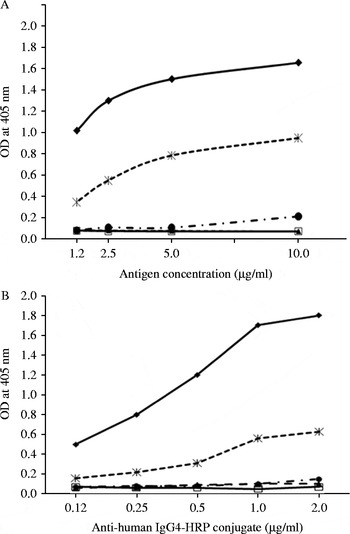
To optimize the antigen concentration, microtitre wells were coated with various concentrations of B. malayi somatic antigen, i.e. 10, 5, 2.5 and 1.25 μg/ml. The assays were performed, as mentioned above, using the control samples, i.e. high-positive control, low-positive control, negative control and bovine serum albumin. Graphs of the OD of samples against each antigen concentration were plotted. The optimal antigen concentration was chosen.
To optimize the concentration of the conjugate, mouse monoclonal antibody to human IgG4–HRP, assays were performed using various concentrations of the conjugate, i.e. 2, 1, 0.5, 0.25 and 0.12 μg/ml. Graphs of the OD of samples against each concentration of the conjugate were plotted. The optimal concentration of the conjugate was selected.
Cut-off point determination
A total of 100 serum samples obtained from parasite-free healthy subjects living outside the endemic areas of lymphatic filariasis were tested in duplicate using the optimal antigen and conjugate concentrations. The mean OD and standard deviation (SD) were calculated.
A receiver operating characteristic (ROC) curve was used to determine the sensitivity and specificity of the assay (Akobeng, Reference Akobeng2007a, Reference Akobengb).
Preparation of the ELISA test
Wells of a 96-well flat-bottomed microplate (Costar, Corning, Union City, California, USA) were coated with B. malayi somatic antigen using the optimal concentration of 0.5 μg/well in carbonate–bicarbonate buffer, pH 9.4, 100 μl/well. The microplate was incubated overnight at 4°C and washed with phosphate-buffered saline (PBS), pH 7.2, containing 1% Tween 20 in preservative. The non-specific binding sites of each well were blocked by incubation with 200 μl of a milk-free solution. The microplate was wrapped in a sealed, foil-lined bag and silica was added.
The 10 × washing solution of PBS, pH 7.2, containing 1% Tween 20, and its preservative were added. The diluents, consisting of PBS, pH 7.2, containing 1% bovine serum albumin (Sigma Chemicals, St. Louis, Missouri, USA), and 0.05% polyoxyethylene sorbitan monolaurate (Tween 20; Sigma Chemicals) were conjugated and the preservative was added. The conjugate solution consisting of an optimized concentration, i.e. 1:1000, of HRP–mouse monoclonal antibody to human IgG4 (HRP–mouse anti-human IgG4; Invitrogen Laboratories, Grand Island, New York, USA) was prepared. The ABTS substrate solution was composed of one vial of ABTS substrate (KPL, Gaithersburg, Maryland, USA). A set of control samples of 2 ml/vial comprised a high-positive control, a cut-off control and a negative control. The in-house test kit was stored refrigerated at 2–8°C.
Assay protocol
The ready-for-use washing solution was prepared by adding 900 ml of sterile distilled water to the 10 × washing buffer. The washing solution was stored at 2–8°C. The assay was performed as follows. A set of control samples, i.e. high-positive control, cut-off control and negative control sample, were added into wells A1, B1 and C1, respectively, at 100 μl/well. The diluents were added into the other wells, at 100 μl/well, and 1 μl of each serum sample was pipetted into the wells, at one serum sample/well. To promote mixing, the plate was shaken for 2 min using a low-frequency vortex mixer before being incubated for 1 h at 37°C. After washing, 100 μl of mouse monoclonal antibody to human IgG4–HRP were added into each well and incubated for another 1 h at 37°C. After washing, 100 μl of ABTS substrate solution were added into each well, and the plate was kept in the dark at room temperature for 10 min.
The assay result was measured and interpreted using two detection modes. First, OD was measured using the ELISA reader at 405 nm. Second, the colour of the test sample's well was compared with the colour of the cut-off control well by the naked eye. The test sample was considered positive if the intensity of the green colour of the sample's well was equal to or higher than the green colour of the cut-off well, whereas the sample was considered negative if the test well showed no colour, or if the intensity of the green colour was less than that of the cut-off well.
Sensitivity and specificity
To determine the sensitivity of the developed test kit, 4 and 30 microfilaraemic cases of bancroftian and brugian filariasis, respectively, were considered true positives and were used as the ‘gold standard test’. A total of 100 serum samples from parasitic-free healthy subjects living in a non-endemic area were considered true negatives. Our cross-reactivity sample comprised 57 subjects infected with other parasites who lived outside lymphatic filariasis-endemic areas.
Gastrointestinal parasitic infections were detected from stools of the 57 subjects using the formalin–ethyl acetate concentration technique. Ascaris lumbricoides was detected in 3 subjects, Strongyloides stercoralis in 33 subjects, hookworm in 2 subjects, Capillaria phillipphinensis in 2 subjects, Ophistorchis viverrini in 4 subjects, Echinostoma spp. in 1 subject and Taenia in 2 subjects. Tissue-dwelling parasites, i.e. Angiostrongylus cantonensis in 3 subjects, Gnathostoma spinigerum in 4 subjects and Cysticercus in 3 subjects, were diagnosed by the detection of specific antibodies against these parasites. Both healthy subjects and other parasite-infected subjects were used to determine the specificity of the test kit. The positive and negative predictive values of the test kit were also determined.
Intra- and inter-assays
These assays were determined by repeated testing of three individual serum samples representing strongly positive, positive and negative levels of antigenic reactivity. Positive controls were also used. The intra-assay variation of the developed test kit was determined by performing 12 precision runs of the same samples at the same time. The reproducibility of results was assessed by calculating coefficient of variation (CV) values [% CV = (SD/mean) × 100]. The inter-assay variation was determined by performing 21 precisions runs on 21 separate days and evaluating the same serum samples in 21 consecutive assay runs. CV values were calculated to compare mean ODs for the same samples on different days.
Stability and reliability of the test kit
To monitor the stability of the test kit, an assay was conducted monthly for 7 months using the high-positive, low-positive as a cut-off, and negative controls provided in the kit package, and tracked over time. The results were based on the manual calculation of means and 2 × SD values of data. A complete record of the results was maintained. A test was considered valid when all the mean OD values of the controls were within the ± 2 SD limits as prescribed.
To compare the two detection modes, fingerprick daytime blood samples were collected from 4365 children living in brugian filariasis-endemic areas in Narathiwart province. The ages of the children ranged from 2 to 6 years and they resided in the seven districts of Sugai-Padi, Sugai-kolok, Tak-Bai, Yi-Ngo, Bacho, JongAilong and Muang, comprising 22 subdistricts and 87 villages. All personnel were provided with hands-on training for test kit usage prior to actually using the test kit. To ascertain the reliability of the test results, the same set of test samples was evaluated in the Department of Parasitology, Faculty of Medicine, Siriraj Hospital, Bangkok.
Data analysis
SPSS 13 (SPSS Inc., Chicago, Illinois, USA) was used for all parametric and non-parametric descriptive measures, which included all means, SDs and correlations where appropriate. A percentage measure of dispersion, the CV, of intra- and inter-assay validation was also calculated. The kappa statistic was used to measure the extent of agreement between the naked-eye determination from the test kit and the output of ELISA.
Results
Detection of antifilarial IgG4 antibodies
Optimization of reagent and condition
Titration curves were plotted between the OD of the control samples and various concentrations of the B. malayi somatic antigen and the reciprocal titre of the HRP conjugate (fig. 1). The antigen concentration of 5 μg/ml and the conjugate diluted at 1:1000 were chosen as optimal for the test kit.
Relationship between optical density (OD) values and (A) antigen concentrations and (B) anti-human immunoglobulin (Ig) G4–horseradish peroxidase (HRP) conjugate concentrations. ( ), high-positive serum; (
 ), low-positive serum; (
 ), negative serum; (
 ), bovine serum albumin; (
 ), substrate blank.

Cut-off determination
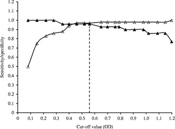
ROC curve analysis was used to determine the sensitivity and specificity of the prototype test kit for the detection of antifilarial IgG4 at various cut-off OD values, i.e. 0.1–1.3, from mean +0.5 SD to mean +10.0 SD. The true positive rate, or sensitivity, was plotted as a function of the false-positive rate, i.e. 1 – specificity, for different cut-off points (fig. 2). Each point on the ROC plot represents a sensitivity–specificity pair corresponding to a particular cut-off OD value. The optimal cut-off value, giving the highest index for both specificity and sensitivity, was 0.56, with a sensitivity of 0.97 and a specificity of 0.97. Therefore the OD of 0.56 was chosen as the cut-off value for the test kit.
Receiver operating characteristic curve of the relationship between sensitivity/specificity of the test kit and cut-off values of the antifilarial immunoglobulin (Ig) G4 antibodies. ( ), optimal cut-off value; (
 ), sensitivity; (
 ), specificity.

Sensitivity and specificity
When compared with the ‘gold standard’ method of microfilariae detection, the sensitivity of the test kit was found to be 97%, as the kit detected antibodies in 33 of the 34 infected cases. Serum samples from 57 patients living outside the endemic areas who were infected with other parasites and serum samples from 100 normal parasite-free subjects were used to determine the specificity of the test kit. The specificity of the developed test kit was found to be 98.7%, as antibodies were detected in only 2 of the 157 subjects. The positive and negative predictive values were 94.2% and 99.3%, respectively.
Intra- and inter-assays
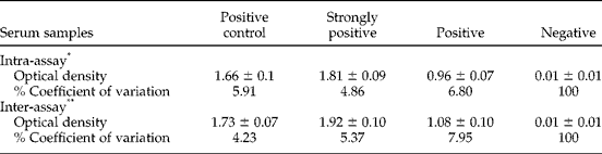
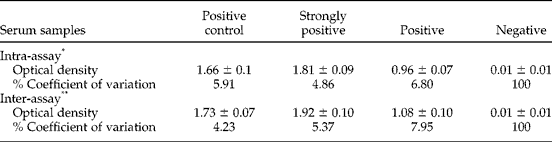
The CVs for intra- and inter-assays were determined by repeated testing of three individual serum samples representing three levels of antigenic reactivity, i.e. strongly positive, positive and negative, and the positive control. Differences in CVs in both assays, with respect to positive variables, were relatively low, being 1.94% and 2.58% in the repeated testing of intra-and inter-assays, respectively (table 1).However, the very high CVs of 100% of both intra- and inter-assays of the negative serum sample reflect a weak or near-zero value of the mean ODs. A better and more appropriate index for the negative serum would be mean ± SD, which for both the intra- and inter-assays would be 0–2% (Servat et al., Reference Servat, Feyssaguet, Blanchard, Morize, Schereffer, Boue and Cliquet2007).
Intra- and inter-assay validation for the ELISA. Mean optical density values (± SD) obtained in validating intra- and inter-assays for the ELISA test using 12 (*) and 21 (**) repeats of test and control sera.
Stability and reliability of the test kit
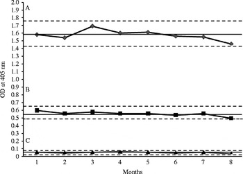
ODs were measured over a 7-month period for the antigenic activity attributed to high-positive, low-positive and negative controls (fig. 3). The mean OD values of the controls were all within the 2 SD limits over time but did manifest a gradual reduction between the sixth and the seventh month of observation. Thus, the stability of the developed test kit is approximately 7 months.
Optical density (OD) of three controls plotted over monthly intervals. –- mean ± 2 standard deviation (mean ± 2SD): (A) high-positive control, (B) low-positive control (cut-off control), (C) negative control.

For field validation, antifilarial IgG4 detection was performed on the blood taken from 4365 children residing in brugian filariasis-endemic areas. The assay was performed by the staff of the Pikhunthong Royal Project and the result was measured and interpreted by comparing by the naked eye the colour of each test sample's well with the colour of the cut-off control well. Additionally, the staff of the Department of Parasitology, Faculty of Medicine, Siriraj Hospital performed an ELISA using an ELISA reader to determine the prevalence of antifilarial IgG4 in the same sample of children. By both procedures, 18 cases of 4365 children were rated positive by the naked-eye reading whereas 11 cases of 4365 children were positive with the ELISA reader. The kappa metric of 0.758, 95% CI 0.584–0.932 (P< 0.0001) was judged as very good in its strength of agreement between these two procedures (Landis & Koch, Reference Landis and Koch1977).
Discussion
For a successful global elimination programme of lymphatic filariasis, a strong system of evaluation and sensitive tools for monitoring are essential. Problems arise when measuring the extent of brugian filariasis, especially when using thick blood smears and night blood collection, which are often inaccurate and inconvenient, respectively. Additionally, unlike the measurement of W. bancrofti, no antigen detection assay is available for B. malayi.
We previously developed an ELISA assay for the detection of isotype antibodies including IgG4 antibodies in residents of a brugian filariasis-endemic area in Narathiwart province, southern Thailand. This assay was sensitive and specific for the diagnosis of brugian filariasis in subjects displaying a wide spectrum of clinical manifestations (Wongkamchai et al., Reference Wongkamchai, Rochjanawatsiriroj, Monkong, Nochot, Loymek, Jiraamornnimit, Hunnangkul and Choochote2006). When compared with similar commercially available products, the cost of this in-house assay is significantly reduced and could readily be used diagnostically, thereby overcoming the disadvantages of the microfilariae detection method.
Nevertheless, the in-house ELISA assay has some shortcomings. It takes a long time to perform, including overnight incubation; it requires the preparation of many reagents; it involves the use of expensive equipment such as the ELISA washer and reader; and it requires trained personnel with appropriate expertise. In resource-poor endemic areas, satisfying these requirements is unsuitable and virtually impossible. Thus, we report here the development and validation of a single-serum-dilution based ELISA test kit with ready-to-use reagents for detecting antifilarial IgG4 antibodies.
The intention of this project was to develop an ELISA assay that could be performed successfully by any laboratory lacking the necessary sophisticated equipment, and by an analyst with no, or only minimal, experience. To fulfil these requirements, the potential test kit must be sufficiently rigorous and robust in demonstrating specificity, sensitivity, reliability/repeatability and validity. The developed test kit, when used to analyse patient serum samples, revealed high sensitivity and specificity. The CVs for both intra- and inter-assay variability were less than 10% for positive samples, while for the negative samples, the CV was relatively high due to the very low OD value, which was near zero. The absolute SDs for both tests were comparable for all positive samples. In general terms, the CV percentage indicated that the developed test kit was precise and consistent. The validity requirement was satisfied in at least two ways. First, the test kit was in 97% agreement with microfilariae detection by blood film examination, and second, the kappa statistic may be interpreted as better than substantial agreement between the results of the test kit and the outcome of ELISA (Viera & Garrett, Reference Viera and Garrett2005).
One possible limitation of the antifilarial antibody assay using crude B. malayi is the cross-reactivity with other nematode parasites, including S. stercoralis (Muck et al., Reference Muck, Pires and Lammie2003). This caveat was negated in our cross-reactivity sample of 57 patients living outside the B. malayi-endemic areas, who were infected with other parasites, including S. stercoralis. Only 2 of 33 cases with S. stercoralis showed infectivity, indicating a negligible influence on the IgG4 antibody response.
Besides being sensitive and specific, an ideal serodiagnostic test, especially for developing countries, should be cheap, simple and easy to perform (Voller & De Savigny, Reference Voller and De Savigny1981). The results presented here demonstrate that the developed test kit is simplified by the ready-to-use reagent provided. Moreover, in the sample application step, diluents fill the 96 wells within 2 min. Then 1 μl of each test serum or plasma sample is added into each well, followed by shaking the plate for 2 min using a vortex mixer. No pre-diluted serum in Eppendorf tubes is needed. Moreover, the assay result can also be interpreted by the naked eye. Thus, this procedure would reduce the number of procedural manipulations needed to physically perform the assay, and eliminate the need for a full set of standard equipment, leading to a greater simplification of the interpretation of the results.
In conclusion, this study presents a process of development and validation of a single-serum-dilution based ELISA test kit with ready-to-use reagents for detecting antifilarial IgG4 antibodies. With a minimum of common laboratory equipment, this assay may be completed successfully by district public health laboratories assessing the burden of lymphatic filariasis in residents of disease-endemic areas.
Acknowledgements
Special thanks are extended to Ms Bungon Sermsart from the Department of Parasitology, Faculty of Medicine, Siriraj Hospital, Mahidol University for technical assistance. Thanks are also offered to the staff of the Filaria division, Disease Control Department, Ministry of Public Health and the staff of the Filaria Project, Phikhunthong Royal Project for the field validation of the developed test kit.
Financial support
This work was supported by grant number 2551-197 from the Thailand Research Council.
Conflict of interest
The ELISA test kits have been produced and used in Thailand.
Ethical standards
The authors assert that all procedures contributing to this work comply with the ethical standards of the relevant national guidelines on human experimentation and with the Helsinki Declaration of 1975, as revised in 2008, and has been approved by the Ethics Committee on Research Involving Human Subjects, from the Faculty of Medicine, Siriraj Hospital, Mahidol University, Thailand.