Introduction
Gamete cryopreservation is an important practice in assisted reproductive technology. In 1953, the earliest human offspring resulting from cryopreserved semen was reported (Bunge and Sherman, Reference Bunge and Sherman1953). Since then, marked progress has been made in field sperm cryobiology, however poor sperm quality in terms of motility, plasma membrane functionality, acrosome integrity and viability is often observed post thaw (Hezavehei et al., Reference Hezavehei, Sharafi, Kouchesfahani, Henkel, Agarwal, Esmaeili and Shahverdi2018).
Despite the first pregnancy being achieved with transfer of embryos derived from frozen oocytes nearly 40 years ago (Chen, Reference Chen1986), oocyte cryopreservation was only considered safe and efficient with the advent of vitrification (Coello et al., Reference Coello, Pellicer and Cobo2018), which is now the gold standard in oocyte freezing for in vitro fertilization (IVF) (Iussig et al., Reference Iussig, Maggiulli, Fabozzi, Bertelle, Vaiarelli, Cimadomo, Ubaldi and Rienzi2019).
Several indications exist for gamete cryopreservation, such as fertility preservation in oncological patients. Oocyte cryopreservation is also an option for women wishing to delay pregnancy, and for oocyte donation treatment, which is a well established treatment for female infertility that allows oocyte recipients to achieve motherhood.
In Brazil, the conduction of oocyte donation cycles for commercial purposes is prohibited. As a consequence, oocyte cryobanks consist of surplus oocytes donated from young patients undergoing IVF treatments (e.g. infertile couples), therefore creating a marked scenario in which vitrified and fresh oocytes from the same donor can be compared in terms of competence and development potential.
The effect of egg and sperm cryopreservation on the outcomes of recipient cycles is uncertain. The objective of this study was to investigate the influence of oocyte and sperm cryopreservation on donated eggs in terms of laboratory and clinical outcomes of intracytoplasmic sperm injection (ICSI) cycles.
Materials and methods
Patients and experimental design
This historical cohort study was performed in a private university-affiliated IVF centre, between January/2016 and January/2018. Data were obtained by chart review of 115 oocyte donor ICSI cycles, and 122 oocyte recipients undergoing 152 ICSI cycles, participating in an egg-sharing donation programme. Extraction of data from the computer database was performed by two independent reviewers (AS and DB).
Cycles were split into four groups based on the origin of oocytes and semen, as follows: the FO/FS Group, recipients in which fresh oocytes were injected with fresh sperm (n = 19); the FO/CrS Group, recipients in which fresh oocytes were injected with cryopreserved sperm (n = 14); the CrO/FS Group, recipients in which cryopreserved oocytes were injected with fresh sperm (n = 85); and the CrO/CrS Group, recipients in which cryopreserved oocytes were injected with cryopreserved sperm (n = 34). The effect of oocyte and semen cryopreservation on recipient ICSI outcomes was investigated.
Donor patients were ≤ 35 years old. All cases with severe spermatogenic changes, including surgically retrieved sperm, repeated implantation failure (considering the transfer of at least four good-quality embryos in woman up to 39 years) and preimplantation genetic diagnosis and karyotype changes were excluded. All recipients included in this study had ovarian factor of infertility, due to premature ovarian failure (n = 9) or advanced maternal age, being already menopausal or presenting with insufficient response to controlled ovarian stimulation (n = 113).
Controlled ovarian stimulation and laboratory procedures
Controlled ovarian stimulation was performed using recombinant follicle-stimulating hormone (r-FSH; Gonal-F®, Serono, Geneva, Switzerland) and pituitary blockage using a gonadotropin-releasing hormone (GnRH) antagonist, cetrorelix acetate (Cetrotide® Merck KGaA, Serono, Geneva, Switzerland). Leuprolide acetate (Lupron; TAP Pharmaceuticals, North Chicago, IL, USA) or recombinant hCG (Ovidrel; Serono, Geneva, Switzerland) was administered to trigger the final follicular maturation in 102 and 13 donors, respectively. The oocytes were collected 35 h later. Mature recovered oocytes were either vitrified or submitted to ICSI following routine procedures (Palermo et al., Reference Palermo, Colombero and Rosenwaks1997).
Oocyte morphology was assessed using an inverted Nikon Diaphot microscope with a Hoffmann modulation contrast system under ×400 magnification (Eclipse TE 300 microscope, Nikon, Tokyo, Japan), just before sperm injection (5–7 h after retrieval). Oocyte dysmorphisms individually recorded were the presence of intracytoplasmic granulation clusters, smooth endoplasmic reticulum clusters, dark cytoplasm, vacuoles, PVS-L, PVS-G, zona pellucida dysmorphisms and polar body fragmentation.
Oocyte vitrification and warming
Both vitrification and the warming procedures were performed using the Cryotop method (Kuwayama et al., Reference Kuwayama, Vajta, Kato and Leibo2005). At 3 h after ovum pickup, vitrification was achieved by exposure of oocytes to the equilibration solution, followed by a 30-s exposure to the vitrification solution.
Individual oocytes were then placed on top of a very fine polypropylene strip attached to a hard-plastic handle. As soon as the oocyte was placed onto the thin polypropylene strip of the Cryotop, it was immediately submerged vertically into liquid nitrogen. Then the thin strip was covered with a hard-plastic cover on top of the Cryotop sheet.
For warming, the polypropylene strip of the Cryotop was immersed directly into the thawing solution at 37°C for 1 min. Oocytes were retrieved and transferred into dilution solution for 3 min and then washed twice in the washing solution for 5 min each. Within 3 h after warming, oocytes were submitted to ICSI following routine procedures (Palermo et al., Reference Palermo, Colombero and Rosenwaks1997).
The tools and solutions required for the vitrification and warming processes were obtained from Kitazato® (Tokyo, Japan).
Semen analysis, cryopreservation and thawing
Semen samples were collected in the laboratory by masturbation, and were analyzed according to WHO guidelines (World Health Organization, 2010). After liquefaction for 30 min, semen samples were evaluated for sperm count, motility and morphology.
For cryopreserved semen samples, the cryoprotectant used was a buffered solution of glycerol 12%, egg yolk 20% and 10 µg/ml gentamycin (freezing medium – TYB with glycerol and gentamicin; Irvine Scientific, Santa Ana, CA, USA). The cryoprotectant was added to the ejaculate and the mixture was left at room temperature for 10 min, and transferred to cryovials. The cryovials were suspended in liquid nitrogen vapours for 10 min and then were immersed into liquid nitrogen.
For thawing, cryopreserved semen samples were kept at room temperature for 10 min and prepared using a two-layered density gradient centrifugation technique (50% and 90% isolate; Irvine Scientific, Santa Ana, CA, USA).
Embryo culture and morphology evaluation
Approximately 16 h after ICSI, fertilization was confirmed by the presence of two pronuclei and the extrusion of the second polar body. The embryos were maintained in a 50-µl drop of culture medium (Global®; LifeGlobal, CT, USA) and covered with paraffin oil in a humidified atmosphere under 6% CO2 at 37°C for 5 days.
The high-quality cleavage-stage embryos were defined as those with all of the following characteristics: 4 cells on day 2 or 8−10 cells on day 3, <15% fragmentation, symmetric blastomeres, the absence of multinucleation, colourless cytoplasm with moderate granulation and no inclusions, the absence of perivitelline space granularity and the absence of zona pellucida dysmorphism. Embryos lacking any of these characteristics were considered to be of low quality.
Full blastocysts onwards, presenting morphologically normal inner cell mass (ICM) and trophectoderm (TE) were defined as high-quality blastocysts. A tightly packed ICM presenting many cells was defined as a high-quality ICM. Similarly, the TE was classified as high quality by the presence of many cells forming a cohesive epithelium.
Endometrial preparation
For recipients, after menses, endometrial development was followed by ultrasound examination, and the patients received 200 µg of transdermal 17β-estradiol every 3 days (Estradot®, Noven Pharmaceuticals, Miami, USA). When the endometrium showed proliferative morphology and thickness of at least 7.5 mm, 600 mg of progesterone (Utrogestan®, Farmoquímica, Rio de Janeiro, Brazil) was vaginally administered per day.
Both 17β-estradiol and progesterone were administered concomitantly after embryo transfer and were suspended for a negative βhCG test. For a positive βhCG test, the 17β-estradiol and progesterone treatments were maintained until weeks 6 and 12 of gestation, respectively.
Embryo transfer and clinical follow-up
One to three embryos were transferred per patient on day 5 of embryo development. Surplus high-quality embryos were cryopreserved. Embryo cryopreservation rate was calculated by dividing the number of embryos cryopreserved per the number of obtained embryos.
All women with a positive test, performed 10 days after transfer, had a transvaginal ultrasound scan 2 weeks later. A clinical pregnancy was diagnosed when the fetal heartbeat was detected. Miscarriage was defined as clinical pregnancy loss before 20 weeks.
Data analysis and statistics
The post hoc computed achieved power was superior to 99%, considering the sample size, the effect size obtained for implantation rate and 5% significance level. The study power was calculated using G*Power 3.1.7 (Franz Faul, Universität Kiel, Germany).
The effect of oocyte cryopreservation on recipient ICSI outcomes was investigated using generalized linear mixed models (GzMM) fit by restricted maximum likelihood, to account for clustering of the data. As: (i) many patients donated oocytes to more than one recipient; (ii) multiple embryos were created per cycle; and (iii) multiple cycles were present per couple, three random effects were added to account for the correlation: (i) between recipients from a same donor; (ii) between embryos within the same recipient cycle; and (iii) between embryos of different cycles of the same recipient.
Mixed effects models were generated with unstructured covariance structure. A Gaussian distribution was assumed and the normal distribution of model residuals was checked to confirm goodness of fit. Final model selection was decided using the Akaike information criterion and Schwarz’s Bayesian criterion. Paternal age was included as covariate in all models. Maternal age and endometrial thickness were included as covariates only in the clinical outcome models (implantation, pregnancy and miscarriage rates). All models were followed by Bonferroni post hoc test for the comparison of means between the four groups.
The implantation rate was calculated by dividing the number of gestational sacs per the number of transferred embryos. Clinical pregnancy rate was calculated per transfer.
The results were expressed as mean differences (MD) with 95% confidence intervals (CI), considering the FO/FS Group as the reference group for the exposure variable, and P-values. P < 0.05 was considered statistically significant.
Data analysis was conducted using SPSS Statistics 21 (IBM, New York, NY, USA).
Results
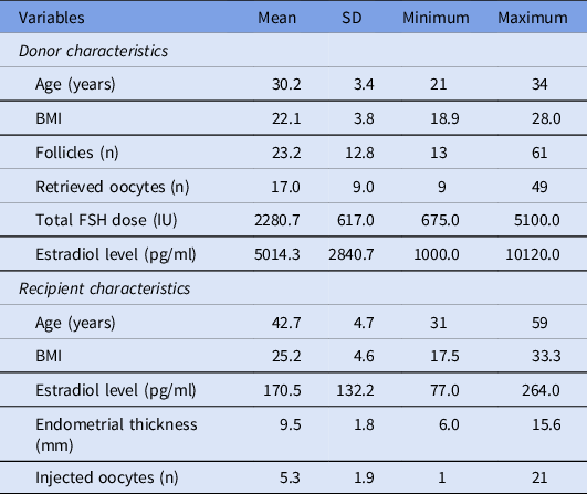
Demographic data concerning donors and recipients are shown in Table 1. Factors of infertility for oocyte donors are described in Figure 1.
Donors and recipients demographic data

Note: BMI, body mass index; ICSI, intracytoplasmic sperm injection; IU, international unit; SD, standard deviation.
Values are mean ± SD, unless otherwise noted.
Distribution of factors of infertility for oocyte donors. PCOS, polycystic ovary syndrome.

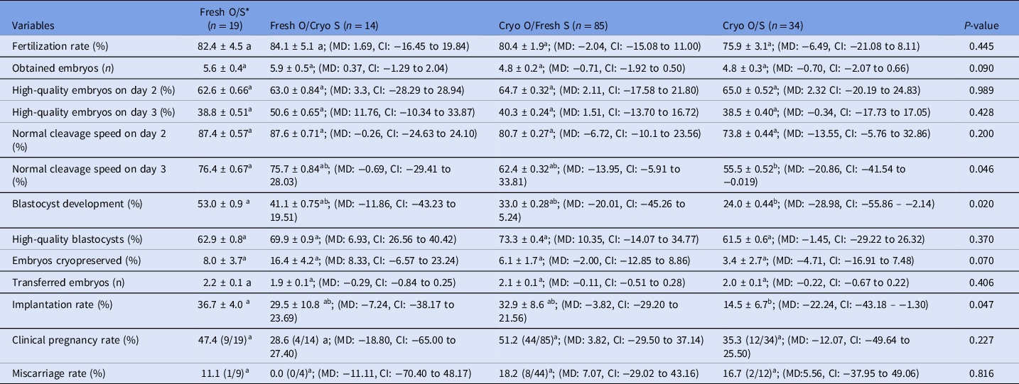
Results from GzMM analysis are shown in Table 2. Normal cleavage speed rate on day 3 was significantly lower in the CrO/CrS Group (55.5%) compared with the FO/FS Group (76.4%, P: 0.046, MD −0.209, CI: −0.436 to −0.019). Blastocyst development rate was significantly lower in the CrO/CrS Group (24.0%) compared with the FO/FS Group (53.0%, P: 0.020, MD −0.290, CI: −0.559 to −0.021). Implantation rate was also significantly lower in the CrO/CrS Group (14.5%) compared with the FO/FS Group 36.7%, P: 0.047, MD −22.241, CI: −43.182 to −1.301). The rates of fertilization, high-quality embryos on days 2 and 3, normal cleavage speed on day 2, high-quality blastocyst, clinical pregnancy and miscarriage were similar between the groups.
GzMM and Bonferroni post hoc result for the comparison of ICSI outcomes using fresh or warmed oocytes and sperm

Note: *Reference group for gamete status.
GzMM, generalized mixed models; ICSI, intracytoplasmic sperm injection; O, oocyte; S, sperm.
Values are mean ± standard error and mean difference (MD) and confidence interval (CI), considering fresh O/S group as the reference group for gamete status, a≠b.
Discussion
The present study compared the outcomes of ICSI in oocytes donation cycles in which warmed or fresh oocytes were injected with warmed or fresh sperm. When warmed oocytes were injected with warmed sperm, the rates of embryo development on day 3, blastocyst development and implantation were significantly lower compared with the fresh use of both oocytes and sperm. There were no statistically significant differences between the four groups in terms of pregnancy outcomes. The small sample size of the study may account for this lack of significance.
Our results were in accordance with those from previous studies that have shown lower fertilization and embryo quality in thawed oocytes compared with fresh oocyte (Chen et al., Reference Chen, Lien, Chang, Tsai, Ho and Yang2004; Chamayou et al., Reference Chamayou, Alecci, Ragolia, Storaci, Maglia, Russo and Guglielmino2006; Magli et al., Reference Magli, Lappi, Ferraretti, Capoti, Ruberti and Gianaroli2010; Braga et al., Reference Braga, Setti, Figueira, Azevedo Mde, Iaconelli, Lo Turco and Borges2016). Vitrified oocytes showed unfavourable morphokinetic differences from time to 2 cells to blastocyst development when compared with fresh oocytes (Cobo et al., Reference Cobo, Coello, Remohi, Serrano, de Los Santos and Meseguer2017). Conversely, other groups have shown similar fertilization, embryo and blastocyst development, and clinical outcomes between fresh and vitrified oocytes (Cobo et al., Reference Cobo, Kuwayama, Perez, Ruiz, Pellicer and Remohi2008; Rienzi et al., Reference Rienzi, Romano, Albricci, Maggiulli, Capalbo, Baroni, Colamaria, Sapienza and Ubaldi2010; Garcia et al., Reference Garcia, Noriega-Portella and Noriega-Hoces2011; Parmegiani et al., Reference Parmegiani, Cognigni, Bernardi, Cuomo, Ciampaglia, Infante, Tabarelli de Fatis, Arnone, Maccarini and Filicori2011; Trokoudes et al., Reference Trokoudes, Pavlides and Zhang2011; Goldman et al., Reference Goldman, Noyes, Knopman, McCaffrey and Grifo2013). Our cohort of oocytes, which was derived from infertile couples, could be responsible for the discrepant findings and the reduced outcomes observed in the present study.
Regarding the effect of cryopreservation on oocyte competence, it has been suggested that defects in oocyte activation and embryo cleavage may occur as a consequence of a cryogenic damage. In fact, especially during oocyte vitrification, multiple aster formation may happen due to exposure to cryoprotectant and high cooling rates. Multiple asters are formed close to the male pronucleus, causing delayed first cleavage and reduced blastocyst development (Hara et al., Reference Hara, Hwang, Kagawa, Kuwayama, Hirabayashi and Hochi2012). Moreover, thawed oocytes show spindle alterations (Rienzi et al., Reference Rienzi, Martinez, Ubaldi, Minasi, Iacobelli, Tesarik and Greco2004) that impair embryo cleavage, and epigenetic changes such as abnormal mitochondrial distribution (Van Blerkom et al., Reference Van Blerkom, Davis and Alexander2000) that induce errors in intracellular calcium signalling post sperm injection, also affecting normal embryogenesis (Jones et al., Reference Jones, Van Blerkom, Davis and Toledo2004).
Even though the spermatozoon is considered less sensitive to cryodamage because of its high membrane fluidity and low water content (Di Santo et al., Reference Di Santo, Tarozzi, Nadalini and Borini2012), several studies have reported deleterious changes in sperm structure and function that may be attributable to ice crystal formation during the freezing of extracellular fluids. Sperm cryoinjury may present as: (i) ultrastructural changes in plasma and mitochondrial membranes, and acrosome (Barthelemy et al., Reference Barthelemy, Royere, Hammahah, Lebos, Tharanne and Lansac1990; Ozkavukcu et al., Reference Ozkavukcu, Erdemli, Isik, Oztuna and Karahuseyinoglu2008), leading to impaired fertilization potential, reduced motility, abnormal morphology and reduced sperm viability; and (ii) compromised DNA integrity (Donnelly et al., Reference Donnelly, McClure and Lewis2001; de Paula et al., Reference de Paula, Bertolla, Spaine, Cunha, Schor and Cedenho2006; Thomson et al., Reference Thomson, Fleming, Aitken, De Iuliis, Zieschang and Clark2009; Zribi et al., Reference Zribi, Feki Chakroun, El Euch, Gargouri, Bahloul and Ammar Keskes2010), leading to altered decondensation of the sperm nucleus once inside the oocyte, therefore reducing fertilization rate. Nevertheless, studies that have compared the outcomes of ICSI with fresh and warmed semen samples showed similar fertilization rates (Kuczynski et al., Reference Kuczynski, Dhont, Grygoruk, Grochowski, Wolczynski and Szamatowicz2001), and similar rates of implantation and pregnancy (Borges et al., Reference Borges, Rossi, Locambo de Freitas, Guilherme, Bonetti, Iaconelli and Pasqualotto2007, Kalsi et al., Reference Kalsi, Thum, Muneer, Pryor, Abdullah and Minhas2011, Kopeika et al., Reference Kopeika, Thornhill and Khalaf2015). In fact, Kuczynski et al. (Reference Kuczynski, Dhont, Grygoruk, Grochowski, Wolczynski and Szamatowicz2001) showed higher ongoing pregnancies with cryopreserved semen samples compared with fresh ejaculated samples. These studies suggested that the freezing–thawing procedure causes more damage in patients with changes in semen quality than that in patients with normal semen but, once fertilization is successful, pregnancy rates are not negatively affected by semen cryopreservation, neither in patients with sperm abnormalities, nor in those with normal semen parameters.
A late paternal effect could be the reason why we found reduced implantation rate in the CrO/CrS Group compared with the FO/FS Group, even though no statistical significance was found when high-quality blastocyst rates were compared between those groups. It could be suggested that although the blastocyst can be morphologically normal, it can carry a hidden abnormality derived from a cryoinjury that will negatively effect a late stage of the reproductive process, such as the embryo implantation. Even though the human oocyte is able to repair paternal DNA abnormalities (Stringer et al., Reference Stringer, Winship, Liew and Hutt2018), it is possible that cryopreservation of both gametes results in a condition in which a cryoinduced sperm DNA fragmentation is misrepaired due to cryoinduced lower oocyte quality, which in turn leads to impaired embryo development and implantation.
Braga et al. (Reference Braga, Setti, Figueira, Iaconelli and Borges2015) showed that low-quality oocytes hinder normal embryo quality and blastocyst development when injected with cryopreserved/thawed sperm.
The strength of this study is the use of a nested statistical analysis that allowed affirmation that the reduced outcomes observed in the CrO/CrS Group compared with all other groups were independent of oocyte donor characteristics, as the statistical analysis autoadjusts the results for potential differences. However, this study also has limitations, such as the retrospective design, small sample size, and the fact that donated oocytes originated from infertile couples undergoing IVF. Nevertheless, oocyte donors were young, and showed adequate ovarian reserve and response to ovarian stimulation.
The results of this study may provide a rationale for increasing efforts to use fresh oocytes in egg-donation cycles. It is easier to attempt a synchronization of donor and recipient cycles, to use fresh oocytes in donation cycles, than to count on the use of fresh sperm, because the general reason for semen cryopreservation is oncological, and a fresh semen sample is not suitable for ICSI. Consequently, the oocytes would be fresh and the semen sample would be frozen, a configuration that seems to result in similar outcomes compared with those obtained with both fresh gametes, or with frozen oocytes injected with fresh sperm.
In conclusion, in an egg-sharing donation programme, embryo developmental competence and implantation rates are reduced when vitrified oocytes are injected with frozen sperm. Efforts must be made so that donor–recipient matching makes it possible to receive fresh eggs, as the use of fresh sperm may be a more difficult condition to manage.
Declarations of interest
None
Ethical approval
All patients signed a written informed consent form, and the study was approved by the local Institutional Review Board (#1.682.433).
Financial support
This research received no specific grant from any funding agency, commercial or not-for-profit sectors.