Since the first reported use of intravascular stents to treat vascular stenoses in children with CHD by Mullins et al, it has become widely accepted as a safe and effective procedure in pediatric cardiology.Reference Ing 1 , Reference Mullins, O’Laughlin and Vick 2 Substantial advancements have been made in treating children with CHD, despite the off-label use of stents that are primarily designed for adults.Reference Ing 1 , Reference Peters, Ewert and Berger 3 The limitations to these now commonly used stents are their large delivery profile, potential to jail side-branches, and disadvantages of hand-crimped stents such as stent slippage and pinhole rupture of balloons. The Bard Valeo peripheral vascular stent (Bard Peripheral Vascular, Tempe, Arizona, United States of America) features an “open-cell” design and triple helical architecture that comes pre-mounted on low-profile balloons. These features make it suitable for use in small children; however, there have been only a few small studies that have reported its use and some include concerns for its poor radial strength and stent distortion.Reference Stern and Baird 4 To our knowledge, this study is the largest series to date that extensively describes the performance of the Valeo stent through our bench testing and clinical experience.
Materials and methods
For our study purposes, the Valeo stents were categorised into two groups: the medium Valeo stent and the large Valeo stent. The medium Valeo stents were pre-mounted on 6–8-mm balloons and were delivered through a 6-Fr sheath. The large Valeo stents were pre-mounted on 9–10-mm balloons and were delivered through a 7-Fr sheath.
Bench testing through serial dilations was performed to analyse stent geometry, which included the following: the maximum diameter of the medium and large stents, the maximum diameter of the side-cells, and the degree of stent length foreshortening. Diameters were measured directly, whereas stent foreshortening was based on fluoroscopic images.
In addition to bench testing, a retrospective analysis of all Valeo stents used at our centre was performed from 1 October, 2012 to 1 October, 2014. Patient data were obtained, including age, weight, biventricular versus single ventricular physiology, stent size, implant location, whether the stent had been re-mounted, and whether a side-cell had been dilated. Pre-stent and post-stent diameter, pressure gradient, stent length (foreshortening), and stent recoil were measured. Vessel diameter increase was calculated from the difference between the post-intervention and pre-intervention minimum vessel diameter, and the percentage increase in diameter was calculated by dividing the change in vessel diameter by the pre-intervention vessel diameter. The pressure gradient was calculated as the difference between the pressures distal and proximal to the stenosis. Gradient reduction was calculated as the difference between the pre-implant and post-implant gradients. Percent foreshortening of the stent was calculated by dividing the difference between the pre-implant and post-implant stent length by the pre-implant stent length. Stent recoil was calculated as the difference between the stent diameter during maximal balloon inflation and after balloon deflation. Percent recoil was calculated by dividing the stent recoil by the stent diameter during maximal balloon inflation. Cases in which the stents were re-mounted onto a different size balloon or in which the stent side-cells were balloon dilated were also reviewed.
Specific measured outcomes were gradient reduction, percent gradient reduction, diameter increase, percent diameter increase, percent foreshortening, and percent recoil. A comparison of these outcomes based on stent implant location was performed using paired t-tests. The ratio of right ventricle systolic pressure to the systemic pressure was also calculated and compared before and after stenting of the pulmonary arteries in biventricular patients. Statistical significance was considered at p-value <0.05.
Results
Bench testing
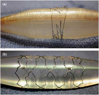
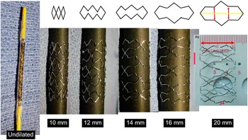
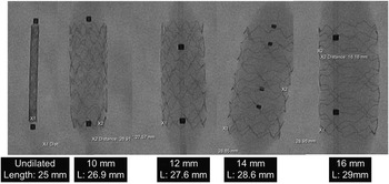
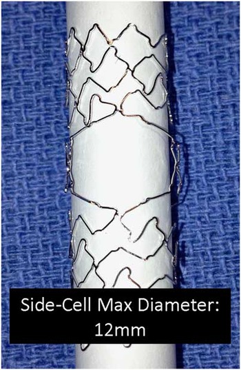
The medium Valeo stents could be maximally dilated to 13 mm before fracturing (Fig 1). The large Valeo stents could be maximally dilated to 20 mm before fracturing (Fig 2). Serial dilation under fluoroscopy did not reveal any stent foreshortening, but rather the stent length actually increased up to 4 mm (Fig 3). The side-cells were maximally dilated to 12 mm (Fig 4).
Medium versus large Valeo stents (Bard Peripheral Vascular, Tempe, Arizona, United States of America) dilated with a 16-mm balloon. ( a ) Depiction of a medium Valeo stent fracturing when dilated beyond 13 mm using a 16-mm balloon. ( b ) Depiction of a large Valeo stent remaining intact with inflation using a 16-mm balloon.

Large Valeo stent (Bard Peripheral Vascular, Tempe, Arizona, United States of America) with maximal diameter and minimal foreshortening. Bench testing results of the large Valeo stent serially dilated to 20 mm with minimal foreshortening.

Large Valeo stent (Bard Peripheral Vascular, Tempe, Arizona, United States of America) with minimal foreshortening. Bench testing results of the large Valeo stent serially dilated up to 16 mm under fluoroscopy with minimal foreshortening in length.

Side-cell maximal diameter of 12 mm. Bench testing results of the side-cell dilation using two 12-mm balloons side-by-side.

Retrospective clinical study
Patient demographics
Between 1 October, 2012 and 1 October, 2014, 81 Valeo stents were placed in 61 patients. The median age was 58.9 months, ranging from 3.7 months to 19.8 years. The median weight was 15.3 kg, ranging from 4.6 to 110 kg. There were 30 small infants weighing <15 kg (49%). There were 24 patients with single ventricle physiology (39%) and 37 patients with biventricular circulation (61%). Specific indications for the use of Valeo stents included small patient size of <15 kg (n=30) and important side-branches adjacent to the stenotic vessel (n=4). The remaining 47 Valeo stents used were chosen at the discretion of the interventional cardiologist.
Pre-implant and post-implant catheterization data
A total of 53 stents were placed in the pulmonary arterial system, 22 in the systemic venous system, that is, in the superior caval vein, inferior caval vein, and femoral veins, five in the aorta, and one in the pulmonary vein. Of the 55 large Valeo stents implanted, 30 (37%) were pre-mounted on 9-mm and 25 (30.9%) on 10-mm balloons. Of the 26 medium Valeo stents implanted, seven (8.6%) were pre-mounted on 6-mm balloons, 14 (17.3%) on 7-mm, and five (6.2%) on 8-mm balloons. The majority of large Valeo stents was placed in the proximal pulmonary arteries, aorta, superior caval vein, and inferior caval vein vessels and was anticipated to need further dilations up to 20 mm at a later stage to accommodate somatic growth. The medium stents were implanted in lobar/distal branch pulmonary arteries, femoral veins, and pulmonary vein vessels that were not anticipated to need further dilations beyond 13 mm.
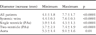
Overall, the mean vessel diameter increased from 4.1±1.8 to 7.7±1.7 mm, p<0.0001 (Table 1). This represents a mean diameter increase of 121.7%. In the systemic veins, the mean diameter increased from 4.4±0.3 to 7.6±0.3 mm. For single ventricle patients with stents placed in the pulmonary arteries, the mean diameter increased from 3.9±1.6 to 4.3±1.3 mm, and for biventricular patients the pulmonary artery mean diameter increased from 3.7±2.1 to 7.4±1.9 mm. In the aorta, the mean diameter increased from 5.3±1.4 to 9.1±1.6 mm. When divided into groups by location, all mean vessel diameter increases were significant with p-values <0.01.
Diameter increase of the Valeo stent.

PAs=pulmonary arteries
For all patients, the diameter increased from 4.1±1.8 to 7.7±1.7 mm, p<0.0001. For stents placed in the systemic veins, the diameter increased from 4.4±0.3 to 7.6±0.3 mm, p<0.0001. For single ventricle patients with stents placed in the PAs, the diameter increased from 3.9±1.6 to 4.3±1.3 mm, p<0.0001, and for biventricular patients with stents placed in the PAs, the diameter increased from 3.7±2.1 to 7.4±1.9 mm, p<0.0001. In the aorta, the diameter increased from 5.3±1.4 to 9.1±1.6 mm, p=0.01. All of these findings are statistically significant
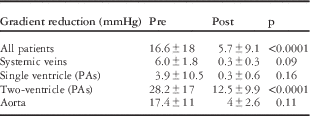
The overall mean gradient reduction for all the patients improved from 16.6±18 to 5.7±9.1 mmHg, p<0.0001. Mean gradient reduction with stents implanted in the systemic veins improved from 6±1.8 to 0.3±0.3 mmHg, p=0.08. In single ventricle patients with stented branch pulmonary arteries, only 22 of 24 had available post-stent gradient data, the mean gradient decreased from 3.9±10.5 to 0.25±0.6 mmHg, p=0.16. Percent gradient reduction was 61.2%. Among biventricular patients with stented branch pulmonary arteries, 31 of 37 had available post-stent gradient data. The mean gradient decreased from 28.2±17 to 12.5±9.9 mmHg, p<0.0001. The percent gradient reduction was 63.7%. The mean pre-intervention right ventricle:left ventricle or right ventricle:femoral artery ratio was 0.73, which decreased to 0.66 after stent implantation, p=0.01. For all patients, including single and biventricular repair, with stented coarctation of the aorta (n=5), the gradient decreased from 17.4±11.0 to 4.0±2.6 mmHg, p=0.11 (Table 2). The percent gradient reduction was 77.1%.
Gradient reduction of the Valeo stent.

PAs=pulmonary arteries
For all patients, the mean gradient reduction decreased from 16.6±18.0 to 5.7±9.1 mmHg, p<0.0001. The gradient decreased from 6±1.8 to 0.3±0.3 mmHg, p=0.08, in the systemic veins, from 3.9±10.5 to 0.25±0.6 mmHg, p=0.16, in the PAs of single ventricle patients, from 28.2±17.0 to 12.5±9.9 mmHg, p<0.0001, in the PAs of double ventricle patients, and 17.4±11.0 to 4.0±2.6 mmHg, p=0.11, in the aorta. All these findings were statistically significant, except for the stents placed in the systemic veins, PAs of single ventricle patients, and the aorta, likely due to insufficient number of patients to add power to the results
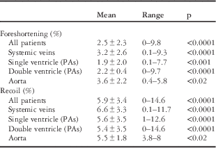
The overall mean percent stent foreshortening was 2.5±2.3%, p<0.0001. The mean percent stent recoil was 5.9±3.4%, p<0.0001 (Table 3). In systemic veins, the mean percent stent length foreshortening was 3.2±2.6%, p<0.0001, and the mean percent recoil was 6.6±3.3%, p<0.0001, for stents placed in the systemic veins. In single ventricle patients with stents in the pulmonary arteries, the mean percent foreshortening was 1.9±2.0%, p<0.001, and the mean percent recoil was 5.6±3.5%, p<0.0001. In biventricular patients with stents placed in the pulmonary arteries, the mean percent foreshortening was 2.2±0.4%, p<0.0001, and the mean percent recoil was 5.4±3.5%, p<0.0001. For stents placed in the aorta, the mean percent foreshortening was 3.6±2.2%, p<0.02, and the mean percent recoil was 5.5±1.8%, p<0.02. These findings for stent foreshortening and recoil remained statistically significant when divided into groups by location.
Foreshortening and recoil of the Valeo stent.

PAs=pulmonary arteries
For all patients, the mean percent foreshortening was 2.5±2.3%, p<0.0001, and the mean percent recoil was 5.9±3.4%, p<0.0001. For stents placed in the systemic veins, the mean percent foreshortening was 3.2±2.6%, p<0.0001, and the mean percent recoil was 6.6±3.3%, p<0.0001. For single ventricle patients with stents in the PAs, the mean percent foreshortening was 1.9±2.0%, p<0.001, and the mean percent recoil was 5.6±3.5%, p<0.0001. For biventricular patients with stents placed in the PAs, the mean percent foreshortening was 2.2±0.4%, p<0.0001, and the mean percent recoil was 5.4±3.5%, p<0.0001. For stents placed in the aorta, the mean percent foreshortening was 3.6±2.2%, p<0.02, and the mean percent recoil was 5.5±1.8%, p<0.02. All these findings were statistically significant
Technical considerations
Several technical considerations require explanation. Re-mounting large Valeo stents onto different balloons before implantation was performed in seven cases. In four instances, the Valeo stents originally pre-mounted on 9-mm balloons were re-mounted onto smaller 6-mm balloons and implanted in small patients who were <15 kg (5.3, 7.9, 11.2, and 16.1 kg). In one case, a large Valeo stent was re-mounted onto a 7-mm balloon for implantation in a 13-kg child. In two instances, a Valeo stent originally pre-mounted onto a 10-mm balloon was re-mounted onto a larger 12-mm balloon before implantation in the pulmonary arteries. The reason for the re-mounting maneuver was specifically to take advantage of the future dilation potential of the large Valeo stent while still matching the distal diameter of the stenotic proximal branch pulmonary artery that required a stent.
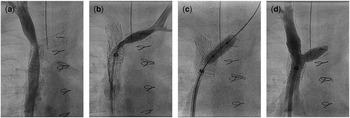
Side-cell dilations were performed in four cases using 3–8-mm diameter balloons. In two cases, a stent was implanted in a left pulmonary artery stenosis adjacent to the left cavopulmonary anastomosis. The third case involved an right pulmonary artery stenosis adjacent to the right upper pulmonary artery branch. The fourth case involved a stent implanted in the superior caval vein stenosis extending across the orifice of the innominate vein in a patient with superior caval vein syndrome (Fig 5). In all the cases, it was anticipated that the stent would jail the adjacent side-branch, and the side-cell of the stent was balloon dilated successfully relieving the potential for the jailed adjacent side-branches to become obstructed. In situations where the flow across a jailed side-branch did not appear to be obstructed, the side-cells were not dilated. There were no technical complications or stent malpositions during any of the procedures.
Side-cell dilation of the Valeo stent (Bard Peripheral Vascular, Tempe, Arizona, United States of America) implanted in superior caval vein (SVC). ( a ) SVC stenosis. ( b ) Status after Valeo stent implantation (10×17 mm) stent placement in SVC. ( c ) Balloon dilation of the side-cell with 6-mm Dorado balloon. ( d ) Status after side-cell balloon dilation of the jailed innominate vein.

Discussion
The Valeo stent is a safe and effective alternative for treatment of vascular stenoses in small children with CHD. The large Valeo stents pre-mounted on 9–10-mm balloons have a maximal diameter of 20 mm, whereas the medium Valeo stents pre-mounted on 6–8-mm balloons have a maximal diameter of 13 mm before fracturing. The low profile and the large maximal diameter of the large Valeo stent make it ideal for implantation into stenotic large vessels such as the proximal branch pulmonary arteries and the superior and inferior caval veins. This stent is also useful in select aortic coarctations in small patients who are not anticipated to grow into large-sized adults. These large stents can be further dilated up to 20 mm diameter to accommodate for somatic growth to adulthood;Reference Shepherd, Connolly and Morgan 8 however, for those patients with aortic coarctations who are anticipated to grow into average-sized or larger adults, we recommend the extra-large stents that are dilatable up to 25-mm diameter, such as the Palmaz XL (Cordis, Fremont, California, United States of America) or the Max LD stents (Covidien, Plymouth, Minnesota, United States of America). The open-cell design of the Valeo stent permits dilation of the jailed side-branches to maintain patency where obstruction from a jailed side-branch is a concern. Similar to other open-cell stents such as the Mega or Max LD (Covidien), the side-cells are dilatable to 12 mm.Reference Peters, Ewert and Berger 3
The pre-mounted form of the Valeo stent is not only convenient for the user but it also minimizes the profile for delivery, which enhances the trackability within tortuous vessels. Furthermore, its mean stent foreshortening of 2.5% is minimal compared with other stents. Published data show that most of the small stents have a foreshortening of 8–10%, medium stents 20–40%, and large stents 22–40%.Reference Peters, Ewert and Berger 3 In many instances, re-mounting the large stents onto smaller balloons allowed for the stent to be placed at a smaller size and maintain its low profile. This is particularly advantageous in smaller patients where sheath size can be limited but serial dilation of the large stent is anticipated during growth into adulthood.
Although its radial strength is generally thought to be less than that of closed-cell stents, it appears to be adequate for the range of diameters used in our cohort with minimum mean stent recoil (5.9%) and is even superior when compared with other medium-to-large stents studies, where the recoil potential was ~15%.Reference Peters, Ewert and Berger 3 Despite the statistical significance found within this study for stent foreshortening and recoil, the differences do not appear to be of clinical significance given the small degree of foreshortening and recoil with the Valeo stent. In a study including 10 patients (12 stents) by Kudumula et al,Reference Kudumula, Noonan, Taliotis and Duke 5 the Valeo stent showed adequate radial strength when implanted in pulmonary arteries and maintained its geometry on medium follow-up (0–30 months), which is consistent with our results. Similar findings were noted in a small case series in which Valeo stents were successfully placed in the pulmonary arteries.Reference Sharma and Goreczny 6 Even if the radial strength is low at larger diameters, further structural support can be achieved by telescoping another stent within the Valeo stent in the future when the patient has grown in size and a larger delivery system is no longer a limitation.
Extensive stent distortion and re-stenosis have been described with the Valeo stent placed in a highly resistant superior caval vein stenosis within a 5-month period.Reference Aiyagari 7 This was not observed in our series, including a Valeo stent that was placed in the superior caval vein.
Gradient reduction of stents placed in the aorta and venous systems did not reach statistical significance. This is likely attributed to the insufficient number of patients within these groups, providing lesser power to the study. In addition, small gradients in venous systems can have greater clinical significance particularly in single ventricle populations.
Limitations to this study were its retrospective design and lack of long-term follow-up. Larger series and longer term follow-up are needed to determine the durability and performance of the stent in adulthood, specifically evaluating the degree of neointimal proliferation, occurrence of stent fractures, patency of jailed side-branches, recurrence of stenosis, and resistance to thrombus formation.
Acknowledgement
None.
Financial Support
This project was supported by a grant from the Hearts Foundation and the Heart Ambassadors of Children’s Hospital Los Angeles.
Conflicts of Interest
None.
Ethical Standards
The authors assert that all procedures contributing to this work comply with the ethical standards of the Belmont Report and with the Helsinki Declaration of 1975, as revised in 2008, and has been approved by the CHLA Institutional Review Board.