Case report
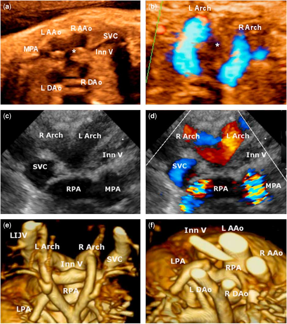
A 25-year-old gravida 2 para 0 woman was referred for fetal echocardiogram at 35 weeks of gestation. In addition to demonstrating tetralogy of Fallot, the echocardiogram revealed an extra vessel between the main pulmonary artery and the superior caval vein that was suspicious for a second aortic arch (Fig 1a and b).
( a ) Fetal echocardiogram, the axial “three-vessel view” demonstrating an additional vessel between the main pulmonary artery (MPA) and the superior caval vein (SVC) suspicious for a second aortic arch. ( b ) Similar colour Doppler flow patterns are seen in both arches. ( c ) Suprasternal coronal view showing two aortic arches passing over a retroaortic innominate vein (Inn V); ( d ) colour Doppler. ( e ) MRI reconstruction demonstrating a double aortic arch and ( f ) the branch pulmonary arteries bifurcating at an unusual superior angle from the MPA. LIJV=left internal jugular vein; L/R AAo=left/right ascending aorta; L/R Arch=left/right aortic arch; L/R DAo=left/right descending aorta; L/R PA=left/right pulmonary artery; *=trachea.

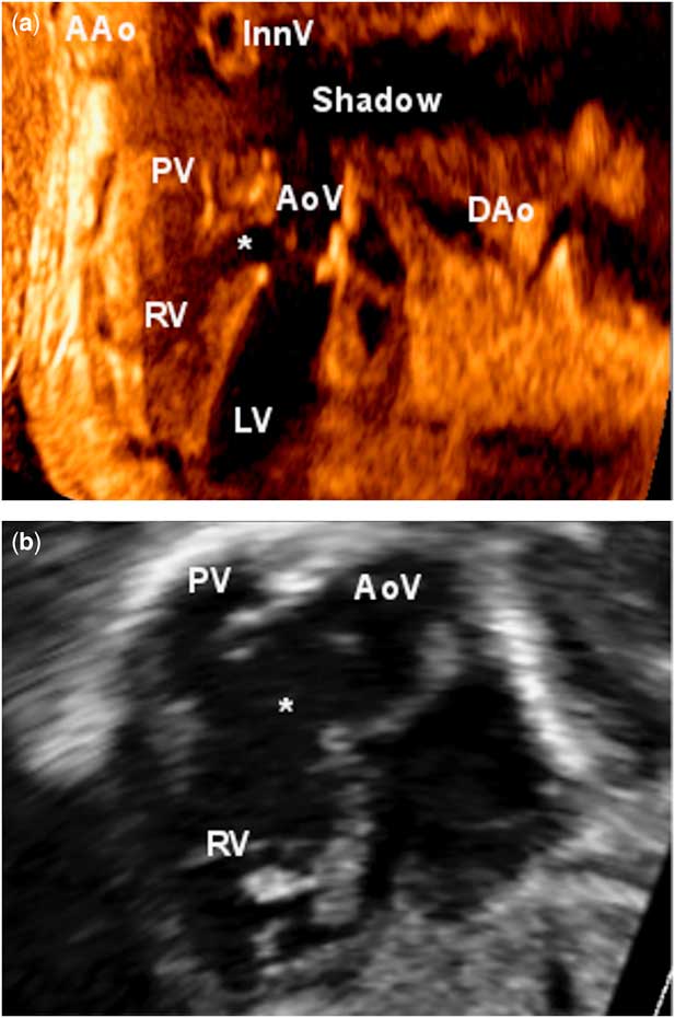
The baby was born at full term without complications. In the hospital, she maintained saturations >95% and displayed no stridor or dysphagia. Her first outpatient transthoracic echocardiogram confirmed tetralogy of Fallot (Fig 2), but the branch pulmonary arteries were difficult to fully define as they were in an unusual plane. Again there appeared to be two aortic arches (Fig 1c and d). Owing to the unusual pulmonary artery and arch anatomy, she was referred for further evaluation with cardiac MRI. This confirmed the presence of a double aortic arch (Fig 1e and f) and also revealed tracheal and oesophageal narrowing between the arches.
( a ) Fetal echocardiogram showing features of tetralogy of Fallot including hypoplastic pulmonary valve (PV), overriding aorta (AoV), and ventricular septal defect. The aorta and innominate vein are also partially visualised, but obscured by a shadow from the sternum. ( b ) Transthoracic echocardiogram subcostal sagittal view demonstrating similar findings. AAo=ascending aorta; DAo=descending aorta; Inn V=innominate vein; LV=left ventricle; RV=right ventricle; *=ventricular septal defect.

In light of the tracheal narrowing, she underwent double arch repair 1 week later. Intraoperative bronchoscopy demonstrated tracheal narrowing just above the carina. Direct inspection showed two similarly sized arches, and the right arch was surgically divided. Her post-operative recovery was uncomplicated. She subsequently underwent tetralogy of Fallot repair at 12 months of age.
Prenatal diagnosis of vascular rings has been described both in case reports and in series. In the largest series of 81 cases, about one-third of those with other congenital malformations had CHD;Reference Li, Luo and Norwitz 1 however, to our knowledge, there have been no reported cases of a prenatal diagnosis of tetralogy of Fallot in association with a double aortic arch, specifically.
As demonstrated in this case report, a double aortic arch can be diagnosed by fetal echocardiogram using the axial three-vessel and trachea view.Reference Trobo, Bravo, Alvarez, Pérez, Gámez and De León-Luis 2 From this view, in a normal fetus, the aortic arch and the ductal arch form a V-shaped structure as they join at the descending aorta.Reference Trobo, Bravo, Alvarez, Pérez, Gámez and De León-Luis 2 In the case of a double aortic arch, these structures along with the second aortic arch instead form a trident shape – the left prong being the ductus arteriosus, the middle prong the left aortic arch, and the right prong the right aortic arch – with the trachea located between the middle and the right prongs.Reference Trobo, Bravo, Alvarez, Pérez, Gámez and De León-Luis 2 As one arch is often atretic and positioned more caudally, however, clear visualisation of all of these vessels simultaneously can be difficult.Reference Trobo, Bravo, Alvarez, Pérez, Gámez and De León-Luis 2 Judicious imaging is therefore required to differentiate a double aortic arch from other anatomical variants such as a right aortic arch with a left ductus arteriosus.
Acknowledgements
The authors thank Dr Jon Detterich for his assistance with the cardiac MRI images.
Financial Support
This research received no specific grant from any funding agency, commercial, or not-for-profit sectors.
Conflicts of Interest
None.
Supplementary Material
To view supplementary material for this article, please visit http://dx.doi.org/10.1017/S1047951116000329