Introduction
Patients with locally advanced glottic laryngeal carcinoma typically present with tumour (T) stage T3 and T4 disease, with or without nodal disease.Reference Mendenhall, Hinerman, Amdur, Mancuso, Villaret, Werning, Halperin, Perez and Brady1, Reference Hartl, Ferlito, Brasnu, Langendijk, Rinaldo and Silver2 This covers a spectrum of disease, from moderately bulky disease sufficient to prevent vocal fold movement (while maintaining an adequate airway), to extensive (more typically) bilateral laryngeal disease, extralaryngeal spread and a compromised airway. Functional consequences differ with each situation. For the former, voice and larynx preservation are still feasible; for the latter, airway management is the priority. Therefore, treatment methods must be considered within the context of disease, functional outcomes, and the potential for salvage should local and/or nodal recurrence occur.
Many studies combine disease characteristics when defining laryngeal carcinoma and analysing outcomes such as survival and recurrence. These studies report outcomes in patients with T3–4 laryngeal carcinoma without differentiation of anatomical site;Reference Gourin, Conger, Sheils, Bilodeau, Coleman and Porubsky3–Reference Reizenstein, Holmberg, Bergqvist, Linder, Ekman and Lödén5 they report glottic carcinomas, but do not distinguish between T1–2 and T3–4 stage disease;Reference Lima, Freitas, Dias, Barbosa, Kligerman and Soares6 or they report stage IV glottic disease, without separating into local and regional diseaseReference Spector, Sessions, Lenox, Newland, Simpson and Haughey7 or T classification.Reference Chen, Schrag, Hao, Flanders, Kepner and Stewart8 Studies either present small patient numbers, or combine patient populations with laryngeal carcinomas. Therefore, there is a paucity of literature that specifically investigates the outcomes of patients with locally advanced T3–4 glottic carcinoma.
The British Medical Journal Best Practice Guide suggests that treatment for advanced T3–4 glottic carcinoma should involve either concurrent chemoradiotherapy or surgery.9 For T3 glottic carcinomas, neither chemoradiotherapy nor surgery offer any further benefit in overall survival and cancer-specific survival.Reference Pfister, Laurie, Weinstein, Mendenhall, Adelstein and Ang10 Irrespective of subsite, patients with T3 laryngeal carcinoma treated surgically, with or without radiotherapy (RT), have higher overall survival compared to patients treated non-surgically with RT, with or without chemotherapy.Reference Hoffman, Porter, Karnell, Cooper, Weber and Langer11, Reference Al-Gilani, Skillington, Kallogjeri, Haughey and Piccirillo12 If the laryngeal cartilage is involved in patients with T4 glottic carcinomas, then a total laryngectomy is recommended over concurrent chemoradiotherapy; however, no preference is given when cartilage is not involved.Reference Hristov and Bajaj13
This review, representing an historical perspective, presents the outcomes specifically for a large number of patients with locally advanced glottic carcinomas, treated by surgery and/or RT in a single centre, in order to evaluate management improvement in the current circumstances.
Materials and methods
Study design
Retrospective data collection was approved by the South-Eastern Sydney Local Health District Ethics Committee. The records of all patients with newly diagnosed advanced stage glottic laryngeal carcinoma who presented to the Prince of Wales Hospital Cancer Centre between 1 May 1967 and 30 June 2015 were reviewed.
Eligibility criteria included: curative treatment of T3 and T4 stage laryngeal carcinoma of glottic origin, squamous cell carcinoma (SCC) histology, age of 18 years or more, and a potential minimum follow-up period of 2 years. All nodal (N) stages were included. All clinical staging (incorporating physical assessment and, where available, imaging), was defined in accordance with the Union for International Cancer Control tumour–node–metastasis classification, seventh edition.Reference Sobin, Gospodarowiczm and Wittekind14 Patients were excluded if referred with recurrent or progressive disease, or distant metastases.
Treatment
Patients were reviewed by the multidisciplinary head and neck clinic at this institution, and treated with surgery only, surgery plus RT, or RT only.
Radiotherapy was delivered with 4 mV photons pre-1996 and with 6 mV post-1996, delivering 200–225 cGy per fraction per day. More recent patients have been treated with intensity-modulated radiation therapy; however, this is a relatively small group. All fields were treated daily, using an appropriate head immobilisation device.
Surgery was the primary treatment for advanced glottic carcinoma, with or without post-operative RT, for patients with obstructive T4 glottic carcinoma. The decision to include neck dissection was based upon disease features and the likelihood of the patient receiving post-operative RT. Patients with lesser volume T3 disease were offered radical RT with surgical salvage, in the belief, based on contemporaneous literature, that this would result in comparable overall survival.
The outcomes pre- and post-1995 were assessed separately to determine whether a patient treated more recently, with access to better imaging and advanced technological approaches, had an improved outcome.
Data collection and follow up
Data were collected from patients’ notes and medical records, and categorised into patient-, disease- and treatment-related information. All patients were routinely assessed every 3 to 4 months post-treatment during the first 2 years, and every 6 to 12 months thereafter. Follow-up information was collected via the New South Wales Registry of Births, Marriages and Deaths, or via the Ryerson Index website (public domain),15 and from medical records, and letters from clinicians and general practitioners.
Outcomes
The primary endpoints were time to local failure and ultimate local failure. Local failure was defined as persistent disease within the larynx after treatment or a local recurrence after a complete response (excluding patients with regional failure only). Ultimate local failure was declared when there was a local failure and salvage treatment was not performed, or salvage treatment was performed but the cancer still recurred.
The secondary endpoints were cancer-specific survival and overall survival. Cancer-specific survival was defined as survival or death in patients without head and neck cancer (no primary, regional recurrence or metastasis). Overall survival represented the fate of the patient, irrespective of cause of death.
Lastly, the tertiary endpoint was time to malignancy development, which was defined by the subsequent development of a new primary.
Statistical analysis
Statistical analysis was conducted using SPSS Statistics software, version 24.0 (IBM, Armonk, New York, USA). For continuous variables, normality was assessed using the Shapiro–Wilk test. Categorical variables (treatment modality, RT treatment length, and local and ultimate local control) were analysed using binomial logistic regression. Odds ratios resulting from binomial logistic regression are reported alongside 95 per cent confidence intervals (CIs). Time-to-event analysis (local and ultimate local failure, cancer-specific survival, and overall survival) was performed using the Kaplan–Meier method, and was measured from the date of the first treatment to the date of failure or last follow up. The five-year survival and control rates are reported alongside a standard error. The log-rank test was used to determine statistical significance between survival curves. A Cox regression was performed to analyse variables that showed significance during univariate Kaplan–Meier analysis. Hazard ratios resulting from Cox regression are reported alongside a 95 per cent CI. The level of significance for all tests was set to p < 0.05. All p-values are two-sided.
Results
Study population
The selection criteria for this study are displayed in Fig. 1. Between May 1967 and June 2015, 1663 patients presented at the Prince of Wales Hospital, in Randwick, New South Wales, Australia, with laryngeal carcinoma. Of these, 169 patients with T3–4 glottic carcinomas were eligible for inclusion in the study. The median follow-up period was 3.9 years (interquartile range, 1.4–10.9 years). As of January 2017, 23 patients (14 per cent) were alive, with a median follow-up period of 7.4 years (interquartile range, 2.7–10.9 years). No patients were lost to follow up.
Eligibility criteria. SCC = squamous cell carcinoma; POWH = Prince of Wales Hospital; T = tumour stage

Baseline characteristics
Table 1 displays the patients’ demographics and disease characteristics. The majority of patients were male (n = 157, 93 per cent), with a male to female ratio of 13:1. The mean age was 64 years (range, 29–92 years), with patients aged 65 years or more experiencing reduced survival (log-rank p < 0.001). Of the patients, 160 (95 per cent) had a smoking history. Six patients (4 per cent) were not fit for surgery.
Patient demographics and disease characteristics

* n = 34;
† n = 35;
‡ n = 100;
** n = 169.
ECOG = Eastern Cooperative Oncology Group;
IQR = interquartile range
Squamous cell carcinoma was confirmed in 156 patients (92 per cent), with the remaining patients having carcinoma not otherwise specified, with 1 unknown carcinoma clinically categorised as SCC. Fig. 2 displays the most common sites of involvement (not just site of origin). Of the patients, 158 (94 per cent) had fixed vocal folds. Regarding T classification, most patients presented with T3 disease (n = 145, 86 per cent). Regarding N classification, N0 disease was the most prevalent (n = 143, 85 per cent).
Sites of involvement. L = left; R = right

There was no statistical significance between patient demographics or disease characteristics and treatment type.
Treatment details
Treatment details are presented in Table 2. One hundred patients (59 per cent) were treated with surgery plus RT (92 were treated with RT post-surgery and 8 were treated with RT pre-surgery) as the planned pre-operative treatment, this being the protocol at that time. There were no differences observed in treatment modality, survival or failure pre-1995 and post-1995.
Treatment details

* n = 34;
† n = 35;
‡ n = 100;
** n = 169.
IQR = interquartile range
Radiotherapy
The median number of treatment days was 40, with a median dose of 55 Gy, in 28 fractions, for those patients treated with surgery plus RT. For those patients treated by RT only, the median number of treatment days was 44, with a median dose of 66 Gy, in 33 fractions. Nine per cent of patients experienced a treatment interruption of more than a week.
Surgery
All eligible patients had operable cancer, with 163 (96 per cent) deemed fit for surgery. Of the patients, 134 (79 per cent) received surgery to their primary tumour, with laryngectomy being the most common surgical procedure (n = 123, 92 per cent) (Table 2).
Treatment outcomes
Treatment outcomes are displayed in Table 3. Thirty-one patients developed local recurrence (including persistent disease), of which 28 progressed to develop a second local recurrence (including persistent disease).
Treatment outcomes

Data represent numbers (and percentages) of cases.
* n = 34;
† n = 35;
‡ n = 100;
** n = 169
Local control
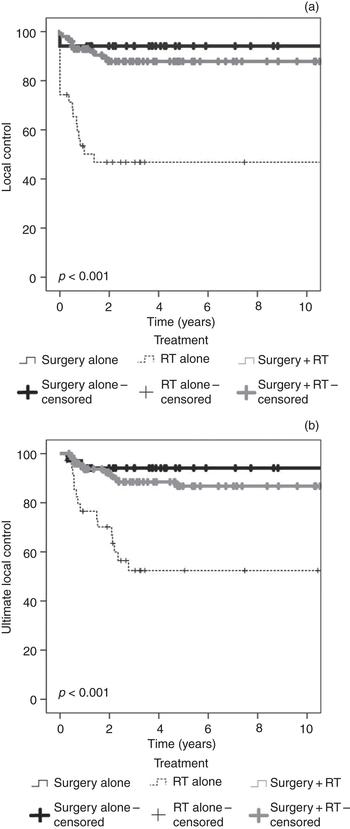
Five-year local control rates were 94.1 per cent for surgery only, 87.9 per cent for surgery plus RT, and 46.8 per cent for RT only groups (log-rank p < 0.001) (Fig. 3a, Table 4). The overall 5-year local control rate for all patients was 80.7 per cent, with a median time to local failure of 4.9 months. Local failure occurred in 31 patients (18 per cent); most cases of failure (18 out of 31, 58 per cent) occurred in patients treated with RT only. Of these 31 patients, almost half (14 out of 31, 45 per cent) did not receive salvage treatment (Table 3).
(a) Local control and (b) ultimate local control by treatment type. RT = radiotherapy

Rates of survival and control by treatment type

* Log-rank test p-value calculated between treatment types from Kaplan–Meier survival analysis.
SE = standard error
Treatment modality and RT treatment length were the only significant univariate predictors for local failure. Patients treated with RT over a period of 41 days or more were 5 times more likely to experience local failure, compared to those treated in less than 41 days (odds ratio = 5.0, 95 per cent CI = 1.9–13.3, p < 0.001). Irrespective of treatment length, patients receiving RT only were 17 times more likely to experience local failure than patients receiving surgery with or without RT (odds ratio = 16.9, 95 per cent CI = 3.5–81.8, p < 0.001).
Ultimate local control
Five-year ultimate local control rates were 94.1 per cent for surgery only, 86.8 per cent for surgery plus RT, and 52.4 per cent for RT only groups (log-rank p < 0.001) (Fig. 3b, Table 4). The overall 5-year ultimate local control rate was 80.9 per cent (Table 4), with a median time to ultimate local failure of 10.6 months. Twenty-eight patients experienced ultimate local failure (17 per cent). Of the eight patients initially treated with RT who underwent surgical salvage, three patients were ultimately free of their cancer (Table 3).
As with local failure, treatment modality and RT treatment length were the only significant univariate predictors for ultimate local failure. The risk of ultimate local failure increased 4-fold if RT treatment length was 41 days or more, compared to those treated in less than 41 days (odds ratio = 4.1, 95 per cent CI = 1.5–11.0, p = 0.005). Regardless of RT treatment length, the likelihood of a patient experiencing ultimate local failure increased 12-fold if they initially received RT only, compared to patients receiving surgery with or without RT (odds ratio = 12.0, 95 per cent CI = 2.5–58.1, p = 0.002). No difference was observed in local control and ultimate local control after adjusting for patients who were unfit for surgery.
Secondary outcomes
A total of 146 patients died; 55 died of head and neck cancer and 91 died of other causes. The five-year cancer-specific survival and overall survival rates were 63.8 per cent and 44.0 per cent, respectively (Table 4). There was no difference in cancer-specific survival or overall survival with treatment type (Fig. 4). The median cancer-specific survival and overall survival time was 1.3 years (95 per cent CI = 0.6–2.1) and 3.5 years (95 per cent CI = 2.7–4.3) respectively.
(a) Overall survival and (b) cancer-specific survival by treatment type. RT = radiotherapy

Univariate analysis identified involvement of the epiglottis and bilateral vocal folds as significant predictors for cancer-specific survival (log-rank p < 0.05). However, during multivariate analysis, Cox regression did not identify epiglottis and bilateral vocal fold involvement as independent predictors for cancer-specific survival.
Bilateral vocal fold involvement was a significant univariate predictor for overall survival (log-rank p = 0.015). Compared to patients with unilateral vocal fold involvement, patients with bilateral vocal fold involvement had significantly reduced median survival time (unilateral – median overall survival = 4.9 years, 95 per cent CI = 3.5–6.2; bilateral – median overall survival = 2.8 years, 95 per cent CI = 1.9–4.4). Bilateral vocal fold involvement is more likely a surrogate for greater volume of disease.
Tertiary outcome
Forty patients (24 per cent) developed a new primary tumour distant from the larynx that was not associated with treatment type. The median time to a second malignancy was 4.8 years (95 per cent CI = 1.6–8.1).
Discussion
This historical and retrospective study focused on the local control and survival of patients with locally advanced T3–4 glottic carcinoma. The pattern of local spread of laryngeal cancer can be vertical and/or horizontal. Vertical spread occurs when the supraglottis or subglottis becomes involved after paraglottic extension (initially involving the ventricle). Horizontal spread occurs when there is extension across to the contralateral fold, into (more typically) the anterior commissure, as well as lateral spread into extralaryngeal structures. Thyroid cartilage penetration has been reported in 44 per cent of patients with extralaryngeal spread.Reference Chen, Muller, Chen, Hudgins, Shin and Khuri16 This is important in defining whether a non-surgical approach can be used, given that the volume of malignancy has been identified as being inversely related to the likelihood of control after RT.Reference Mendenhall, Parsons, Mancuso, Pameijer, Stringer and Cassisi17 These approaches must be evaluated when considering treatment for local control or laryngeal preservation.
Spector et al. reported on 96 patients presenting with stage IV glottic carcinoma.Reference Spector, Sessions, Lenox, Newland, Simpson and Haughey7 All but seven patients underwent total laryngectomy, with differing combinations of neck dissections and post-operative RT. A locoregional control rate of 75 per cent was reported, with 18 per cent recurring at the primary site.Reference Spector, Sessions, Lenox, Newland, Simpson and Haughey7 Additionally, Lima et al. reported on 43 patients with T3–4 glottic carcinoma who underwent subtotal laryngectomy and reconstruction; the local recurrence rate was 7 per cent.Reference Lima, Freitas, Dias, Barbosa, Kligerman and Soares6 Furusaka et al. found that super-selective chemotherapy and chemoradiotherapy was able to provide a 5-year laryngeal preservation rate of 71 per cent in 64 patients with T3–4 glottic cancer, with a laryngeal preservation rate of almost double for T3 versus T4 carcinoma (93 per cent vs 49 per cent).Reference Furusaka, Matsuda, Tanaka, Matsuda and Ikeda18
In a small series spanning 10 years, Kada and colleagues reported no local recurrences in 33 patients with advanced laryngeal SCC (of glottic and subglottic origin), 29 of which were T3–4 tumours.Reference Kada, Hirano, Tateya, Kitamura, Ishikawa and Kanda19 All patients with T3–4 staged SCC were treated with total laryngectomy and neck dissection. While no local recurrences occurred, two patients with glottic origin experienced distant metastasis.Reference Kada, Hirano, Tateya, Kitamura, Ishikawa and Kanda19 Delaere et al. interestingly reported on 17 patients (9 presenting with recurrences) who had unilateral advanced glottic disease managed by extended hemilaryngectomy and tracheal auto-transplantation as a two-stage procedure, with only one patient experiencing treatment failure locally.Reference Delaere, Goeleven, Poorten, Hermans, Hierner and Vranckx20 A study by Timmermans et al., of 182 patients with T3–4 laryngeal cancer, did not report outcome data by site; nevertheless, they also found no difference in overall survival by T stage (T3 vs T4) or by treatment modality.Reference Timmermans, de Gooijer, Hamming-Vrieze, Hilgers and van den Brekel4 Harris et al. scrutinised the Surveillance, Epidemiology and End Results database, whereby 6797 patients with advanced laryngeal cancer were evaluated.Reference Harris, Bhuskute, Rao, Farwell and Bewley21 Unfortunately, there was no separation of glottic origin from supraglottic origin regarding local outcome. The conclusion, however, was that patients with T3–4 laryngeal cancer have a greater chance of survival with primary surgery.Reference Harris, Bhuskute, Rao, Farwell and Bewley21
Canis and colleagues have reported on patients with T3 and T4 glottic carcinoma treated with transoral laser microsurgery, with post-operative chemoradiotherapy used in 18 per cent of T3 patients and in 33 per cent of T4 patients.Reference Canis, Martin, Ihler, Wolff, Kron and Matthias22, Reference Canis, Ihler, Martin, Wolff, Matthias and Steiner23 Local control rates were reported as 72 per cent and 67 per cent for T3 and T4 disease respectively. Transoral laser surgery has been used in many facilities, with or without neck dissection and RT, with reported local control rates of up to 75 per cent in 117 patients.Reference Hinni, Salassa, Grant, Pearson, Hayden and Martin24 However, this was in a population of patients with advanced laryngeal cancer (with subgroups not reported), with all patients pathologically and not clinically staged, thus making it difficult to compare to non-surgically managed patients, as in our series.
In the current series of 169 patients, 21 per cent were treated with RT only. The five-year local control and ultimate local control rates among these patients were 46.8 per cent and 52.4 per cent respectively. These rates are significantly lower than those for patients treated by surgery with or without RT (p < 0.001). All but one patient with T4 disease was treated by surgery with or without RT, which may explain the trend toward improved local control for patients with T4 disease compared to those with T3 disease. The decision to use post-operative RT was based on the presence or absence of close or positive margins, and the nature of nodal disease if neck dissection was performed. There was no difference in the parameters relevant to those patients who had post-operative RT for the primary tumour versus those treated because of the neck disease. Thus, those patients who underwent surgery alone had favourable pathological features sufficient to warrant no further treatment.
In a review of the literature for T3 cancers treated by RT only, Mendenhall et al. recorded initial local control rates ranging from 36 per cent to 77 per cent, with surgical salvage being performed to generate ultimate local control rates of 70–80 per cent.Reference Mendenhall, Hinerman, Amdur, Mancuso, Villaret, Werning, Halperin, Perez and Brady1 In a series of 109 patients with previously untreated T3–4 glottic SCC, Hinerman et al. reported a 5-year local control rate of 67 per cent and ultimate local control rate of 89 per cent in patients with T3 glottic SCC, and a cancer-specific survival rate of 83 per cent in patients with stage III disease treated with primary RT.Reference Hinerman, Mendenhall, Morris, Amdur, Werning and Villaret25 The same review reported no evidence of recurrent disease rates of 66 per cent for patients with T3 disease treated by laryngectomy, compared to 48 per cent in a series of patients treated by RT with surgical salvage.Reference Al-Mamgani, Tans, van Rooij and Levendag26 MacKenzie et al. reported disease control above the clavicles in 65 per cent of patients with T3–4 glottic primaries treated with radical RT and surgical salvage.Reference MacKenzie, Franssen, Balogh, Gilbert, Birt and Davidson27 Croll et al. reported comparable figures with the same radical RT and surgical salvage approach.Reference Croll, Gerritsen, Tiwari and Snow28 Lyhne et al. reported on the outcomes of the Danish Head and Neck Cancer Group ‘DAHANCA 6’ study, which investigated fractionation patterns, covering comparable time frames, similar doses used and the absence of chemoradiotherapy.Reference Lyhne, Primdahl, Kristensen, Andersen, Johansen and Andersen29 The local control outcomes reported by Lyhne et al. for T3–4 glottic carcinomas are comparable with those achieved at our institution. The outcomes thus reported here represent a ‘real world’ depiction.
Surgical intervention remains the treatment modality that yields the highest rates of local control, and total laryngectomy is still the appropriate treatment for disease obstructing a large volume of the airway. However, where laryngeal preservation is a consideration, Hartl et al. noted that while high-level evidence favours combined chemoradiotherapy, or altered fractionation, compared to RT only as non-surgical strategies, levels of evidence for optimal treatment selection remain low.Reference Hartl, Ferlito, Brasnu, Langendijk, Rinaldo and Silver2 Further, while Forastiere et al. have demonstrated high rates of laryngeal preservation in randomised studies of non-surgical intervention for patients with glottic and supraglottic laryngeal carcinoma (particularly using concurrent chemoradiotherapy), higher rates of non-cancer deaths were also noted.Reference Forastiere, Zhang, Weber, Maor, Goepfert and Pajak30 Other studies have also reported decreased rates of overall survival in patients with laryngeal carcinoma, attributed to the shift away from surgical approaches.Reference Hoffman, Porter, Karnell, Cooper, Weber and Langer11, Reference Connor, Pattle, Kerr and Junor31 In this current series, there were insufficient patients with concurrent chemoradiotherapy to evaluate separately, and therefore a comparative comment cannot be made.
As well as the treatment delivered, various patient and disease factors can also influence the outcome. These include: treatment in a high-volume centre,Reference Chen, Fedewa, Pavluck and Ward32 insurance status,Reference Kwok, Langevin, Argiris, Grandis, Gooding and Taioli33 volume (on imaging) of disease being treatedReference Mendenhall, Parsons, Mancuso, Pameijer, Stringer and Cassisi17 and the impact of patient co-morbidities.Reference Smee, De-Loyde, Broadley and Williams34 Bøje et al. reported that up to 36 per cent of head and neck cancer patients may have significant co-existing co-morbidities that can independently influence treatment outcomes.Reference Bøje, Dalton, Grønborg, Primdahl, Kristensen and Andersen35
• Locally advanced glottic carcinomas should be reported separately to supraglottic carcinomas
• Concurrent chemoradiotherapy is the standard, with less than 10 per cent improvement in local control and worsening of toxicity
• Non-treatment factors influence outcomes
• Non-surgical approaches are not advocated where the airway is threatened
• This audit of practice led to a re-evaluation of decision making and recommended delivery
• It can act as a comparator for practice at other community-based centres, and be compared with multicentre studies for outcome evaluation
While most series report more favourable outcomes for T3 compared to T4 disease,Reference Hartl, Ferlito, Brasnu, Langendijk, Rinaldo and Silver2 the results of the current study do not reflect this, which in fact demonstrates a trend toward better local control in patients with T4 disease. This may be attributable to the treatment modalities utilised; most patients with T4 disease (96 per cent) received primary treatment involving surgery, and 20 per cent of patients with T3 disease received RT only. It has been previously established that treatment for T3 glottic carcinomas involving surgery with or without RT has superior outcomes compared to non-surgical approaches,Reference Hoffman, Porter, Karnell, Cooper, Weber and Langer11 and therefore the lower survival outcome of patients with T3 disease observed in this study is likely attributable to the primary treatment modality. However, Vengalil et al. also commented that selected patients with small volume T4 glottic carcinoma, with minimal cartilage involvement, may still be considered for RT or chemoradiotherapy rather than laryngectomy as a method for laryngeal preservation.Reference Vengalil, Giuliani, Huang, McNiven, Song and Xu36
This series represents one of the largest single-centre experiences with locally advanced laryngeal SCC of glottic origin; however, it is not without limitations. While this study aimed to provide a historical measure of the management of locally advanced glottic SCC, the study spans many decades, over which considerable changes in diagnostic imaging and evaluation, and surgical and RT techniques have occurred. However, all patients were clinically staged, and the multidisciplinary head and neck team attending these patients was consistent over time. A proportion of patients in this study were treated prior to: the widespread introduction of computed tomography scans and magnetic resonance imaging; improvements in surgical techniques (including the use of lasers); the introduction of intensity-modulated RT; and the use of concurrent chemoradiotherapy as a method for laryngeal preservation. Therefore, while direct comparisons between this population and other current studies are difficult, our results can be used as a guide for measuring progress in technology and treatment, and to compare long-term outcomes.
Acknowledgements
This paper would have not been possible without funding from the Head and Neck Cancer Research Fund at the Prince of Wales Hospital. The publication pays tribute to the many members of the Prince of Wales Hospital Multidisciplinary Head and Neck Team, including our radiation oncologists, ENT and head and neck surgeons, dietician, speech pathologists, and our head and neck nurse, Jody Buchman. The work of the Prince of Wales Hospital Radiation Oncology Research Team is much appreciated.
Competing interests
None declared