Introduction
Entomopathogenic fungi are common and widespread pathogens of virtually every insect order and are often used for biological control (Jaronski Reference Jaronski2010). Although these pathogens infect insects living in a wide array of habitats, their ability to control pest populations is often context dependent (Roy et al. Reference Roy, Steinkraus, Eilenberg, Hajek and Pell2006; Boomsma et al. Reference Boomsma, Jensen, Meyling and Eilenberg2014). Fungal entomopathogen life cycles are closely linked to climate, with favourable abiotic conditions sometimes leading to massive epizootics by enhancing pathogen development and infectivity (Vega et al. Reference Vega, Chandler, Goettel, Pell and Wajnberg2010). For example, free living stages of entomopathogens, particularly fungal conidia, are highly influenced by a variety of abiotic factors including temperature, moisture, and solar radiation (Hajek and St. Leger Reference Hajek and St. Leger1994). Consequently, mycosis (i.e., fungal infection) is often sporadic with massive insect die-offs occurring in some years and virtually no signs of mycosis in others (Pickford and Riegert Reference Pickford and Riegert1964; Erlandson et al. Reference Erlandson, Johnson and Olfert1988; Hajek et al. Reference Hajek, Larkin, Carruthers and Soper1993).
Grasshoppers are major agricultural pests in arid ecosystems whose populations can be limited by entomopathogenic fungi depending on prevailing environmental conditions (Dempster Reference Dempster1963; Carruthers et al. Reference Carruthers, Ramos, Larkin, Hostetter and Soper1997; Jaronski Reference Jaronski2010; Kistner and Belovsky Reference Kistner and Belovsky2014). Fungal mycoses in grasshoppers occur more frequently under moist conditions (Hajek and St. Leger Reference Hajek and St. Leger1994; Boomsma et al. Reference Boomsma, Jensen, Meyling and Eilenberg2014) and massive epizootics are often linked to precipitation (Skaife Reference Skaife1925; Pickford and Riegert Reference Pickford and Riegert1964; Carruthers et al. Reference Carruthers, Ramos, Larkin, Hostetter and Soper1997). Naturally occurring epizootics can dramatically reduce pest grasshopper densities, but the impact of these pathogens varies both spatially and temporally (Dempster Reference Dempster1963; Erlandson et al. Reference Erlandson, Johnson and Olfert1988). Several previous studies demonstrated that temperature, sunlight, and moisture can affect immediate efficacy and persistence of mycoinsecticides in the environment thereby limiting their ability to control pest grasshopper populations (Goettel et al. Reference Goettel, Johnson and Inglis1995; Inglis et al. Reference Inglis, Johnson and Goettel1997; Klass et al. Reference Klass, Blanford and Thomas2007a, Reference Klass, Blanford and Thomas2007b; Jaronski Reference Jaronski2010).
In this study we examined how abiotic factors influence host-pathogen outcomes of the pest grasshopper, Camnula pellucida (Scudder) (Orthoptera: Acrididae), and its obligate fungal entomopathogen, Entomophaga grylli (Fresenius) (Entomophthorales: Entomophthoraceae) pathotype 1, in a North American intermountain bunch-grass prairie. Entomophaga grylli pathotype 1 is one member of a species complex of obligate grasshopper fungal entomopathogens (Goettel et al. Reference Goettel, Johnson and Inglis1995). Pathotype 1 is endemic to North America and only infects grasshoppers in the subfamily Oedipodinae (Carruthers et al. Reference Carruthers, Ramos, Larkin, Hostetter and Soper1997). The life cycle of this entomopathogen is directly linked to its environment (MacLeod et al. Reference MacLeod, Cameron and Soper1966; Carruthers et al. Reference Carruthers, Feng, Ramos and Soper1988a) with massive epizootics occurring after heavy rainfalls in some years (Pickford and Riegert Reference Pickford and Riegert1964; Erlandson et al. Reference Erlandson, Johnson and Olfert1988). Pathotype 1 overwinters as resting spores that germinate under the proper temperature and soil moisture conditions (Carruthers et al. Reference Carruthers, Ramos, Larkin, Hostetter and Soper1997). Following germination, resting spores produce specialised spores, germ conidia, which are forcibly injected onto the soil surface and lower canopy where they infect hosts by direct contact. Once inside the host haemocoel, the fungus multiplies rapidly, digesting the inner tissues to develop and replicate (MacLeod et al. Reference MacLeod, Cameron and Soper1966). Within 7–10 days, the infected grasshopper climbs to the top of the plant canopy where it dies grasping the foliage (i.e., summit disease), usually in mid-afternoon to ensure the best possible conditions for conidia production and subsequent release during the cooler night (Carruthers et al. Reference Carruthers, Ramos, Larkin, Hostetter and Soper1997; Roy et al. Reference Roy, Steinkraus, Eilenberg, Hajek and Pell2006). With adequate moisture, the fungus will sporulate shortly after host death, with conidiophores exiting the host body and forcibly ejecting conidia into the local environment (Carruthers et al. Reference Carruthers, Larkin, Firstencel and Feng1992). While conidia are highly infectious, they are quickly killed by overheating, desiccation, and ultraviolet radiation (Carruthers et al. Reference Carruthers, Feng, Ramos and Soper1988a). Entomophaga grylli conidia also have limited dispersal and travel only a few metres in still air due to their large physical size and pear-like shape (Wilding Reference Wilding1970; MacLeod and Müller-Kögler Reference MacLeod and Müller-Kögler1973). However, if conidia land on a moist non-host substrate they may germinate and produce secondary conidia to further increase the probability of contacting a viable host (MacLeod et al. Reference MacLeod, Cameron and Soper1966). If current conditions are not favourable for conidia production then the fungus may produce dormant resting spores (Carruthers et al. Reference Carruthers, Ramos, Larkin, Hostetter and Soper1997). Since E. grylli pathotype 1 can produce both long lived, resilient resting spores as well as infectious but ephemeral conidia, it can remain dormant for years before ideal abiotic (i.e., rainfall) and biotic (i.e., high grasshopper densities) conditions for massive epizootics coincide (Carruthers et al. Reference Carruthers, Larkin and Soper1988b, Reference Carruthers, Ramos, Larkin, Hostetter and Soper1997). Naturally occurring E. grylli epizootics are sporadic throughout the C. pellucida range with the entomopathogen causing massive population declines in some years and virtually no impact on grasshopper densities in other years (Pickford and Riegert Reference Pickford and Riegert1964; Erlandson et al. Reference Erlandson, Johnson and Olfert1988; Carruthers et al. Reference Carruthers, Larkin, Firstencel and Feng1992; Kistner and Belovsky Reference Kistner and Belovsky2014).
Entomophaga grylli regulation of C. pellucida remains difficult to predict given the interaction of abiotic and biotic components (Belovsky and Joern Reference Belovsky and Joern1995). For instance, E. grylli infections can sometimes elicit a behavioural response in which grasshoppers, including C. pellucida, warm their internal body temperatures above thermal limit of E. grylli (Carruthers et al. Reference Carruthers, Ramos, Larkin, Hostetter and Soper1997), thereby completely eliminating entomopathogenic fungi (Carruthers et al. Reference Carruthers, Larkin, Firstencel and Feng1992; Inglis et al. Reference Inglis, Johnson and Goettel1997) or increasing their survival time (Elliot et al. Reference Elliot, Blanford and Thomas2002). However, higher temperatures that could be used by C. pellucida to reduce fungal infection can also reduce food availability via desiccation of plants. Since grasshopper populations in bunch-grass prairies are food limited and compete exploitatively for food resources (Scharff Reference Scharff1954; Belovsky and Joern Reference Belovsky and Joern1995; Belovsky and Slade Reference Belovsky and Slade2000; Branson Reference Branson2008), rising temperatures has also been found to increase intraspecific competition when C. pellucida densities are high (Belovsky and Slade Reference Belovsky and Slade1995; Laws and Belovsky Reference Laws and Belovsky2010).
To date, few studies have manipulated abiotic conditions in field experiments with grasshopper fungal entomopathogens (but see Inglis et al. Reference Inglis, Johnson and Goettel1997). To address this shortcoming, we manipulated pathogen exposure, temperature, and moisture for the pathogen in field caged grasshoppers to better predict their effects on grasshopper–fungal entomopathogen interactions. We also tested treatment effects on the local plant community in terms of grass biomass, grass nitrogen content, and soil moisture given that any additional changes in grasshopper food resources could act as confounding variables in our experiment.
Materials and methods
Study site and organisms
This experiment was conducted at the National Bison Range, Montana, United States of America (47°21.040′N, 114°10.190′W), at an elevation of 832 m. This site is primarily bunch-grass prairie dominated by C3 grasses: Poa pratensis Linnaeus (Poaceae) and Pascopyrum smithii (Rydberg) Barkworth and Dewey (Poaceae). Dominant forbs include Aster falcatus Lindley (Asteraceae), Achillea millefolium Linnaeus (Asteraceae), and Erigeron Linnaeus (Asteraceae) species. The gramnivorous clear-winged grasshopper, C. pellucida, is common at the site and is univoltine, overwintering as eggs that hatch in late May through early June (Pfadt Reference Pfadt1994). Entomophaga grylli pathotype 1 is common at this site and can dramatically decrease C. pellucida populations in some years (Kistner and Belovsky Reference Kistner and Belovsky2013, Reference Kistner and Belovsky2014).
Although we did not confirm the E. grylli pathotype used in our study with molecular analyses, E. grylli pathotype 1 is an obligate entomopathogen of grasshoppers in the subfamily Oedipodinae, with C. pellucida being a preferred host (Carruthers et al. Reference Carruthers, Larkin, Firstencel and Feng1992, Reference Carruthers, Ramos, Larkin, Hostetter and Soper1997). Pathotype 1 produces both conidia and resting spores, and conidia production was observed in this and previous E. grylli/C. pellucida studies at this site (Kistner and Belovsky Reference Kistner and Belovsky2013). Further, pathotype 2 infects grasshoppers of the subfamily Melanoplinae and produces only resting spores. Pathotype 3 is endemic to Australia and is able to infect members of both subfamilies. Although pathotype 3 was released in North Dakota in 1989–1991, it failed to become established (Bidochka et al. Reference Bidochka, Walsh, Ramos, St. Leger, Silver and Roberts1996). This evidence strongly suggests that the entomopathogen used in our study was E. grylli pathotype 1. Voucher specimens were deposited at the University of Notre Dame Environmental Research Center in South Bend, Indiana, United States of America.
Experimental design
Grasshopper populations were established in aluminum screen cages placed over natural vegetation (basal area: 0.5 m2; height: 1 m). Each cage was buried in the ground and secured with stakes; the top of the cage was closed with binder clips allowing easy access (Belovsky and Slade Reference Belovsky and Slade1995). Cages were spaced about 1.5 m apart with attempts to place cages over areas of similar vegetation. Before stocking cages with grasshoppers, all cages were sterilised to control for level of pathogen exposure (see Kistner and Belovsky Reference Kistner and Belovsky2014, for procedure).
Three treatments were employed to manipulate C. pellucida and E. grylli interactions: (1) pathogen exposure (E. grylli infected grasshopper cadaver present or absent in cage); (2) temperature (removable greenhouse over cage present or absent to provide higher ambient temperature); and (3) moisture supplementation for the pathogen (water pillows present or absent in cage). We used a 2×2×2 fully crossed design with four replicate cages for each treatment combination for a total of 32 cages. Treatments were randomly assigned to cages. Ten laboratory-reared third-fourth instar grasshoppers were stocked in each cage (Kistner and Belovsky Reference Kistner and Belovsky2014), which coincided with intermediate midsummer densities observed at our site (Belovsky and Slade Reference Belovsky and Slade1995). In pathogen-exposed treatments, an E. grylli infected fifth instar female cadaver (one per cage) was tied on a wire 0.6 m above the ground to mimic the effects of E. grylli summit disease (Carruthers et al. Reference Carruthers, Ramos, Larkin, Hostetter and Soper1997). These infectious cadavers were placed in cages in the evening before stocking to increase chances of fungal sporulation under cool humid nighttime conditions (MacLeod et al. Reference MacLeod, Cameron and Soper1966; Carruthers et al. Reference Carruthers, Feng, Ramos and Soper1988a; Sawyer et al. Reference Sawyer, Ramos, Poprawski, Soper and Carruthers1997). All E. grylli infected C. pellucida cadavers were collected from an irrigated hay field in Charlo, Montana where an E. grylli outbreak was observed on 27 June 2013. Only cadavers exhibiting E. grylli characteristic summit disease symptoms were used in the experiment (Carruthers et al. Reference Carruthers, Ramos, Larkin, Hostetter and Soper1997).
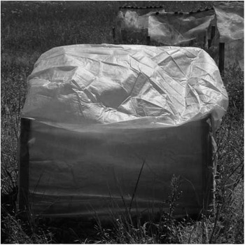
To manipulate temperature, removable greenhouse covers constructed of polypropylene (Frost Protek Plant Cover Inc., Fort Collins, Colorado, United States of America) were secured over cages. These light weight, porous covers (30 g/m2) allow air, ultraviolet rays, and rainfall to penetrate (Fig. 1). Temperature treatments can impact different aspects of the physical environment inside the cages such as humidity or soil moisture (Laws and Belovsky Reference Laws and Belovsky2010). To assess the effect of warming on evaporation covers were removed for <30 minutes every week and soil moisture readings were taken weekly with a handheld moisture reader (OMEGA Engineering Inc., Stamford, Connecticut, United States of America). In addition, covers were removed for <30 minutes every two days and the number of surviving grasshoppers in experimental cages were counted. Otherwise, covers remained on throughout the duration of the experiment. Temperature and relative humidity readings within experimental cages were taken hourly using universal serial bus data loggers (n=16 with two loggers per treatment; Measurement Computing Inc., Norton, Massachusetts, United States of America).
Example of removable greenhouse cover used in the experiment. Temperature is manipulated by enclosing cages under porous polypropylene covers.

To manipulate the effect of moisture on the pathogen, cricket water pillows (Zillia Products, Franklin, Wisconsin, United States of America) were placed at the bottom of cages. These small silicate gel pillows can hold 30 g of water each and can retain water for more than 24 hours even in very arid environments (McCluney and Sabo Reference McCluney and Sabo2009). Water pillows supplied moist habitat for E. grylli colonisation without impacting the plant community, thereby isolating the effects of moisture on E. grylli mycosis in our experimental grasshoppers (Carruthers et al. Reference Carruthers, Feng, Ramos and Soper1988a). Water supplementation, mimicking natural precipitation, increases plant biomass at our site, which could obviate the negative effects of this entomopathogen as grasshoppers benefit from increased food resources (Belovsky and Slade Reference Belovsky and Slade1995; Kistner and Belovsky Reference Kistner and Belovsky2014). Treated cages received five water pillows (one pillow/0.1 m2), which were rehydrated daily.
Relative pathogen abundance was estimated by placing five 75×25 mm glass microscope slides on the cage ground (one slide/0.1 m2) within each pathogen-exposed cage (Carruthers et al. Reference Carruthers, Feng, Ramos and Soper1988a). Slides were collected and replaced with new ones every two days. Slides were stained with lacto fuchsin (AEML, Pompano Beach, Florida, United States of America) in the field to avoid conidia loss during transport. Slides were then placed in a cooler and later examined under a compound microscope for the presence of conidia. Entomophaga grylli conidia were easily distinguished from other particles by their unique pear-like shape (MacLeod and Müller-Kögler Reference MacLeod and Müller-Kögler1973). Total number of conidia was summed across the five slides by sampling date.
The experiment ran from 28 June through 10 August 2013 when plant senescence and cold nights began to kill grasshoppers. Individual grasshoppers were counted every two days. Cadavers clinging high in the vegetation were noted as mortality from E. grylli infection, as this behaviour is uniquely characteristic of E. grylli (Carruthers et al. Reference Carruthers, Ramos, Larkin, Hostetter and Soper1997). These individuals were left in place in the event that they would continue disease transmission (Sawyer et al. Reference Sawyer, Ramos, Poprawski, Soper and Carruthers1997). All other cadavers were collected, frozen, and later stained with lacto fuchsin to determine E. grylli presence under a microscope (Sánchez-Peña Reference Sánchez-Peña2005). Only recovered or observed cadavers were included in the mortality analysis (16% of all experimental individuals were never recovered). Mortality due to E. grylli infection was the sum of cadavers observed clinging high in the vegetation plus the number of other cadavers later found to be infected.
To assess treatment effects on the plant community, grass biomass and nitrogen content were determined from living (green) shoots clipped at ground level. Shoots were dried at 60 °C for 48 hours before weighing (Belovsky and Slade Reference Belovsky and Slade1995). Nitrogen content, an estimate of grasshopper food quality (Belovsky and Slade Reference Belovsky and Slade2000; Laws and Belovsky Reference Laws and Belovsky2010), was measured with an elemental CHN analyser (ECS 4010; Costech Analytical Instruments, Valencia, California, United States of America). Analyses were only run on grasses because C. pellucida preferentially feed on grasses (Pfadt Reference Pfadt1994).
Statistical analyses
Treatment effects on mean daily temperature, mean daily relative humidity, and weekly soil moisture readings within experimental cages were analysed using repeated measures (ANOVA). Percentage soil moisture and relative humidity values were transformed using arcsine square-root before analysis (Zar Reference Zar1999). Treatment effects on the number of conidia were analysed using a generalised linear model (GLM) with a quasipoission distribution and log function. We employed a GLM with a binominal distribution and logit function to test the effect of warming and water-pillow supplementation on the proportion of grasshoppers whose deaths were attributable to E. grylli infection. Parametric survival models for pathogen-exposed and pathogen-free treatments compared abiotic treatment effects in the absence of the pathogen. Survival models employed a Weibull distribution with warming and water-pillow supplementation as predictive factors (Crawley Reference Crawley2013) and were run using the survival analysis package in R 3.2.0 (Therneau Reference Therneau2013). Differences in grass biomass and % nitrogen content across treatments were analysed using GLMs. Generalised linear models and repeated measures ANOVAs were performed using the stats package in R 3.2.0 (R Development Core Team 2015).
Results
Effects of treatments on abiotic conditions within experimental cages
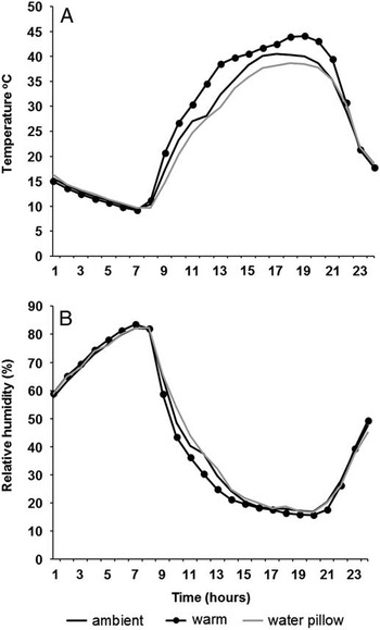
Treatment effects on mean daily temperature and relative humidity levels (Table 1) were driven by day-time changes with treatments having no noticeable effect on night conditions (Fig. 2). Greenhouse covers increased mean daily temperature by ~4.6 °C (F=152.14; df=1, 12; P<0.001; Table 1), whereas water pillows had no effect (F=0.56; df=1, 12; P=0.47). Mean daily temperature varied by day (F=638.34; df=19, 228; P<0.001) with a significant day×warming interaction (F=1.85; df=19, 228; P=0.02). Mean daily relative humidity decreased under warming by 1.6% (F=27.10; df=1, 12; P<0.001; Table 1). Relative humidity also varied by day (F=435.79; df=19, 228; P<0.001) with a significant day×warming interaction (F=2.98; df=19, 228; P<0.001). Water pillows had no significant effect on mean daily relative humidity levels (F=0.10; df=1, 12; P=0.75; Table 1), but there were significant day×water pillow (F=3.43; df=19, 228; P<0.001), and warming×water pillow×day (F=2.60; df=19, 228; P<0.001) interactions. The decrease in soil moisture over time was significant (F=0.61; df=2, 24; P<0.001), with soil moisture levels in warmed and ambient cages being roughly equal (differing by 1.1%). By the third week of July, the lack of precipitation and increased daily temperatures yielded 0% soil moisture readings in all treatment cages.
Average hourly (A) temperature and (B) relative humidity for each abiotic treatment plotted against time. Data were averaged over the course of the study (n=43 days).

Daily mean, maximum, and minimum temperatures (°C) and relative humidity % (RH) inside cages for each of the three treatments over the course of the study (n=43 days).

Mycosis in field caged grasshoppers
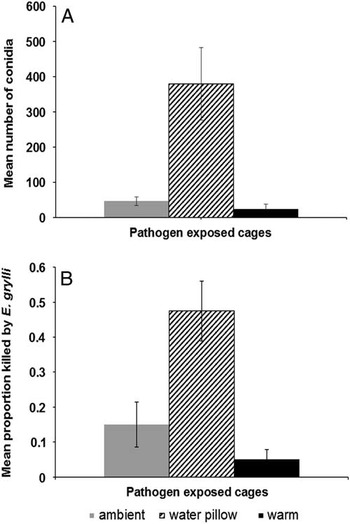
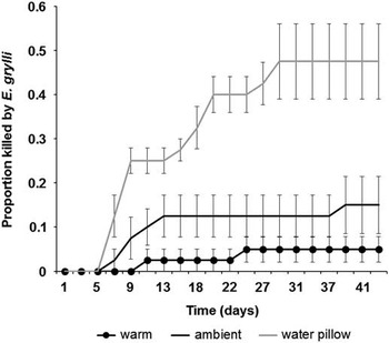
Water-pillow supplementation increased conidia levels, as estimated from microscope slides, by eightfold (F=32.52; df=1, 318; P<0.001; Fig. 3A). Warming decreased conidia levels by 49% (F=32.52, df=1, 318; P<0.001) due to conidia desiccation or infected grasshoppers producing resting spores. The warming×water-pillow interaction was not significant (F=1.81; df=1, 318; P=0.18). Warming decreased grasshopper mortality from E. grylli by 67% (χ2=9.22; df=1, 156; P=0.002), whereas water-pillow supplementation resulted in a threefold increase in mortality from E. grylli (χ2=14.57; df=1, 156; P<0.001; Fig. 3B). The warming×water-pillow interaction was not significant (χ2=0.006; df=1, 156; P=0.94). Mycosis caused by E. grylli peaked in July and was no longer detected by early August (Fig. 4). When water pillows were added, mortality from E. grylli peaked three times in a single month, while the other treatments exhibited only one minor mycosis event (Fig. 4).
Number of conidia recovered from pathogen-exposed cages (A) and the final proportion of grasshoppers exposed to Entomophaga grylli that died from mycosis (B) across different abiotic treatments. Data are reported as means±standard error.

Disease progress for caged pathogen-exposed grasshopper populations across different abiotic treatments over the course of the experiment. Data are reported as means±standard error.

Grasshopper survival
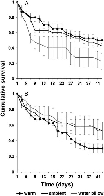
Survival of pathogen-exposed and pathogen-free grasshoppers responded differently to abiotic treatments (Fig. 5). Despite low mortality rates from E. grylli infections, warming did not increase survival time of pathogen-exposed grasshoppers compared with those left under ambient conditions (χ2=0.47; df=1; P=0.49; Fig. 5A). Unsurprisingly, water-pillow supplementation shortened pathogen-exposed grasshopper survival time by half (χ2=7.18; df=1; P<0.01) and the warming×water-pillow interaction was not significant (χ2=0.04; df=1; P=0.84). For pathogen-free grasshoppers, warming shortened survival time by 43.36%, but the effect was not significant (χ2=3.51; df=1; P=0.06; Fig. 5B). Water-pillow supplementation had no significant effect on survival time of pathogen-free grasshoppers (χ2=1.34; df=1; P=0.25). The warming×water-pillow interaction was not significant (χ2=0.36; df=1; P=0.55).
Grasshopper survival (±standard error) in pathogen-exposed (A) and pathogen-free (B) populations across different abiotic treatments.

Effects of treatments on grass quantity and quality
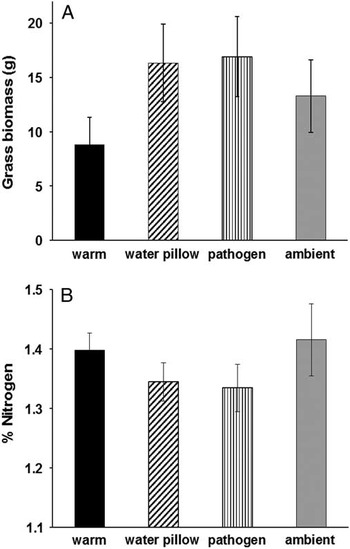
Treatments had no notable effects on grass biomass and nitrogen content within experimental cages compared with cages left under ambient conditions. Warming, water pillow, and pathogen exposure treatments did not alter grass biomass (F=4.04; df=1, 24; P=0.06; F=1.23; df=1, 24; P=0.28; F=2.27; df=1, 24; P=0.14; respectively; Fig. 6A). The warming×pathogen exposure (F=0.01; df=1, 24; P=0.93), warming×water pillow (F=0.85; df=1, 24; P=0.37), water pillow×pathogen exposure (F=0.70; df=1, 24; P=0.41) and warming×water pillow×pathogen exposure interactions were not significant (F=0.11; df=1, 24; P=0.74). Similarly, warming, water pillow, and pathogen exposure treatments had no effect on grass nitrogen content (χ2=0.01; df=1, 24; P=0.91; χ2=0.02; df=1, 24; P=0.88; χ2=0.02; df=1, 24; P=0.90; respectively; Fig. 6B). The warming×pathogen exposure (χ2=0.01; df=1, 24; P=0.91), warming×water pillow (F=0.01; df=1, 24; P=0.92), water pillow×pathogen exposure (χ2=0.02; df=1, 24; P=0.89) and warming×water pillow×pathogen exposure interactions were not significant (χ2=0.01; df=1, 24; P=0.91).
Grass biomass (A) and percent nitrogen content (B) are plotted for each treatment. Data are reported as means±standard error.

Discussion
The warming and water-pillow treatments altered both mycosis caused by E. grylli and pathogen-exposed grasshopper survival. While both temperature and water-pillow manipulations affected mycosis, moisture was the key abiotic factor driving fungal entomopathogen–grasshopper outcomes in our system. Warmed pathogen-exposed grasshoppers did not exhibit enhanced survival despite significant reductions in mortality from E. grylli, indicating a compensatory response to increased temperatures (Kistner and Belovsky Reference Kistner and Belovsky2014). Warming and water-pillow supplementation did not affect pathogen-free grasshopper survival time or grass biomass and nitrogen content so we could not attribute our findings to changes in food quantity or quality.
Moisture was the key driver of E. grylli mycosis in our experimental cages with only water-pillow supplemented cages exhibiting epizootics. This outcome is unsurprising given that conidia production, survival time, and infectivity are directly linked to moisture (Carruthers et al. Reference Carruthers, Larkin and Soper1988b; Sawyer et al. Reference Sawyer, Ramos, Poprawski, Soper and Carruthers1997). Furthermore, E. grylli epizootics often occur after heavy rainfalls (Pickford and Riegert Reference Pickford and Riegert1964; Erlandson et al. Reference Erlandson, Johnson and Olfert1988; Carruthers et al. Reference Carruthers, Ramos, Larkin, Hostetter and Soper1997) and E. grylli prevalence tends to be higher in irrigated rangelands compared with non-irrigated grasslands (Kistner and Belovsky Reference Kistner and Belovsky2013). Thus, warmed and ambient pathogen-exposed treatments exhibited low levels of conidia, which ultimately had little impact on overall grasshopper survival. In contrast, grasshoppers in cages receiving water pillows exhibited significant reductions in survival time as a result of increased conidia production. We suspect that the water pillows acted as a catalyst for secondary E. grylli conidia production. When a conidium spore lands on a moist non-host substrate, it may germinate and produce secondary conidia, which infect the host by contact in the same manner as conidia ejected from an infected host cadaver (Carruthers et al. Reference Carruthers, Ramos, Larkin, Hostetter and Soper1997). Trace amounts of conidia were found on the water pillows and grasshoppers often frequented them over the course of the experiment (E.J.K., personal observation). In turn, an individual using these water pillows as a water source could have an increased risk of infection since moisture enhances the infection process once viable conidia contact a host (MacLeod et al. Reference MacLeod, Cameron and Soper1966; Carruthers et al. Reference Carruthers, Feng, Ramos and Soper1988a). While our water-pillow treatment did isolate the effects of moisture on E. grylli–grasshopper interactions, it did not accurately represent natural environmental conditions. Entomophaga grylli epizootics are linked to precipitation, which also directly influences plant community dynamics at our site (Belovsky and Slade Reference Belovsky and Slade1995). Therefore, these results should be approached with caution since precipitation also affects grasshopper population dynamics by altering food availability (i.e., plant biomass) and food quality (i.e., nitrogen content) (Belovsky and Slade Reference Belovsky and Slade1995; Ovadia and Schmitz Reference Ovadia and Schmitz2004; Branson Reference Branson2008). Future studies should provide additional water to the plant community as well as E. grylli so that both top–down and bottom–up processes in this fungal entomopathogen–grasshopper system may be better understood.
For pathogen-exposed grasshoppers, warming greatly reduced mycosis resulting in <1% of warmed grasshoppers dying from E. grylli infections. Two hypotheses might explain the mechanism by which warming reduces mycosis caused by E. grylli. These hypotheses are not mutually exclusive and each may contribute. First, higher temperatures and lower humidity levels are known to cause E. grylli pathotype 1 to produce mainly dormant resting spores instead of infectious conidia (Carruthers et al. Reference Carruthers, Feng, Ramos and Soper1988a, Reference Carruthers, Ramos, Larkin, Hostetter and Soper1997). Conidia levels in warmed cages were 50% lower than those cages left at ambient conditions indicating that the few grasshoppers that did die of E. grylli infections were producing little if any infectious conidia. Second, an infected grasshopper may be able to reduce (Elliot et al. Reference Elliot, Blanford and Thomas2002) or completely eliminate fungal entomopathogen infections (Carruthers et al. Reference Carruthers, Larkin, Firstencel and Feng1992; Inglis et al. Reference Inglis, Johnson and Goettel1997; Klass et al. Reference Klass, Blanford and Thomas2007b) via behavioural thermoregulation or fever (Hajek and St. Leger Reference Hajek and St. Leger1994; Roy et al. Reference Roy, Steinkraus, Eilenberg, Hajek and Pell2006). Carruthers et al. (Reference Carruthers, Larkin, Firstencel and Feng1992) found that C. pellucida can reduce or completely eliminate E. grylli pathotype 1 infections if its internal body temperature is maintained between 38 °C and 40 °C for just four hours each day. The mean day-time temperatures within our warmed cages were well within the termal limit of E. grylli (35 °C) with highs ranging between 38 °C and 41 °C. Unfortunately, it was impossible to distinguish behavioural fever from normal thermoregulatory behaviour since all grasshoppers bask as a part of their normal daily activities. Even so, more pathogen-exposed individuals were observed basking during the afternoon when temperatures reached levels detrimental to E. grylli compared with pathogen-free grasshoppers, which tended to spend more time in the lower canopy during the same time periods (E.J.K., personal observation). Our findings are consistent with Inglis et al. (Reference Inglis, Johnson and Goettel1997) who found that shading increased mycosis caused by Beauveria bassiana (Balsamo) Vuillemin (Hypocreales: Clavicipitaceae), an ecologically similar fungal entomopathogen, in field caged grasshoppers due to decreased temperature and light exposure.
Interestingly, despite reductions in mycosis caused by E. grylli, warmed pathogen-exposed grasshoppers did not exhibit increased survival time compared with pathogen-exposed grasshoppers left at ambient conditions. We hypothesise that warmed pathogen-exposed grasshoppers became more resource limited as individuals that would have otherwise died from E. grylli continued to diminish food resources among surviving individuals. This outcome makes intuitive sense since grasshopper populations in bunch-grass prairies are food limited (Scharff Reference Scharff1954; Belovsky and Slade Reference Belovsky and Slade1995; Branson Reference Branson2008) and supports previous E. grylli research at this site (Kistner and Belovsky Reference Kistner and Belovsky2014). Consequently, intraspecific competition for food replaced E. grylli as the key factor influencing grasshopper survival. Similar results have been found for predator effects on grasshopper populations at our study site (Belovsky and Slade Reference Belovsky and Slade1993, Reference Belovsky and Slade2010).
While our removable greenhouses did not significantly reduce overall survival time of pathogen-free grasshoppers, mean adult densities recovered at the end of the study were 35% lower in the greenhouse treatment compared with the control (i.e., ambient). Previous research on grasshopper responses to temperature manipulations found that warming can decrease C. pellucida survival especially at higher densities under strong intraspecific competition (Laws and Belovsky Reference Laws and Belovsky2010). From 1995 to 2013, observed mid-summer nymphal grasshopper densities at our site ranged from 2.71 to 73.75/m2 (G.E.B., unpublished data). Given that we only examined temperature effects on an intermediate population density of 20 grasshoppers/m2, it is possible that we may be underestimating potential interactions between temperature and intraspecific competition at our site. To better address the role of temperature in fungal entomopathogen–grasshopper outcomes, future studies should evaluate rising temperature impacts over a range of population densities.
In conclusion, the results of this study suggest that E. grylli pathotype 1 epizootics will likely decrease in both frequency and severity under ongoing climate change in western Montana, which has become much warmer and drier over the past century (Lesica and Kittelson Reference Lesica and Kittelson2010; Pederson et al. Reference Pederson, Graumlich, Fagre, Kipfer and Muhlfeld2010). Subsequently, reductions of entomopathogenic fungi under increasingly arid conditions (Harvell et al. Reference Harvell, Mitchell, Ward, Altizer, Dobson and Ostfeld2002) could lead to more frequent and severe grasshopper outbreaks in bunch-grass prairies. However, release from pathogen pressure may not necessarily result in more severe grasshopper pest outbreaks since warming may also enhance intraspecific competition within surviving grasshopper populations (Belovsky and Slade Reference Belovsky and Slade1995; Laws and Belovsky Reference Laws and Belovsky2010). Because our study examined grasshopper and fungal entomopathogen responses to summer abiotic manipulations at a single grassland ecosystem, strong generalisations regarding the effects on climate change on fungal entomopathogen–grasshopper interactions are not possible. Additional studies across different temporal and spatial gradients are greatly needed to enhance our understanding of fungal entomopathogen–grasshopper outcomes under future climate change (Klass et al. Reference Klass, Blanford and Thomas2007a, Reference Klass, Blanford and Thomas2007b). Research examining the impact of extreme climatic events on mycosis caused by both naturally occurring entomopathogens and commercially available mycoinsecticides is also warranted.
Acknowledgements
The authors thank the National Bison Range personnel for permitting the research. Janet Kistner and Edward Kistner provided field assistance. Nathan Payne helped analyse grass samples. They acknowledge Shayna Sura, David Flagel, and Clayton Thomas for valuable feedback in preparation of this manuscript. Funding was provided by a research grant from the Orthopterists’ Society.