Introduction
Significant progress has been made in the in vitro embryo production of domestic cats and wild felids in the last 40 years (Pope, Reference Pope2019a, Reference Pope2019b). However, the developmental capacity after embryo transfer (ET) remained low compared with other species as bovine and murine (Veraguas et al., Reference Veraguas, Echeverry, Castro and Rodriguez-Alvarez2017a; Veraguas et al., Veraguas et al., Reference Veraguas, Saez, Aguilera, Echeverry, Gallegos, Saez-Ruiz, Castro and Rodriguez-Alvarez2020a). In vivo developmental capacity decreased even more when felid embryos were generated using SCNT or interspecific SCNT (iSCNT) (Gómez et al., Reference Gómez, Pope, Ricks, Lyons, Dumas and Dresser2009). Removal of the zona pellucida has been used to enhance the production of cloned embryos and live offspring by handmade cloning and embryo aggregation in different species (Rodríguez-Alvarez et al., Reference Rodríguez-Alvarez, Sharbati, Sharbati, Cox, Einspanier and Castro2010; Gambini et al., Reference Gambini, Jarazo, Olivera and Salamone2012; Buemo et al., Reference Buemo, Gambini, Moro, Hiriart, Fernández-Martín, Collas and Salamone2016). Embryo aggregation enhances the developmental capacity of domestic cat cloned embryos and embryos from cheetah, tiger and kodkod generated using iSCNT (Moro et al., Reference Moro, Jarazo, Buemo, Hiriart, Sestelo and Salamone2015a, Reference Moro, Hiriart, Buemo, Jarazo, Sestelo, Veraguas, Rodriguez-Alvarez and Salamone2015b; Veraguas et al., Reference Veraguas, Aguilera, Echeverry, Saez-Ruiz, Castro and Rodriguez-Alvarez2020b). However, in vitro culture of domestic cat embryos without the zona pellucida affects the gene expression pattern at the blastocyst stage (Veraguas-Davila et al., Reference Veraguas-Davila, Cordero, Saez, Saez-Ruiz, Gonzalez, Saravia, Castro and Rodriguez-Alvarez2021). Domestic cat blastocysts cultured without the zona pellucida had reduced expression of SOX2 and NANOG and increased expression of BAX (Veraguas-Davila et al., Reference Veraguas-Davila, Cordero, Saez, Saez-Ruiz, Gonzalez, Saravia, Castro and Rodriguez-Alvarez2021). This did not affect the in vitro development of ZF domestic cat embryos, but no implantations were achieved after ET (Veraguas-Davila et al., Reference Veraguas-Davila, Cordero, Saez, Saez-Ruiz, Gonzalez, Saravia, Castro and Rodriguez-Alvarez2021). Furthermore, ZF domestic cat blastocysts had an increased expression of trophectoderm genes YAP1 and EOMES (Veraguas-Dávila et al., Reference Veraguas-Dávila, Saéz-Ruíz, Álvarez, Saravia, Castro and Rodríguez-Alvarez2022). Similarly, in ZF mouse blastocysts ICM and TE had altered expression of differentiation genes and the embryo implantation rate was lower compared with ZI embryos (Fan et al., Reference Fan, Huang, Wu, Bai, Kawahara and Takahashi2022). This indicates that the removal of the zona pellucida affects the expression of pluripotency and differentiation genes, along with the implantation capacity of in vitro-produced embryos in some species.
Mechanisms involved in maternal recognition and implantation of domestic cat embryos are barely understood. The molecular signals that allow maternal recognition during the development of felid embryos have been scarcely studied. Furthermore, different studies have hypothesized that an intact zona pellucida must be present in domestic cat embryos to ensure implantation after ET (Kanda et al., Reference Kanda, Oikawa, Nakao and Tsutsui1995; Denker, Reference Denker2000; Pope, Reference Pope2014; Veraguas-Davila et al., Reference Veraguas-Davila, Cordero, Saez, Saez-Ruiz, Gonzalez, Saravia, Castro and Rodriguez-Alvarez2021). The surface of the zona pellucida in domestic cat oocytes contains numerous spherical and elliptical pores of different sizes (Lunn and Wright, Reference Lunn and Wright2009). The porous nature of this matrix allows the penetration of specific molecules; this does not depend on the relative size of the molecules but on other biochemical and physicochemical properties (Prasad et al., Reference Prasad, Skinner, Carino, Wang, Cartwright and Dunbar2000). Possibly, the zona pellucida of domestic cat embryos has a crucial role in the regulation of molecular signals during maternal recognition and implantation.
Most miRNAs inhibit protein synthesis by repressing translation, promoting mRNA deadenylation or inducing mRNA scission (Krol et al., Reference Krol, Loedige and Filipowicz2010). For this reason, miRNAs have a crucial role in cell-to-cell communication (Gross et al., Reference Gross, Kropp and Khatib2017a). Extracellular miRNAs can be detected in all the biological fluids participating in several physiological and pathological processes (Turchinovich et al., Reference Turchinovich, Samatov, Tonevitsky and Burwinkel2013). Furthermore, all types of in vitro-cultured cells release miRNAs into the medium by binding to apolipoproteins, inside extracellular vesicles (EVs) or during apoptosis (Hawke et al., Reference Hawke, Watson and Betts2021b; Gong et al., Reference Gong, Yu, Wang, Wang, Liu, Paul, Millard, Xiao, Ashraf and Xu2017; Zhang et al., Reference Zhang, Feng, Xu, Wang, Cui, Li, Wang, Teng, Hao, Wan, Tan, Wang and Zhou2016). Different miRNAs have been involved in embryo–maternal communication, some of them are released by endometrial cells and others by preimplantation embryos (Abu-Halima et al., Reference Abu-Halima, Häusler, Backes, Fehlmann, Staib, Nestel, Nazarenko, Meese and Keller2017; Gross et al., Reference Gross, Kropp and Khatib2017b; Capalbo et al., Reference Capalbo, Ubaldi, Cimadomo, Noli, Khalaf, Farcomeni, Ilic and Rienzi2016). The miRNAs expressed by preimplantation embryos are involved in trophectoderm formation, invasion, and implantation (Doridot et al., Reference Doridot, Miralles, Barbaux and Vaiman2013; Paul et al., Reference Paul, Sadek and Mahesan2019). Significant differences in the expression profile of miRNAs have been detected in implanted blastocysts compared with non-implanted ones, and between receptive and non-receptive endometrium (Liu et al., Reference Liu, Niu, Li, Pang, Chiu and Yeung2016; Paul et al., Reference Paul, Sadek and Mahesan2019). Furthermore, several miRNAs described in the spent culture medium of in vitro-produced embryos have been proposed as biomarkers of embryo quality or predictors of in vivo development (Kropp et al., Reference Kropp, Salih and Khatib2014; Kropp and Khatib, Reference Kropp and Khatib2015). Different miRNAs have been related to in vitro fertilization (IVF) failure and embryo degeneration (miR-25), morula to blastocyst transition (miR-130), blastocyst formation (miR-24, miR-199a-5p, miR-196, miR-302), trophoblast differentiation, invasion, and migration (miR-21, miR-96, miR-29, miR-155), and implantation and immune response (Let7/miR-98) (Liang et al., Reference Liang, Wang and Wang2017; Reza et al., Reference Reza, Choi, Han, Song, Park, Hong and Kim2019). Despite that, the evaluation of miRNAs expressed by domestic cat preimplantation embryos has not been reported.
Embryonic miRNAs can traverse the zona pellucida carried by EVs or binding to apolipoproteins (Kim et al., Reference Kim, Lee, Lee and Jun2019; Hawke et al., Reference Hawke, Ahmed, Watson and Betts2021a). The zona pellucida, along with other extra-embryonic coats, works as a mailbox between embryos and the maternal environment (Denker, Reference Denker2000; Herrler and Beier, Reference Herrler and Beier2000). We hypothesize that in vitro culture of domestic cat embryos without the zona pellucida might affect the expression of miRNAs. The abnormal expression of specific miRNAs might be one of the factors that affect the implantation capacity of these embryos. For these reasons, the objective of this study was to compare the expression of specific miRNAs in domestic cat blastocysts cultured with or without the zona pellucida and in the spent culture medium of both groups.
Materials and methods
All chemical reagents were purchased from Sigma-Aldrich Chemicals Company (St. Louis, MO, USA), except for those otherwise indicated.
Ethics statement
All procedures that involved animal manipulation were approved by the Ethics Committee (Comité de Bioética de la Facultad de Ciencias Veterinarias de la Universidad de Concepción). Certificate of approval no. CBE-08-2020.
Animals
Healthy female and male domestic cats aged between 6 months and 5 years were selected as oocyte and sperm donors for in vitro embryo production.
Experimental design
Two experimental groups were done. (1) Domestic cat embryos generated by IVF and cultured in vitro (Zona intact; ZI group). (2) Domestic cat embryos generated by IVF and cultured in vitro without the zona pellucida (Zona free; ZF group). The ovaries from domestic cats were collected by ovariohysterectomy. Immature cumulus–oocyte complexes (COCs) were collected from these ovaries by slicing and then subjected to in vitro maturation (IVM). Subsequently, the in vitro matured COCs were subjected to IVF using epididymal sperm. Only in the ZF group, the zona pellucida of presumptive zygotes was removed. Then ZF embryos were cultured in vitro using the well-of-the-well system (WOW). In addition, ZI embryos were cultured normally in four-well dishes. Embryos were cultured in vitro for 7 days. The cleavage, morula and blastocyst rates were evaluated. The total cell number of blastocysts was estimated. Finally, the expression of pre-miRNAs and mature miRNAs (miR-21, miR-24, miR-25, miR-29, miR-98, miR-103, miR-130, miR-155, miR-191, miR-196, miR-199, miR-302 and miR-96) was evaluated in blastocysts by RT-qPCR. In addition, only mature miRNAs were evaluated in spent medium. The geometric mean of miR-103 and miR-191 was used as an internal control.
Ovariohysterectomy and COCs collection
The ovaries of female domestic cats were collected by ovariohysterectomy. Anaesthesia was induced using an (i.m.) dose of 0.5 mg/kg xylazine (Xilazina 2%, Virbac Chile) and 5 mg/kg ketamine (Ketamina 100; Chemie Chile) and maintained by administering xylazine/ketamine (i.v.) at the same dose. Atipamezole (Antisedan, Zoetis) was used to eliminate the effects of xylazine once the surgical procedure ended, using a unique dose of 0.02 to 0.05 ml ovaries were transported in a sterile solution containing 0.9% NaCl and 0.1% gentamycin (Gentamicina 10%, Veterquimica, Chile) at 38.5°C. Immature COCs were recovered from the ovaries by slicing in a 100 mm Petri dish containing 10 ml of medium-199 with Earle’s salts supplemented with 0.18 mM HEPES, 5% fetal bovine serum (FBS) and 50 μg/ml gentamycin (He199) at 38.5°C. Only grade I and grade II immature COCs were subjected to IVM.
In vitro maturation
In vitro maturation of immature COCs was performed in four-well dishes containing 500 µl of medium-199 with Earle’s salts supplemented with 0.3% fraction-V BSA, 0.1 IU/ml FSH-LH (Pluset, Serono, Italy), 1 μg/ml 17β-estradiol, 0.36 mM sodium pyruvate, 2 mM glutamine, 2.2 mM calcium lactate, 20 ng/ml epidermal growth factor (EGF), 10 µl/ml insulin, transferrin, selenium (ITS) and 50 μg/ml gentamycin (IVM-199) in an humidified gas atmosphere with 5.0% CO2, at 38.5°C for 24–26 h.
Sperm collection
Only male domestic cats older than 10 months of age were used as sperm donors. The anaesthesia procedure was the same as that described for ovariohysterectomy. Additionally, lidocaine (Lidocalm, 2%, Dragpharma, Chile) was administered in the genital area as local anaesthesia. Testes were transported in sterile 0.9% NaCl solution with 0.1% gentamycin at room temperature. The caudal portions of the epididymis were cut into small pieces of ∼1 mm in a 100 mm Petri dish containing 10 ml of He199 supplemented with 0.3% Fraction-V BSA instead of FBS (He199-BSA) at 38.5°C. The medium with the sperm was passed through a sterile nylon filter (40 µm) into a 50 ml sterile tube. The filtrate was centrifuged at 1500 rpm for 5 min, and the pellet was resuspended in 5 ml of 20% Andromed medium (diluted in sterile distilled water according to the manufacturer’s instructions; Minitube, Tiefenbach, Germany) and refrigerated at 4°C for 24 h (Veraguas et al., Reference Veraguas, Saez, Aguilera, Echeverry, Gallegos, Saez-Ruiz, Castro and Rodriguez-Alvarez2020a).
In vitro fertilization
Once IVM was finished, COCs were washed in TALP medium supplemented with 6 mg/ml BSA, 0.36 mM sodium pyruvate, 1 mM glutamine, 2.2 mM calcium lactate, 1% MEM nonessential amino acids (NEAA), 0.5% MEM essential amino acids (EAA), 0.01 mg/ml heparin sodium salt and 50 μg/ml gentamycin (TALP-IVF). Refrigerated sperm were allowed to swim up for 30 min in He199-BSA at 38.5°C (Veraguas et al., Reference Veraguas, Saez, Aguilera, Echeverry, Gallegos, Saez-Ruiz, Castro and Rodriguez-Alvarez2020a). The supernatant was collected and then centrifuged at 1500 rpm for 5 min. The pellet was collected and resuspended in TALP-IVF. For IVF, 20–30 COCs were placed in four-well dishes containing 500 µl of TALP-IVF and co-incubated with 1.5 to 2.5 × 10⁶ spermatozoa/ml, in a humidified atmosphere of 5% CO2 in air, at 38.5°C, for 24 h. Subsequently, cumulus cells were removed from the presumptive zygotes using a 0.5 mg/ml hyaluronidase solution and vortexed for 6 min.
In vitro embryo culture
In the ZI group, presumptive zygotes were cultured in supplement SOF medium (Veraguas et al., Reference Veraguas, Cuevas, Gallegos, Saez-Ruiz, Castro and Rodriguez-Alvarez2018). However, FBS was replaced by ITS (10 µl/ml) to avoid the presence of contaminating miRNAs carried by serum-EVs. The culture was done in four-well dishes containing 500 µl of SOF medium supplemented with 0.37 mM trisodium citrate, 2.77 mM myo-inositol, essential and nonessential amino acids (final concentration 1×), 50 µg/ml gentamycin, 10 µl/ml ITS, 20 ng/ml EGF and 3 mg/ml essentially fatty acid-free BSA (SOF-B); 10–20 embryos were placed into each well.
In the ZF group, the zona pellucida from presumptive zygotes was removed by incubation in 2 mg/ml of pronase for 4 min, at 37°C. Subsequently, presumptive zygotes were washed three times in He199 supplemented with 30% FBS (He199-30) to remove pronase, and then washed three times in SOF-B to eliminate FBS. The ZF embryos were cultured in four-well dishes using the WOW system (Vajta et al., Reference Vajta, Korösi, Du, Nakata, Ieda, Kuwayama and Nagy2008). In the WOW system, several microwells were made in the culture dish, and each ZF embryo was individually cultured in one microwell to prevent the disaggregation of blastomeres (Vajta et al., Reference Vajta, Korösi, Du, Nakata, Ieda, Kuwayama and Nagy2008). In this study, the microwells were created using an aggregation needle (DN-09N, BLS Ltd). Eighty microwells were made in a four-well dish, and up to 20 ZF embryos were cultured per well.
In both experimental groups, on day 2 of in vitro culture (IVC), cleavage embryos were selected and the rest were discarded. At day 5 of IVC, morulae were selected, and the remaining embryos were discarded. The morulae were cultured in medium-199 with Earle’s salts supplemented with 0.37 mM trisodium citrate, 2.77 mM myo-inositol, essential and nonessential amino acids (final concentration 1×), 50 µg/ml gentamycin, 3 mg/ml essentially fatty acid-free BSA, 10 µl/ml ITS and 20 ng/ml EGF (M199-IVC). The culture was carried out in a humidified atmosphere of 5% CO2, 5% O2 and 90% N2 at 38.5°C, for 7 days. The cleavage, morula and blastocyst rates were estimated on days 2, 5 and 7, respectively. For sample collection, only embryos at the blastocyst stage on day-7 of culture were selected.
Morphological evaluation of the blastocysts
Blastocysts were fixed in a 3% glutaraldehyde solution for 72 h at 4°C. Fixed blastocysts were stained with 5 μg/ml Hoechst 33342 for 20 min. Stained embryos were placed on a slide with a drop of glycerin and then covered with a coverslip. Visualization was achieved using the EVOS FL Auto Cell Imaging System (Thermo Fisher Scientific).
Gene expression analysis
RNA extraction and gDNA digestion in blastocyst samples
For the analysis of pre-miRNAs and miRNAs, ZI and ZF day-7 blastocysts were pooled within their respective groups. Ten pools were made in both groups; pools had five day-7 blastocysts. The samples were stored at −80°C.
Total RNA extraction of blastocysts was performed using the Ambion® Cells-to-cDNA™ II Kit (Thermo Fisher Scientific, Austin, TX, USA) as previously described (Veraguas et al., Reference Veraguas, Cuevas, Gallegos, Saez-Ruiz, Castro and Rodriguez-Alvarez2018). Blastocyst samples were washed three times in cold PBS to eliminate the remaining culture medium. For this, 100 µl of PBS was added and then samples were centrifuged at 11,000 rpm for 5 min. Next, 50 µl of lysis buffer was added and the samples were incubated at 75°C for 10 min. For gDNA digestion, the samples were treated with DNase I (0.04 U/µl) and incubated at 37°C for 15 min and at 75°C for 5 min.
cDNA synthesis of pre-mature miRNAs in the blastocyst samples
The cDNA synthesis for the pre-miRNAs analysis was done using the SuperScript™ IV First-Strand Synthesis System (Invitrogen, Thermo Fisher Scientific). Next, 10 µl of total RNA was used in a 20-µl final reaction containing: 50 ng/µl random primers hexamers and 10 mM of each dNTP. The mixture was incubated at 65°C for 5 min and kept at 4°C for at least 1 min. Then, the reaction was completed by adding 4 µl 5× SS IV buffer, 1 µl 100 mM DTT, 1 µl ribonuclease inhibitor and 1 µl SuperScript™ IV reverse transcriptase (200 U/ μl). The mixture was centrifuged and then incubated at 23°C for 10 min, 55°C for 10 min and 80°C for 10 min. The cDNA samples were kept at −20°C until qPCR was performed.
miRNA extraction in medium samples
For this, 400–500 µl of culture medium was collected only in the wells in which the morulae reached the blastocyst stage on day 7 of IVC. miRNA extraction of medium samples was carried out using the mirPremier® microRNA Isolation Kit (SNC10). This kit uses a purification column system, which allows to concentrate medium samples in a small volume (30–40 µl). The samples were first centrifuged at 10,000 g for 5 min to remove cellular debris; 300–400 µl of supernatant were transferred and used in the next steps. The lysis mix supplement with 2-mercaptoethanol (1 µl/ml) was added to the medium samples in 1:1 (vol/vol) and then samples were incubated at 55°C for 5 min. Samples were centrifuged at 15,000 g for 5 min to remove gDNA and large RNA, the supernatant was transferred, and the pellet was discarded. Then, a 1.1 volume of 100% ethanol was added to the samples and mixed by vortexing. The binding, wash, and dry column steps were carried out according to the manufacturer’s instructions. Once the dry column step was carried out, 30–40 µl of dilution buffer were added to the columns and then these were centrifuged. Finally, 30–40 µl miRNAs samples were obtained and these were stored at −80°C.
cDNA synthesis of miRNAs in medium and blastocyst samples
To convert miRNAs into cDNA, the MystiCq™ microRNA cDNA Synthesis Mix was used (MIRRT) according to the manufacturer’s instructions. For this we used: 7 µl of total RNA from blastocyst samples or 7 µl of purified miRNA from medium samples. A final reaction of 20-µl volume was made. First, the poly(A) tailing reaction was done with 2 µl of poly(A) tailing buffer (5×) and 1 µl of poly(A) polymerase; the mix was centrifuged and then incubated at 37°C for 60 min, and 70°C for 5 min. Finally, the first-strand cDNA synthesis reaction was done by adding: 9 µl of the MystiCq microRNA cDNA reaction mix, and 1 µl of ReadyScript reverse transcriptase. The mixture was centrifuged and then incubated at 42°C for 20 min, and 85°C for 5 min. The cDNA samples were stored at −20°C.
Real-time quantitative polymerase chain reaction (qPCR)
Gene expression analysis was performed by real-time qPCR using the standard curve method as previously described (Veraguas et al., Reference Veraguas, Gallegos, Castro and Rodriguez-Alvarez2017b). Standard curves were generated using 2 µl of PCR products for each specific miRNA. These PCR products were purified previously using the E.Z.N.A gel extraction kit (Omega, BioTek, Santiago, Chile) and quantified using a spectrophotometer (Epoch, BioTek Instruments, Inc., Winooski, VT, USA). The standard curves were generated using eight 10-fold dilutions from the PCR products. For qPCR analysis, each sample was loaded in duplicate (technical replicates). PCR was performed using 2 µl of cDNA from samples, 1 µl of primers (10 pmol each, forward and reverse), 5 µl of the KiCqStart SYBR Green ReadyMix, Low ROX (Sigma-Aldrich) and 2 µl of betaine solution (5 M; B0300) in a final volume of 10 µl. The reaction was run on an MX3000P Real-Time PCR device (Agilent, Santa Clara, CA, USA). Only experiments with an efficiency within the range 90–110% and a correlation coefficient of at least 0.9 were used for analysis. The software NormFinder was used to select the most stable miRNAs for their use as internal controls (Andersen et al., Reference Andersen, Jensen and Ørntoft2004).
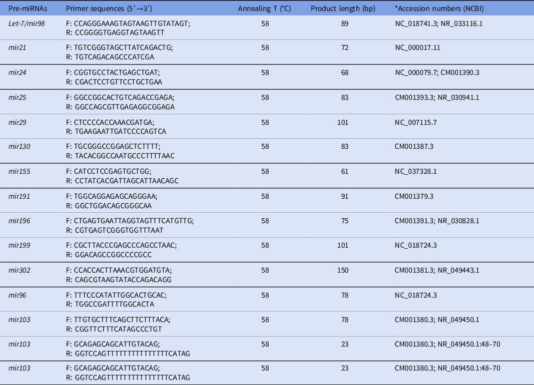
The primers used for the analysis of pre-miRNAs were designed using the RNAcentral database (https://rnacentral.org). These sequences were also verified in the NCBI database for the Felis catus species (https://www.ncbi.nlm.nih.gov/genome/?term=felis+catus) and compared against other species. The primers and PCR conditions are shown in Table 1.
Primer sequences and RT-qPCR conditions used for the expression analysis of pre-microRNAs

* Two accession numbers indicate that sequences alignment was done, a common sequence between domestic cat (Felis silvestris catus) and different species was uses for primer design: dog (Canis lupus familiaris); human (Homo sapiens); zebrafish (Danio rerio); horse (Equus caballus); bovine (Bos taurus); mouse (Mus musculus).
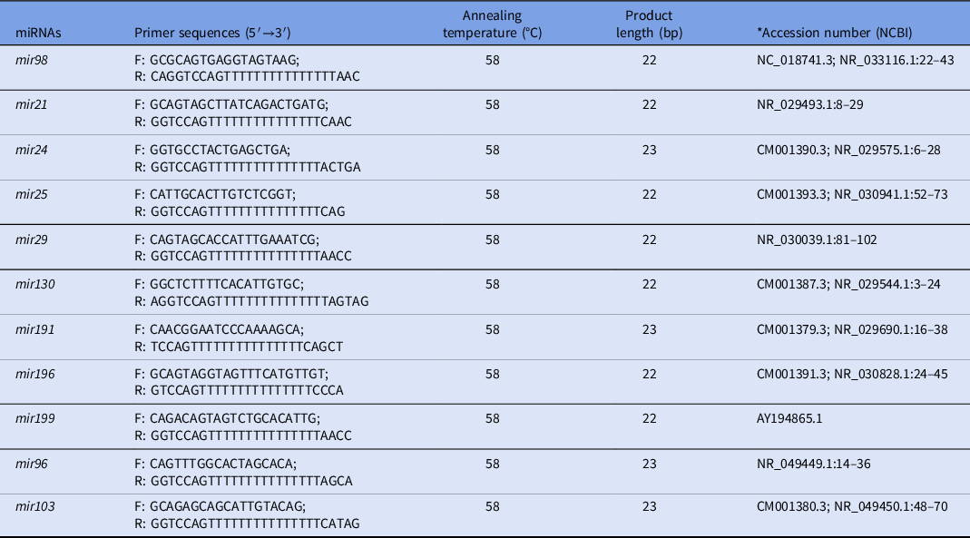
To identify the sequences of miRNAs, sequence alignment was carried out between the sequences of pre-miRNAs from Felis catus and the sequences of miRNAs from different species using the NCBI database (Table 2). To design miRNAs primers, the software miRprimer was used (Busk, Reference Busk2014). This software designed miRNA-specific forward and reverse primers, which allowed the use of SYBR green for quantification by RT-qPCR (Forero et al., Reference Forero, González-Giraldo, Castro-Vega and Barreto2019; Balcells et al., Reference Balcells, Cirera and Busk2011). The primers for miRNAs and their PCR conditions are shown in Table 2.
Primer sequences and RT-qPCR conditions used for the expression analysis of microRNAs

* Two accession numbers indicate that sequences alignment was done, a common sequence between domestic cat (Felis silvestris catus) and different species was uses for primer design: dog (Canis lupus familiaris); human (Homo sapiens); zebrafish (Danio rerio); horse (Equus caballus); bovine (Bos taurus); mouse (Mus musculus).
Statistical analysis
The Wilcoxon nonparametric test was used to evaluate in vitro development and gene expression analysis between the ZI and ZF groups. The t-test was used to evaluate the total cell number of blastocysts between groups. The statistical software InfoStat was used for these analyses (2020 InfoStat/L Version; University of Cordoba, Argentina).
Results
In vitro development of domestic cat embryos cultured with or without the zona pellucida
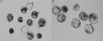
In the ZF group, presumptive zygotes were cultured in microwells to prevent disaggregation of blastomeres. No statistical differences were found in cleavage, morula and blastocyst rates between the embryos cultured in the ZI and ZF groups (P > 0.05) (Table 3). This indicates that the culture system used in this study ensures the in vitro development of ZF domestic cat embryos (Figure 1).
In vitro development of domestic cat embryos cultured with (ZI) and without (ZF) zona pellucida for 7 days.

* N: number of replicates. No significant differences were found between groups (P > 0.05).
In vitro development of domestic cat embryos. (A) Domestic cat blastocysts generated by IVF and in vitro cultured for 7 days (20×). (B) Domestic cat blastocysts generated by IVF and in vitro cultured without the zona pellucida for 7 days (20×).

Morphological evaluation of domestic cat blastocysts cultured with and without the zona pellucida
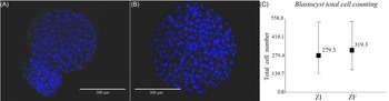
Day-7 blastocysts from the ZI and ZF groups were fixed and stained for total cell counting. Ten blastocysts were evaluated in both experimental groups. No statistical differences were observed between the total cell number of blastocysts (mean ± standard deviation, SD) from the ZI (279.3 ± 129.9) and ZF groups (319.3 ± 126.0) (P > 0.05; Figure 2).
Total cell counting of blastocysts. (A) Hatching domestic cat blastocyst fixed and stained with Hoechst (20×). (B) ZF domestic cat blastocysts fixed and stained with Hoechst (20×). C) Total cell number of blastocysts (mean; min/max) from the ZI (279.3; 143/535) and ZF groups (319.3; 172/539).

Expression analysis of pre-miRNAs and miRNAs in domestic cat blastocysts cultured with and without the zona pellucida
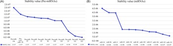
The expression of all the pre-miRNAs and miRNAs selected in this study was analyzed using the software NormFinder. According to this, miR-103 and miR-191 had the most stable expression among samples (Figure 3). For this reason, the geometric mean of pre-miR-103 and pre-miR-191, along with miR-103 and miR-191 were selected as internal controls for the analysis of pre-miRNAs and miRNAs, respectively.
Internal control analysis. Stability value (mean) of pre-miRNAs (A) and miRNAs (B) evaluated in ZI and ZF blastocysts by RT-qPCR. The lower values indicate a higher stability.

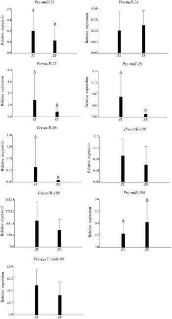
The relative expression of pre-miRNAs was analyzed first. No expression of pre-miR-155 and pre-miR-302 was detected in blastocysts from the ZI and ZF groups. No statistical differences were observed in the relative expression of pre-miR-24, pre-miR-130, pre-miR-196 and pre-let7/miR-98 between blastocysts from the ZI and ZF groups (P > 0.05). The relative expression of pre-miR-21, pre-miR-25, pre-miR-29 and pre-miR-96 was higher in blastocysts from the ZI group than in those from the ZF group (P < 0.05). However, pre-miR-199 was highly expressed in blastocysts from the ZF group compared with their ZI counterparts (P < 0.05; Figure 4).
Relative expression analysis (mean ± SD) of pre-miRNAs. pre-miR21, pre-miR24, pre-miR25, pre-miR29, pre-miR96, pre-miR130, pre-miR196, pre-miR199 and pre-let7/miR98 were evaluated in day-7 blastocysts from the ZI and ZF groups. The geometric mean of pre-miR103 and pre-miR191 was used as an internal control. (A, B) Different superscripts indicate significant differences between groups (P < 0.05).

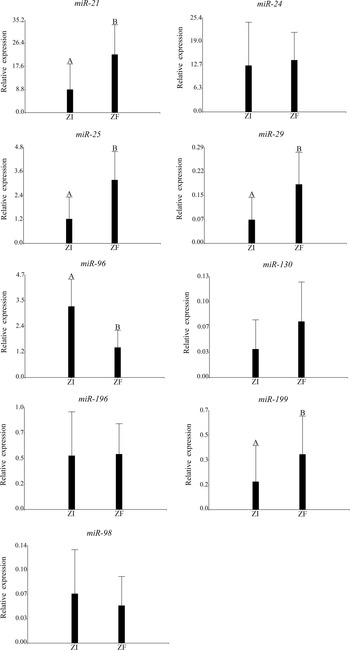
Regarding the relative expression of miRNAs, no statistical differences were observed in the relative expression of miR-24, miR-130, miR-196, and miR-98 between blastocysts from the ZI and ZF groups (P > 0.05). However, the relative expression of miR-21, miR-25, miR-29 and miR-199 was higher in ZF blastocysts than in their ZI counterparts (P < 0.05). Furthermore, the relative expression of miR-96 was lower in ZF blastocysts than in those from the ZI group (Figure 5).
Relative expression analysis (mean ± SD) of miRNAs in ZI and ZF blastocysts. miR-21, miR-24, miR-25, miR-29, miR-96, miR-130, miR-196, miR-199 and miR-98 were evaluated in day-7 blastocysts from the ZI and ZF groups. The geometric mean of miR-103 and miR-191 was used as an internal control. (A, B) different superscripts indicate significant differences between groups (P < 0.05).

Expression analysis of miRNAs in the spent medium of embryos cultured with and without the zona pellucida
The presence of miR-103 was scarcely detected in the spent culture medium of ZI and ZF embryos. For this reason only miR-191 was used as internal control in this type of sample.
No statistical differences were found in the relative expression of miR-24, miR-29, miR-96, miR-130, miR-196 and miR-199 between the spent culture medium from ZI and ZF embryos (P > 0.05). However, miR-21, miR-25 and miR-98 were highly expressed in the spent culture medium from ZF embryos (P < 0.05; Figure 6).
Relative expression analysis (mean ± SD) of miRNAs in spent culture medium. The levels of miR-21, miR-24, miR-25, miR-29, miR-96, miR-130, miR-196, miR-199 and miR-98 were evaluated in the spent culture medium of day-7 blastocysts from the ZI and ZF groups. The expression of miR-191 was used as an internal control. (A, B) different superscripts indicate significant differences between groups (P < 0.05).

Discussion
The results of our study demonstrated that in vitro culture of domestic cat embryos without the zona pellucida until the blastocyst stage resulted in the overexpression of miR-21, miR-25, miR-29 and miR-199, and a reduced expression of miR-96 compared with blastocysts cultured with an intact zona pellucida. This is in accordance with the significant differences observed in the relative expression of pre-miR-21, pre-miR-25, pre-miR-29, pre-miR-96 and pre-miR-199 between ZF and ZI blastocysts. Furthermore, the presence of different miRNAs was detected in spent culture medium, with higher levels of miR-21, miR-25, and miR-98 in spent medium of ZF blastocysts. It was described that miRNAs are capable of passing through the zona pellucida, which indicates a possible regulatory role of the zona pellucida (Hawke et al., Reference Hawke, Ahmed, Watson and Betts2021a). In this study, the differences observed in the expression of miRNAs in the blastocysts and their spent medium, could be caused by the absence of the zona pellucida. In addition, the evaluation of these miRNAs might reveal important information about embryo–maternal crosstalk.
In the present study miR-103 and miR-191 were the most stable genes according to the NormFinder analysis and were selected as internal controls. Mahdipour et al. (Reference Mahdipour, van Tol, Stout and Roelen2015) described similar results in bovine oocytes and early embryos in which miR-103 and miR-93 were the most stable genes. In addition, in porcine oocytes miR-191 was one of the most stable genes according to the geNorm and BestKeeper software (Mahdipour et al., Reference Mahdipour, van Tol, Stout and Roelen2015). This allowed us to perform the specific miRNA analysis.
miR-21 is involved in oocyte maturation and embryo development (Dehghan et al., Reference Dehghan, Mohammadi-Yeganeh, Rezaee and Salehi2021). An overexpression of miR-21 in COCs increases the in vitro fertilization, cleavage and blastocyst formation (Dehghan et al., Reference Dehghan, Mohammadi-Yeganeh, Rezaee and Salehi2021). In addition, the expression of miR-21 significantly increases after implantation (Lv et al., Reference Lv, Yu, Wang, Yi, Zeng and Xiao2018). Inhibition of miR-21 in embryos suppresses in vitro and post-implantation development (Lv et al., Reference Lv, Yu, Wang, Yi, Zeng and Xiao2018). In addition, overexpression of miR-21 in preimplantation embryos has been related to an increase in blastocyst formation by regulation of the apoptosis process (Shen et al., Reference Shen, Han, Zhang, Cui and Kim2009; Zhang et al., Reference Zhang, Shi, Liu, Cao, Tian, Jia, Zhen, Liu and Gao2014). Overexpression of miR-21 has been associated with the upregulation of BCL2L1 and the downregulation of CASP3 (Shen et al., Reference Shen, Han, Zhang, Cui and Kim2009). Furthermore, the induction of miR-21 in mesenchymal stem cells inhibits the expression of SOX2, regulating proliferation and differentiation (Trohatou et al., Reference Trohatou, Zagoura, Bitsika, Pappa, Antsaklis, Anagnou and Roubelakis2014). In the present study, miR-21 was higher in ZF domestic cat blastocysts and in their spent culture medium. Despite that, we did not observe an increase in blastocyst formation rate. We previously described a lower expression of SOX2 and an increased expression of BAX in ZF domestic cat blastocysts (Veraguas-Davila et al., Reference Veraguas-Davila, Cordero, Saez, Saez-Ruiz, Gonzalez, Saravia, Castro and Rodriguez-Alvarez2021). Despite the higher expression of BAX, no alteration was found in the expression of CASP3 (Veraguas-Dávila et al., Reference Veraguas-Dávila, Saéz-Ruíz, Álvarez, Saravia, Castro and Rodríguez-Alvarez2022). The overexpression of miR-21 might be related to lower expression of SOX2 and to the regulation of CASP3 in ZF domestic cat blastocysts.
The presence of miR-25 has been detected in spent culture medium of bovine blastocysts and degenerated embryos (Kropp et al., Reference Kropp, Salih and Khatib2014). Degenrated embryos and their spent medium had an overexpression of miR-25 compared with embryos that reached the blastocyst stage (Kropp et al., Reference Kropp, Salih and Khatib2014). Furthermore, overexpression of miR-25 in circulating exosomes has been associated with embryonic mortality in cows (Pohler et al., Reference Pohler, Green, Moley, Gunewardena, Hung, Payton, Hong, Christenson, Geary and Smith2017). In the present study, miR-25 was highly expressed in ZF domestic cat blastocysts and in their spent medium without affecting the blastocyst formation rate. However, this might be related to the reduced implantation rate of the ZF domestic cat blastocysts (Veraguas-Davila et al., Reference Veraguas-Davila, Cordero, Saez, Saez-Ruiz, Gonzalez, Saravia, Castro and Rodriguez-Alvarez2021).
The miR-29 family has an important role in the expression of pluripotency genes and cell reprogramming (Liang et al., Reference Liang, Nie, Guo, Niu, Shin, Ock and Cui2018; Hysolli et al., Reference Hysolli, Tanaka, Su, Kim, Zhong, Janknecht, Zhou, Geng, Qiu, Pan, Jung, Cheng, Lu, Zhong, Weissman and Park2016; Wang et al., Reference Wang, Zhou, Wan, Yang, Chen, Wang, Zhou, Liu, Ling and Zhang2016). An overexpression of miR-29a or miR-29b during somatic cell reprogramming increases the expression of pluripotency genes such as OCT4 and SOX2 (Liang et al., Reference Liang, Nie, Guo, Niu, Shin, Ock and Cui2018; Hysolli et al., Reference Hysolli, Tanaka, Su, Kim, Zhong, Janknecht, Zhou, Geng, Qiu, Pan, Jung, Cheng, Lu, Zhong, Weissman and Park2016). Furthermore, miR-29a is overexpressed in rat uterus during implantation and binds to the pro-apoptotic genes BAK1 and BMF leading to inhibition of apoptosis in endometrial cells (Xia et al., Reference Xia, Jin, Cao, Hu and Ma2014a). In domestic cat embryos generated by IVF an increased expression of OCT4 is related to enhanced in vitro development (Veraguas et al., Reference Veraguas, Gallegos, Velasquez, Castro and Rodriguez-Alvarez2017, Reference Veraguas, Saez, Aguilera, Echeverry, Gallegos, Saez-Ruiz, Castro and Rodriguez-Alvarez2020a). In accordance with this, no differences were observed in the relative expression of OCT4 between ZF and ZI domestic cat blastocysts, and both groups had a similar blastocyst rate (Veraguas-Davila et al., Reference Veraguas-Davila, Cordero, Saez, Saez-Ruiz, Gonzalez, Saravia, Castro and Rodriguez-Alvarez2021). In this study, overexpression of miR-29 observed in ZF domestic cat blastocysts was not related to an increase in blastocyst rate or an increase in the expression of pluripotency genes. However, it might be related to the regulation of apoptosis.
Regarding miR-199, it has been described that in vitro-produced embryos have lower expression of miR-199a-5p compared with in vivo-produced ones, leading to a reduced developmental capacity of blastocysts and to higher fetal mortality (Tan et al., Reference Tan, Wang, Zhang, Miao, Yu, An and Tian2016). In the present study, the higher expression of miR-199 in ZF domestic cat blastocysts was not related to an increase in blastocyst formation rate.
miR-96 is involved in the implantation process by promoting luteal cell survival and progesterone synthesis by luteinized granulosa cells (Mohammed et al., Reference Mohammed, Sontakke, Ioannidis, Duncan and Donadeu2017). Inhibition of miR-96 increases apoptosis and decreases progesterone production (Mohammed et al., Reference Mohammed, Sontakke, Ioannidis, Duncan and Donadeu2017). Furthermore, miR-96 is highly expressed in stromal cells during pregnancy and is upregulated in implantation sites of mouse uterus (Yang et al., Reference Yang, Xie, Wu, Geng, Li, Xu, Liu and Pan2017). In the present study, the relative expression of miR-96 was lower in ZF blastocysts than in their ZI counterparts. Correct expression of miR-96 in domestic cat embryos might be important for luteal cell survival and implantation. For this reason, a reduced expression of miR-96 could be related to the reduced implantation capacity of ZF domestic cat blastocysts.
miR-98 is a member of the let-7 family, which is involved in embryo implantation (Liu et al., Reference Liu, Pang, Cheong, Ng, Lao, Lee and Yeung2012). It has been reported that different members of the let-7 were overexpressed in dormant blastocysts, which suggests a negative role for embryo implantation (Liu et al., Reference Liu, Pang, Cheong, Ng, Lao, Lee and Yeung2012). Similarly, the upregulation of miR-98 reduces ESC proliferation and increases apoptosis by inhibition of BCL-XL (Xia et al., Reference Xia, Jin, Cao, Shi and Ma2014b). Furthermore, treatment of endometrial epithelial cells with miR-98 downregulated immune system-related genes (Nakamura et al., Reference Nakamura, Kusama, Ideta, Kimura, Hori and Imakawa2019). This might indicate a possible role in the regulation of the maternal immune system during embryo–maternal crosstalk (Nakamura et al., Reference Nakamura, Kusama, Ideta, Kimura, Hori and Imakawa2019). Additionally, expression of miR-98 has been detected in EVs released by in vitro-produced embryos (Andrade et al., Reference Andrade, Bomfim, Del Collado, Meirelles, Perecin and da Silveira2019; Nakamura et al., Reference Nakamura, Kusama, Ideta, Kimura, Hori and Imakawa2019). miR-98 has been associated with downregulation of tight junction mRNAs, which are crucial during embryo implantation (Andrade et al., Reference Andrade, Bomfim, Del Collado, Meirelles, Perecin and da Silveira2019). For this reason, the expression of miR-98 in EVs has been related to a delay in embryo implantation. In the present study, miR-98 was highly expressed in spent culture medium of ZF blastocysts. The high presence of miR-98 in spent medium might be a negative indicator of developmental capacity, and it could be related to the reduced implantation rate of ZF domestic cat blastocysts (Veraguas-Davila et al., Reference Veraguas-Davila, Cordero, Saez, Saez-Ruiz, Gonzalez, Saravia, Castro and Rodriguez-Alvarez2021). Additionally, miR-21, miR-29c and let-7 have been involved in embryonic stem cell differentiation into trophectoderm cells (Liang et al., Reference Liang, Wang and Wang2017; Viswanathan et al., Reference Viswanathan, Mermel, Lu, Lu, Golub and Daley2009). The higher expression of miR-21, miR-29 and miR-98 observed in ZF blastocysts and in their spent medium might be related to the increased expression of YAP1 and EOMES previously reported in these embryos (Veraguas-Dávila et al., Reference Veraguas-Dávila, Saéz-Ruíz, Álvarez, Saravia, Castro and Rodríguez-Alvarez2022).
In conclusion, in vitro culture of domestic cat embryos without the zona pellucida increases the relative expression of miR-21, miR-25, miR-29 and miR-199, and reduces the expression of miR-96 in the blastocyst stage. Additionally, miR-21, miR-25 and miR-98 were highly expressed in spent culture medium of ZF blastocysts. This demonstrates a possible regulatory role of the zona pellucida in the expression and release of embryonic miRNAs. In addition, these miRNAs might be related to altered expression of pluripotency and differentiation genes previously observed in ZF domestic cat blastocysts.
Acknowledgements
We express our appreciation to the veterinary medical team Darling Ruíz Saéz, María Consuelo Álvarez and Yazmín Vásquez Araya for their help in the surgical procedures. In memoriam: The first author of this article wants to thank Daniel Veraguas Bordoli. Thanks for always believing in me.
Financial support
This work was supported by ANID FONDECYT Postdoctorado 3200352, de la Agencia Nacional de Ciencia y Tecnología (ANID), Chile.
Declaration of interest
The authors declare that there are no conflicts of interest to report.
Ethical standards
All the procedures were in accordance with the ethical standards of animal welfare and were approved by the Comité de Bioética de la Facultad de Ciencias Veterinarias de la Universidad de Concepción.