The advances made over the latter half of the 20th century in the treatment of infants and neonates with congenitally malformed hearts have been truly spectacular. Perhaps the most noteworthy advance, and arguably the greatest paradigmatic change, came with the appreciation that it was possible to offer surgical treatment for those born with hypoplastic left heart syndrome.Reference Norwood, Lang and Hansen1 At the time, it was also accepted that, although the prognosis was not as dismal for those born with hypoplasia of the right ventricle in the setting of an intact ventricular septum as for those with hypoplastic left ventricles, results of surgical treatment were far from universally successful. It is perhaps paradoxical, therefore, that results should now be so encouraging for those with hypoplastic left hearts, but still somewhat depressing for those born with hypoplasia of the right ventricle and intact ventricular septum.Reference Freedom, Jaeggi, Perrin, Yoo and Anderson2 Not all patients, nonetheless, with hypoplastic right ventricles have a bad prognosis. The differentiating feature, as we will show, lies in the morphology at the pulmonary ventriculo-arterial junction,Reference Anderson, Anderson and Zuberbuhler3 but this anatomical finding is closely associated with the presence of fistulous communications between the cavity of the right ventricle and the coronary arterial circulation. It is these latter communications that underscore the so-called “right ventricular-dependent circulation”,Reference Freedom and Harrington4 which has long been known to carry a grave prognostic significance.Reference Freedom, Anderson and Perrin5 Fistulous communications between the ventricular cavity and the coronary arterial circulation are also known to exist in the setting of hypoplastic left heart syndrome, albeit not being nearly as obvious during gross anatomical investigations,Reference Freedom, Anderson and Perrin5, Reference O’Connor, Cash, Cottrell, Johnson and Noonan6 but now observed on occasion during foetal echocardiography.Reference Sauer, Gittenberger-de Groot and Geishauser7 In this review, we discuss the morphological features of the fistulous communications when seen in the setting of hypoplastic right and left ventricles with intact ventricular septum.
Pulmonary atresia with intact ventricular septum
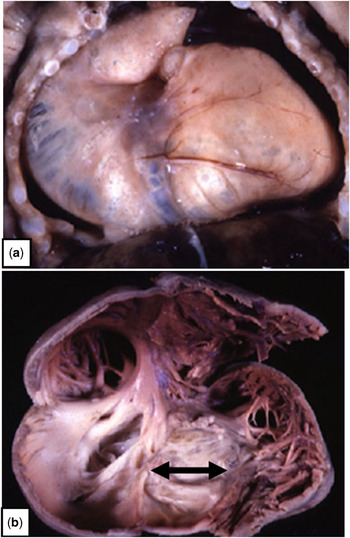
The very fact that the ventricular septum is intact in this entity points to its pathogenesis as acquired disease during foetal life, a conclusion further endorsed by the close morphological similarities between hearts having pulmonary atresia with an intact ventricular septum and those with critical pulmonary stenosis. Even within patients born with pulmonary atresia and an intact ventricular septum, however, there is a marked variability in the morphological features. This variability depends, in the first instance, on whether the right ventricular cavity is hypoplastic or dilated. Cases with right ventricular dilation form a particular subset of patients, with the most florid examples presenting with so-called “wall-to-wall” hearts (Fig 1).Reference Freedom, Jaeggi, Perrin, Yoo and Anderson2 These patients have perhaps the worst prognosis of those within this group, the poor outcome reflecting the fact that the size of the heart squeezes out the lungs, so that although the lungs are morphologically normal, they are grossly hypoplastic, and unable to perform their normal function. The huge size of the right atrium and ventricle reflects the gross tricuspid incompetence occurring during foetal life.Reference Oberhoffer, Cook and Lang9 Patients with dilated right ventricles, however, constitute but a small subset of those with pulmonary atresia and intact ventricular septum, and are never, to the best of our knowledge, associated with fistulous communications between the right ventricular cavity and the coronary arterial circulation.
The upper panel (a) shows a so-called “wall-to-wall” heart produced by pulmonary atresia with intact ventricular septum. The size of the heart has squeezed out the lungs. The lower panel (b) shows the heart itself, the tricuspid valve exhibiting Ebstein’s malformation (double-headed arrow), and the gross incompetence during foetal life producing the marked dilation of both the right ventricle and the right atrium.

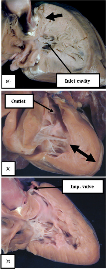
The fistulous communications are found in a subset of patients with hypoplastic right ventricular cavities. In this subset, accounting for the majority of cases, the degree of hypoplasia of the ventricular cavity can still vary markedly. The fistulous communications, broadly speaking, are found in those with the smallest right ventricular cavities. The size of the ventricular cavities is dependent on the degree of right ventricular mural hypertrophy, with this almost certainly being related to the time during gestation when the pulmonary valve becomes atretic. Within the overall spectrum of mural hypertrophy, it is possible to recognise three major anatomical subsets (Fig 2). In the worst subset, the mural hypertrophy is so extreme as to obliterate entirely the right ventricular outflow tract, such that a muscular plate interposes between the cavity of the ventricle and that of the pulmonary trunk. The hypertrophy is also sufficient to effectively exclude the apical trabecular cavity (Fig 2a). In the least afflicted subset, the pulmonary valve is recognisable as an imperforate fibrous plate (Fig 2c). Between these extremes is the subset in which the mural hypertrophy reduces the extent of the apical trabecular ventricular component, and narrows the infundibulum, albeit that the infundibulum remains recognisable as a tract leading towards the ventriculo-arterial junction (Fig 2b). The changes are well understood on the basis of the tripartite structure of the right ventricle. In the patients with the largest ventricular cavities, the mural hypertrophy overall is minimal, and all three ventricular components are well seen. In the intermediate subset, the mural hypertrophy has effectively obliterated the ventricular outlet component, so that the cavity is represented by the apical trabecular and inlet components. In the worst examples, as discussed, the hypertrophy has obliterated also the larger part of the apical trabecular component, so that the ventricular cavity, in essence, is represented only by the inlet component. All hearts still possess all three ventricular components, but with increasing obliteration of their cavities according to the extent of mural hypertrophy.Reference De Leval, Bull, Stark, Anderson, Taylor and Macartney10
The hearts show the spectrum of decreasing cavity size with increasing mural hypertrophy in the setting of pulmonary atresia with intact ventricular septum. The upper panel (a) shows the situation in which the hypertrophy has obliterated both the outlet and the best part of the apical trabecular component. In the middle panel (b), the hypertrophy (double-headed arrow) obliterates the apical component, but spares the outlet and inlet components. The lower panel (c) shows the anatomy associated with an imperforate (Imp.) pulmonary valve. All three components of the ventricular cavity are relatively well formed.

In general terms, it is in those patients with the ventricular cavity represented only by the inlet component, and with muscular atresia at the ventriculo-arterial junction, that it is most frequent to find fistulous communications with the coronary arterial tree. The association between fistulous communications and grossly hypoplastic right ventricular cavities is found in autopsy series examined from specimens obtained in both foetalReference Sandor, Cook, Sharland, Ho, Potts and Anderson11 and postnatalReference Anderson, Anderson and Zuberbuhler3 life. The communications have long been recognised, albeit initially described in terms of “sinusoids”, or “spongy myocardium”.Reference Dusek, Ostadal and Duskova12 The investigation of Gittenberger-de Groot et alReference Gittenberger-de Groot, Sauer, Bindl, Babic, Essed and Buhlmeyer13 did the most to clarify the relationship between the intertrabecular spaces within the hypertrophied ventricular walls and the epicardial arteries themselves, although an important investigation had also been published by the group from Auckland, New Zealand,Reference Calder, Co and Sage14 with this group recently updating their experience to emphasise the clinical significance of the communications.Reference Calder, Peebles and Occleshaw15
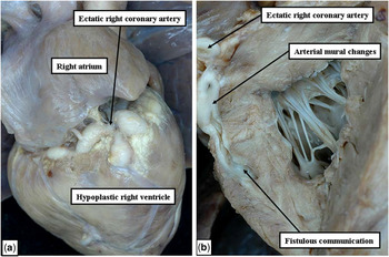
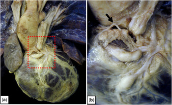
The critical feature is the communications between the right ventricular cavity and the epicardial coronary arteries. These fistulous communications are present in the setting of systemic right ventricular pressure, usually with a competent tricuspid valve, so the flow of blood in the presence of significant communications is typically from the right ventricle to the coronary arterial tree, underscoring the so-called right ventricular-dependent circulation.Reference Freedom and Harrington4, Reference Freedom, Anderson and Perrin5 The fistulous communications are readily revealed by angiographic investigations,Reference Dyamenahalli, McCrindle and McDonald16 and by echocardiographic interrogation, again in both foetal and postnatal life.Reference Sandor, Cook, Sharland, Ho, Potts and Anderson11, Reference Dyamenahalli, McCrindle and McDonald16 The abnormal coronary arteries are obvious at autopsy examination (Fig 3), with the flow of blood into the coronary arterial tree producing significant disease of the proximal segments of the coronary arteries themselves (Fig 4). The changes in the walls of the coronary arteries were initially considered to be inflammatory in origin. We now know that this is not the case.Reference Freedom, Anderson and Perrin5 Instead, the changes are the consequence of myointimal hyperplasia.Reference Freedom, Anderson and Perrin5 In the most severe cases, the proximal coronary arteries can show severe stenosis, or even complete occlusion of the lumens (Fig 5). Indeed, in rare cases both right and left coronary arteries lose their connections with the aortic root.Reference Lenox and Briner17 It is the obstructive lesions in the proximal coronary arteries that underscore the myocardial ischaemia known to be associated with the presence of the fistulous communications. Myocardial rupture, secondary to acute myocardial infarction, has been reported in the occasional patient.Reference Hubbard, Girod, Caldwell, Hurwitz, Mahoney and Waller18 It is the fistulous communications, therefore, that indicate the likelihood of a poor prognosis in neonates born with pulmonary atresia with intact ventricular septum, these lesions being a feature of the subset of patients having muscular rather than valvar atresia, and typically being found in those with the most hypoplastic right ventricular cavities.Reference Anderson, Anderson and Zuberbuhler3, Reference Freedom, Anderson and Perrin5
The panels (a–b) show the ectatic right coronary artery in a patient with pulmonary atresia and intact ventricular septum (a), with dissection revealing the fistulous communication between the hypoplastic ventricular cavity and the abnormal right coronary artery (b).

The panels (a–b) show the consequence of fistulous communications with the coronary arteries in the heart from a patient with pulmonary atresia and intact ventricular septum. Panel a shows the overview, with the boxed area enlarged in panel b. The right coronary artery (arrow) is atretic at its aortic origin, and the anterior interventricular artery is thickened and abnormal.

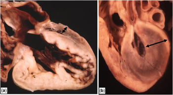
The panels (a–b) show the fundamental difference in the architecture of the left ventricular wall when there is absence of the left atrioventricular connection (a) as opposed to mitral stenosis (b) in the setting of hypoplastic left heart syndrome. The left ventricular wall is appreciably thicker, and “blighted” (double-headed arrows) when there is flow of blood into the left ventricle in the presence of mitral stenosis (b).

Hypoplastic left heart syndrome
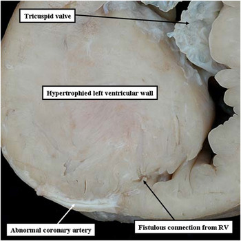
Like pulmonary atresia with intact ventricular septum, hypoplastic left heart syndrome represents cardiac disease acquired during foetal life. In addition, as with the right-sided malformations, there is also marked anatomical heterogeneity to be found among those patients falling within the definition of the syndrome. In those with hypoplastic left ventricles, the major difference reflects the formation of the left atrioventricular junction, which in turn has significance for the architecture of the left ventricular walls. In a minority of patients, there is complete absence of the left atrioventricular connection, and in another small subset, the mitral valve is formed but imperforate (Fig 5a). In these patients, there has never been flow of blood into the left ventricle, which in consequence has a grossly hypoplastic cavity, in some instances only being discovered following careful dissection guided by the location of the anterior and inferior interventricular arteries. In these instances, although the ventricular cavity is grossly hypoplastic, its walls are relatively thin, and the endocardial lining is normal. In the larger subset of patients, the mitral valve is patent, with either aortic atresia or critical aortic stenosis (Fig 5b). In these latter cases, there has been flow of blood into the hypoplastic ventricle during foetal life, and the ventricular walls are hypertrophied, typically with formation of a thick layer of gross endocardial fibroelastosis. It is these patients with hypertrophied ventricular walls and fibroelastosis that the late Robert Freedom considered to have “blighted myocardium”. He had opined that such patients might have a poorer outcome after surgical treatment than those with mitral atresia, albeit that initially little evidence was forthcoming to support his prognostications. It had also been shown, however, that the patients with mitral stenosis rather than atresia also had microscopic fistulous communications with the coronary arterial circulation.Reference O’Connor, Cash, Cottrell, Johnson and Noonan6, Reference Sauer, Gittenberger-de Groot and Geishauser7 The presence of the fibroelastosis likely prevents the development of large connections with the epicardial coronary arteries, as is seen in pulmonary atresia with intact ventricular septum; but increasing experience shows that, on occasion, such overt communications can be found.Reference Roberson, Cui, Cuneo, Van Bergen, Javois and Bharati8 We have recently encountered such connections in an explanted heart from a patient with hypoplastic left heart syndrome. It proved possible to probe a fistulous communication to the inferior interventricular artery from the cavity of the right ventricle, and sectioning the heart revealed a diseased artery (Fig 6). The left ventricular myocardium was grossly hypertrophied, albeit that overt fistulous connections were not identified from the cavity of the left ventricle, which was very restricted. It may well be, nonetheless, that as predicted by Robert Freedom, the myocardium can be “blighted” in the setting of hypoplastic left heart syndrome, and that such findings could indicate a poorer prognosis.
The image shows a section through the ventricular septum in an explanted heart from the patient with hypoplastic left heart syndrome. There is an overt fistulous communication from the cavity of the right ventricle to the diseased inferior interventricular artery.

Conclusions
Fistulous communications between the ventricular cavities and the coronary arterial tree can be found in the presence of hypoplasia of both the right and left ventricle when the ventricular septum is intact. The communications are obvious in the setting of pulmonary atresia with intact ventricular septum, and are well-known to carry a poor prognostic significance. Fistulous communications may prove to be equally important in the setting of hypoplastic left heart syndrome, albeit not as easy to define morphologically.