I. INTRODUCTION
Total reflection X-ray fluorescence analysis (TXRF) is a powerful analytical method and provides several advantages compared with conventional energy dispersive X-ray fluorescence analysis (EDXRF) (Wobrauschek, Reference Wobrauschek2007). The principle of TXRF is that an incoming X-ray beam hits a smoothly polished surface (e.g. a quartz reflector) below the critical angle of incidence (in the mrad range) and is hence totally reflected. The liquid sample is placed in the center of the reflector as a droplet of several microliters and then dried forming a thin film. Due to the small angle of incidence the primary radiation only penetrates a few nm into the substrate leading to a drastically reduced background. As the sample is excited twice by the incoming and totally reflected X-ray beam the fluorescence signal doubles in comparison with conventional EDXRF. The small angle of incidence also leads to the advantage that the detector can be positioned very closely to the surface of the sample substrate leading to an increase of the solid angle seen by the detector. These advantages provide a much better signal-to-noise ratio as compared with other EDXRF techniques leading to detection limits for TXRF of a few picograms (10−12 g) in absolute masses when using laboratory equipment with X-ray tube excitation. When operating TXRF at a synchrotron facility detection limits can be reduced further down to the femtogram (10−15 g) range (Wobrauschek, Reference Wobrauschek2007). With TXRF, the analysis of a wide range of elements up to uranium (U, Z = 92) is possible. However, the analysis of light elements such as C (Z = 6), N, O, F, Na, Mg, and Al (Z = 13) is extremely challenging. Due to the low characteristic energies of the mentioned elements, all measurements have to be performed in vacuum in order to avoid absorption by air. To obtain high fluorescence intensities the energy of the excitation source should be close to the absorption edges of the elements in question. Possible anode materials are chromium (Cr-Kα at 5.4 keV) and copper (Cu-Kα at 8.0 keV) using the K-lines for excitation. Furthermore, the L-lines of molybdenum (Mo-Lα at 2.3 keV) and rhodium (Rh-Lα at 2.7 keV) can be used for the excitation of light elements. In order to detect C, N, O, and F a special detector with an ultrathin polymer window (UTW) is a necessary prerequisite. The UTW is a thin polymer sheet (typically 300 nm) supported by a silicon grid. When operating UTW detectors, the background is raised strongly by photo, Auger, and Compton electrons. Therefore, a magnetic trap has to be positioned in front of the UTW. Unfortunately, this results in an increased distance between sample and detector. Detectors with UTW are extremely light sensitive and thus have to be operated only in a lightproof vacuum chamber. However, it will be shown in this work that a conventional silicon drift detector (SDD) with a 25 μm beryllium window and an active area of 80 mm2 is also suitable for the detection of Na, Mg, and Al as long as these elements are present in the samples in sufficient quantities (>100 ng).
Due to fundamental X-ray physics the intensity of the K-shell fluorescence radiation shows the following behavior:
 $${I_K}\lpar E\rpar \, {\rm \propto }{I_0}\lpar E\rpar \, {\tau _K}\, \omega \, {\rm \epsilon }\, \lpar \Omega /4\pi \rpar \, \lpar m/A\rpar$$
where I 0(E) is the intensity of the incoming primary radiation, τ K (E) the K-shell photoabsorption coefficient, ω the fluorescence yield, ε the detector efficiency, Ω the solid angle of the detector and (m/A) the sample areal mass density. As the fluorescence yield ω is a constant given by nature and generally low for light elements (in the 10−2 range for Na, Mg, and Al) (Krause, Reference Krause1979), TXRF analysis of low Z elements can be optimized by adjusting the other parameters. I 0(E) can be increased using the high-intensity radiation provided by a synchrotron. τ K (E) is generally low for anode materials of conventional X-ray tubes. It can be increased using special windowless X-ray tubes with low Z anodes such as Si-K, W-M, Al-K or Mo-L, and an optimized take-off angle. Furthermore, an appropriate excitation energy, which is close to the absorption edges of the elements of interest, can be chosen at synchrotron sources because of the tunability of the incoming beam energy. The detector efficiency ε can be optimized using detectors with ultrathin polymer entrance windows. Finally, an optimum solid angle Ω can be achieved by optimizing the experimental setup, i.e. by increasing the active area of the detector and using the shortest possible distances between source, sample, and detector. Using synchrotron radiation and a detector with ultrathin window, combined with an optimized geometry, even the detection of boron (B, Z = 5) becomes possible and has been reported e.g. by Streli et al. (Reference Streli, Pepponi, Wobrauschek, Beckhoff, Ulm, Pahlke, Fabry, Ehmann, Kanngießer, Malzer and Jark2003).
The aim of the present work is to show the influence of different excitation sources on the detection of light elements. To achieve optimum excitation, the energy difference between the primary radiation and the absorption edge of the respective element has to be as small as possible, thus for multielement analysis a compromise results as only few anode materials are available. For quantification and determination of detection limits Na, Mg, Al, S, K, Ca, and Ti were considered. Interestingly, the use of compact air-cooled low-power X-ray tubes in comparison with the bulky water-cooled high-power tubes showed considerably good detection limits in almost the same range. Thus, the development of compact tabletop vacuum TXRF spectrometers with a performance equal to chambers operated by conventional water-cooled tubes is an important goal for the future.
II. EXPERIMENTAL
For this work, the Atominstitut TXRF Standard Vacuum Chamber (Rieder et al., Reference Rieder, Ladisich, Wobrauschek, Streli and Kregsamer1993) was used. This chamber is operated under rough vacuum (about 1 mbar) achieved by a membrane pump. The X-ray entrance and exit windows are made of an 8 μm Kapton sheet. The chamber is equipped inside with a W/C multilayer monochromator (2 d = 4 nm), which can also be used as a high-energy cut-off reflector. The sample carrier is loaded into a sample stage, which can be moved automatically into the measurement position. The measurement position can be adjusted by translation and rotation stages via remote control. The sample stage can be adjusted, so that carriers of different thicknesses can be used. In the present work only quartz reflectors with 30 mm diameter and a thickness of 3 mm were chosen.
The spectrometer can be operated with various X-ray tubes as well as with different detectors, which can be easily attached to the chamber from outside. In order to compare different source-detector combinations and to investigate their effects on the analysis of light elements, two sets of experiments have been performed. A view of the actual setup can be seen in Figure 1. The source detector-combinations investigated in this work are shown in Table I.
(Color online) Atominstitut TXRF Standard Vacuum Chamber. Panel (a) shows a top view of the chamber. On the left side different X-ray sources can be attached to the chamber, either via the tube housing shown in the picture or via special adapter pieces. The detector feed-through is shown on top of the photograph. Monochromator unit and sample stage can be seen inside the vacuum chamber. Panel (b) shows a close-up view of the detector feed-through, and on (c) the Mo anode low-power tube connected to the chamber via an adapter piece can be seen.

Source–detector combinations applied in the experiments of this study. Two water-cooled high-power diffraction tubes were compared with two air-cooled low-power tubes. Furthermore, two different silicon drift detectors were used, one with an ultrathin polymer window but only 7 mm2 active area, the other one with a 25 μm beryllium window, and 80 mm2 active area.

For the first measurement series three different samples were prepared. Two samples consisted of a multielement standard with Na, Mg, Al, S, K, Ca, and Ti and had 50 ng and 100 ng of these elements, respectively. The third sample contained 200 ng of the three light elements Na, Mg, and Al only. All the three samples were prepared using the Nanoliter Application Unit constructed by the Atominstitut X-ray Laboratory (Wastl et al., Reference Wastl, Stadlbauer, Prost, Horntrich, Kregsamer, Wobrauschek and Streli2013). With this device it is possible to produce samples consisting either of one central droplet or a pattern of multiple droplets on defined positions (r, φ), which can be reached via computer-controlled stepper motors. The multielement samples were produced from a laboratory-prepared 100 ppm solution containing Na, Mg, Al, S, K, Ca, and Ti, each consisting of a single central droplet with a volume of 500 nl and 1 μl, respectively. The light element sample was a 2 μl droplet of a laboratory-prepared 100 ppm solution of Na, Mg, and Al.
For the experiments, three different X-ray sources were attached to the vacuum chamber. Two water-cooled high-power X-ray diffraction tubes with Cr (1900 W, operated at 30 kV and 30 mA) and Cu (1500 W, operated at 40 kV and 20 mA) anodes, respectively were used. The line focus of these two tubes was chosen. As a third excitation source an air-cooled Mo anode low-power X-ray tube (50 W, operated at 50 kV and 1 mA) with point focus (about 100 × 100 μm2) was attached to the chamber. In this case, the multilayer was used as a high-energy cut-off reflector (Knoth and Schwenke, Reference Knoth and Schwenke1978). A section of the Mo bremsstrahlung continuum up to about 10 keV together with Mo-L lines were hence used as the third excitation mode. The Mo-L lines should help to excite the light elements Na, Mg, and Al. Using a 7 mm2 SDD with a 300 nm ultrathin AP1.3 polymer window, all the three samples were measured for 1000 s each in every excitation mode. The signal from the detector was processed with an analog signal processor. The detector entrance window was positioned about 9 mm away from the front of the sample as the magnetic trap required so much space. The spectra were evaluated using the QXAS software package (IAEA, 1995) and detection limits were calculated for each element and excitation condition.
The second experimental setup was intended to show the possibility of the detection of elements from Na upwards by means of a conventional large-area (80 mm2) SDD with a 25 μm beryllium window instead of an UTW. An advantage of this setup is that no magnetic trap was required and the distance between sample and detector could be reduced to about 3 mm. As excitation source, a 35 W Rh anode air-cooled low-power X-ray tube with line focus (50 × 1500 μm2), operated at 50 kV and 0.6 mA was attached to the vacuum chamber. L-line excitation was used to optimize the analysis of Na, Mg, Al, and S, making the lightest elements (Na, Mg, and Al) visible for the detector. To this end the multilayer was adjusted to a maximum intensity of the Rh-L lines. Nevertheless, Rh-K lines were still visible and were used for the excitation of K, Ca, and Ti. Samples containing 100 ng of each element were produced from the above-mentioned 100 ppm multielement solution and from a similar solution containing only Na, Mg, Al, S, and Ti. Each sample was measured for 200 s. The spectra were evaluated with QXAS and the detection limits were calculated and extrapolated to 1000 s.
III. RESULTS
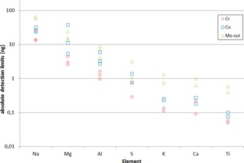
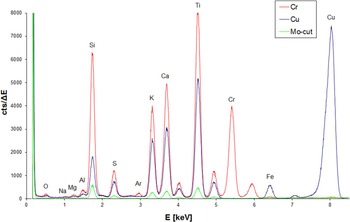
Due to the small active area of the detector (7 mm2) as well as owing to the relatively large distance between sample and detector (9 mm), the detection limits obtained in the first experiment were in the high-picogram range for S, K, Ca, and Ti and amounted to several nanograms for Na, Mg, and Al. Best detection limits were achieved for the excitation with Cr (Table II, Figure 2), as the energy of the Cr-Kα radiation is closer to the absorption edges of the elements in question compared with Cu-Kα. Higher peak intensities as well as a lower background in the area of interest could be obtained in comparison with the excitation with Cu, thus leading to better detection limits for Cr-Kα excitation. Furthermore, it could be shown that the Mo cut-off excitation mode was also helpful for the detection of Na, Mg, and Al, nevertheless the Na signal could only be detected for the 100 and 200 ng samples. Additionally, in all of the spectra, oxygen (O, Z = 8) coming from the quartz carrier, could be detected. In the Cr excitation mode, even a carbon (C, Z = 6) peak was visible in the spectrum. Spectra are shown in Figures 3 and 4. Table I shows the detection limits calculated from the 50 ng sample for each excitation mode.
(Color online) Detection limits of all measured samples. Na, Mg, and Al could be evaluated for all three concentrations (50, 100, and 200 ng), while S, K, Ca, and Ti were present only in the 50 and 100 ng samples. With Mo cut-off excitation, Na was below the limit of detection for the 50 ng sample.

(Color online) Overlay of spectra from the 50 ng multielement sample. Cr excitation leads to the highest intensities over an excellent background. Cu also provides good background conditions, but much weaker peak intensities. The intensities obtained in the Mo cut-off excitation mode are about an order of magnitude lower, but the detection of Mg and Al was possible, because the spectrum is nearly background-free.

(Color online) Overlay of spectra from the 200 ng sample containing only Na, Mg, and Al. It can be seen that the peaks for the three elements are clearly resolved. In every spectrum, oxygen is visible. With Cr excitation a small carbon peak is also visible.

Detection limits (DL) (1000 s) in ng calculated for the 50 ng multielement sample, obtained with a 7 mm2 silicon drift detector with ultrathin window, comparing different excitation sources (900 W Cr, 800 W Cu, and 50 W Mo cut-off). It can be seen that the excitation with Cr leads to the best results. Mg and Al could be detected with all the three excitation sources, whereas Na was below the detection limit for the Mo cut-off mode.

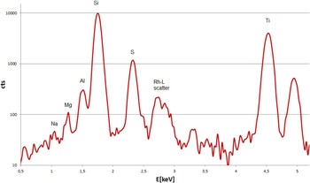
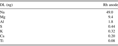
Detection limits for the second setup are shown in Table III. As mentioned above, Rh-L lines contributed to the excitation of Na, Mg, Al, and S. Thus, the peaks of Na, Mg, and Al could be clearly resolved even with a detector without UTW (Figure 5). Detection limits are comparable with those obtained in the first experiment and are displayed in Table III.
(Color online) Spectrum of the 100 ng Na, Mg, Al, S, and Ti multielement sample. Peaks of Na, Mg, and Al are clearly visible, even though a detector with 25 μm beryllium window was used.

Detection limits (DL) (1000 s) in ng obtained from the 100 ng multielement sample (Na, Mg, Al, S, K, Ca, and Ti) with Rh excitation and an 80 mm2 silicon drift detector with a 25 μm beryllium entrance window.

IV. DISCUSSION
In the first experiments, excitation with a Cr anode tube led to highest peak intensities as well as to lowest detection limits for all elements analyzed. Cr also offers the best excitation of the three lightest elements (Na, Mg, and Al) leading to clearly resolved peaks, even for the lowest sample mass investigated (50 ng). In comparison, with the Cu anode, having a lower photoelectric cross-section, lower intensities and higher detection limits, especially for Na, Mg, and Al, were obtained. We could also demonstrate that excitation with bremsstrahlung provided by a 50 W low-power X-ray tube with Mo anode is sufficient for the analysis of light elements, but detection limits for Na, Mg, and Al are higher due to the lower power of the exciting radiation.
The second experiments showed that the detection of elements from Na upwards is also possible with a SDD with a 25 μm beryllium window when using L-line excitation and optimized experimental condition. This is on the one hand because of the large active area of the detector (roughly a factor 11 compared with the UTW). On the other hand, the worse efficiency because of the 25 μm beryllium window, resulting in an intensity loss, was compensated by the reduced distance between sample and detector, as no magnetic trap is required, yielding a factor of 9 in intensity. Future improvements are expected, if large-area UTW detectors and X-ray tubes with extremely thin windows allowing the efficient transmission of the L-lines, e.g. Mo-L, Rh-L, are available.
As detectors with low efficiency were used in this work and the distance between sample and detector was relatively large, the obtained detection limits were comparatively high. A study on light element analysis has been carried out by the Atominstitut X-ray group on a WOBISTRAX vacuum spectrometer with optimized geometry in terms of an extremely short distance from the source to the sample, a Cr anode tube operating at 30 kV/50 mA (1500 W) and a 10 mm2 SDD with 8 μm beryllium window. Detection limits of 900 pg for Na, 156 pg for Mg, and 42 pg for Al were obtained (Streli et al., Reference Streli, Wobrauschek, Pepponi and Zoeger2004). In a recent publication of the Atominstitut X-ray group, detection limits of 34 pg for P and 19 pg for S were achieved using a Cr anode X-ray tube (30 kV/30 mA) and a 30 mm2 SDD with UTW (Rauwolf et al., Reference Rauwolf, Vanhoof, Tirez, Maes, Ingerle, Wobrauschek and Streli2014).
The performance of the two low-power tubes in this work has proven that the construction of a tabletop vacuum TXRF spectrometer for the analysis of light elements is realistic. With an optimized geometry, i.e. shortest possible distances between source and sample as well as between sample and detector, and the use of a large-area SDD with UTW, detection limits comparable with the above-mentioned values should be achievable.