Significant outcomes
This study identifies numerous genomic regions that may harbour genes responsible for autosomal recessive intellectual disability.
Limitations
There is a risk that some of the homozygous genomic regions are false-positive.
Introduction
Intellectual disability (ID) or mental retardation (MR) is an unresolved health-care problem that severely impacts both the affected individuals and their families. For most ID cases, the underlying aetiology is genetic in origin; therefore, it is frequently referred to genetic centres. The mild form of ID is characterised by intelligence quotient (50–70), which could be solely of genetic origin. However, severe forms of ID (intelligence quotient>50), incidence of 0.4%, are thought to be caused by environmental or genetic factors (Reference Ropers1).
In the last few years, it has become obvious that genetic factors such as chromosomal abnormalities, smaller genomic aberrations, as well as point mutations play key roles in the aetiology of ID. The epidemiological study by Rauch et al. (Reference Rauch, Hoyer and Guth2) demonstrated that 15% of the cases of ID are caused by cytogenetic anomalies and 15% are caused by submicroscopic aberrations. Mutations on the X-chromosome are the cause in about 10% of the cases (Reference Ropers and Hamel3). This suggests that the aetiology remains unsolved in more than half of the cases of ID, and of which the majority of these cases are due to autosomal mutations. To date, >100 genes on the X-chromosome have been linked to ID, and these genes are responsible for X-linked ID in ~40% of patients (Reference Lubs, Stevenson and Schwartz4). However, in populations where consanguineous marriages are common, autosomal recessive (AR) causes are responsible for the vast majority of cases, and even in out-bred populations it is estimated that 13–24% of ID may be due to AR causes (Reference Musante and Ropers5). A recent review listed 40 known genes for non-syndromic autosomal recessive ID (NS-ARID); however, it estimates that there may be an excess of 2500 autosomal ID genes in total, with the majority being recessive (Reference Musante and Ropers5). As such, a systematic strategy is required to identify AR causes of ID (Reference Najmabadi, Motazacker and Garshasbi6–Reference Kuss, Garshasbi and Kahrizi8). Large consanguineous families are a great resource for providing the necessary information to map loci, based on analysis in a single family, and thus represent the best starting point.
The prevalence of AR non-specific ID or NS-ARID is unknown, despite empirical data from mouse models suggesting that most genetic defects are inherited as recessive forms (Reference Ropers1,Reference Priest, Thuline, Laveck and Jarvis9,Reference Bartley and Hall10). To date, only ~40 genes for NS-ARID have been identified (Reference Musante and Ropers5). In countries where consanguinity is high, there is a marked increase in morbidity and mortality among infants. In Pakistan, a country with a population of 172.8 million people, there is a 62.7% rate of consanguinity, of which 84.0% are marriages between first cousins (Reference Hussain and Bittles11), making it an ideal population in which to base a search for NS-ARID genes.
The aim of this study is to describe gene mapping studies in 11 consanguineous families with ID from Pakistan.
Materials and methods
Eleven families with a minimum of two affected individuals were recruited in this study and originated from various areas within the Province of Sindh, Balochistan, and Azad Jammu and Kashmir, Pakistan. Approval of the study was obtained from Institution Review Boards and Ethical Committee at the Centre for Addiction and Mental Health, Toronto, Canada, and National University of Sciences and Technology, Islamabad, Pakistan. Families were visited at their homes and were informed about the study. Written informed consent was obtained from the parents/guardian of each family for their participation in this study, and for photography.
All affected individuals were clinically evaluated by a neurologist, ophthalmologist, and a general physician. Specific attention was paid to neurological, morphological, ophthalmological, dermatological, and skeletal examinations. The intelligence quotient level of all affected individuals from each family was evaluated with the help of standard questionnaires. Parents were interviewed about the prenatal and neonatal medical history of the probands and mothers during pregnancy. The pedigree was constructed and the genealogical relationships within families were cross-checked by interviewing different family members. A volume of 5 ml venous blood was drawn from all available affected and unaffected siblings, as well as their parents. DNA was extracted according to the standard method (Reference Lahiri, Bye, Nurnberger, Hodes and Crisp12). The total number of 44 affected individuals including 34 males and 10 females were subjected for molecular genetics analysis. Syndromic cases such as Down’s syndrome were excluded. One family, PJMR4, was excluded because one of the two affected individuals was found to have a chromosomal abnormality. In order to exclude any cases caused by Fragile X expansion/methylation mutations, DNAs from the mothers for each pedigree were checked for premutations by polymerase chain reaction amplification of the CGG repeat at FMR1.
In total, 15 healthy and 44 affected individuals were genotyped, using either Affymetrix 250K NspI (Affymetrix, Santa Clara, CA, USA) or Affymetrix® CytoScan™ HD Array (Affymetrix), and for one family using Illumina CoreExome beadchip arrays (Illumina Inc., San Diego, CA, USA). The platform used was consistent within each family. Marker density was ~262 000 single nucleotide polymorphism (SNPs) for the Affymetrix NspI array. For the Illumina CoreExome array, ~260 000 markers were used for the analysis.
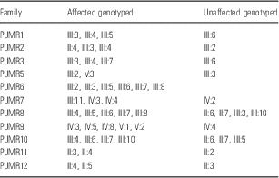
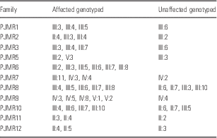
Table 1 provides information about the individuals who were genotyped in each family. It should be noted that, for PJMR5, individual III:8 has ID with clear syndromic presentation, and was thus excluded from analysis.
Genotyped individuals in each family
The data were analysed using the dChip software (Reference Lin, Wei, Sellers, Lieberfarb, Wong and Li13) and HomozygosityMapper (Reference Seelow, Schuelke, Hildebrandt and Nurnberg14) to identify homozygosity-by-descent (HBD) regions. In addition, dChip was used to detect the presence of copy-number variants (CNVs) for the Affymetrix array data and Illumina’s GenomeSuite with cnvPartition 3.2.0 for Illumina CoreExome data.
Results
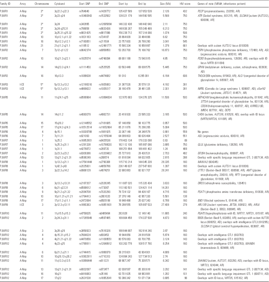
Affected individuals included in this study manifested mild-to-severe forms of ID. All cases were non-syndromic. Fragile X syndrome was excluded for all families by genotyping mothers for the presence of pre-mutations. The microarray data analysis showed 45 HBD regions (>0.9 Mb) that were homozygous and haploidentical across all the affected siblings in the family but not in the genotyped unaffected siblings. However, some of these regions are situated almost adjacent to each other, and may in fact represent single HBD regions, and thus this list may be condensed to 39 regions if this is taken into consideration (Table 2).
HBD results for Pakistani ID families

AD, autosomal dominant; AR, autosomal recessive; bp, base pair; HBD, homozygosity-by-descent; HM, HomozygosityMapper; ID, intellectual disability; QTL, quantitative trait locus; SNP, single nucleotide polymorphism; XL, X-linked.
HM was used to provide the analysis. Genotypes across putative HBD regions were checked to confirm haploidentity across affected, but not unaffected individuals, and to confirm boundaries of the HBD region. Only HBD regions over 0.9 Mb in length are shown. Arrays used are listed as A-Nsp for Affymetrix 250K NspI array, and I-CE for Illumina CoreExome BeadChip array. Genomic co-ordinates from GRCh37/hg19 are used. For genes of note (potential overlapping phenotype with ID) listed within HBD regions, reported inheritance pattern is given as AR, AD, or XL.
* These HBD regions may be part of a single large HBD region.
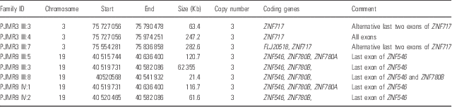
CNV analyses for PJMR1, 2, 5, 6, 7, 8, 10, and 12 were performed using the dChip software (Reference Lin, Wei, Sellers, Lieberfarb, Wong and Li13). Analyses showed no homozygous or heterozygous CNVs that segregate with ID in the families, and no rare CNVs (not overlapping with calls in the database of genomic variants, DGV) disrupting any known ID genes or overlapping with any known deletion or duplication syndromes where ID is a clinical feature. CNV analyses for PJMR3 and 9 were performed using Illumina GenomeSuite software. Analysis for PJMR3 showed no homozygous CNVs segregating with the phenotype in the family. A single gain CNV was seen in the three affected patients genotyped, but not in the unaffected individuals (see Table 3). This CNV spans several exons of a zinc finger gene, ZNF717. However, both gain and loss CNVs at this locus are common in the general population according to the DGVs (http://dgv.tcag.ca). Analysis for PJMR9 showed no homozygous CNVs segregating with the phenotype. PJMR9 had a single gain CNV that was seen in all five affected individuals but not in the unaffected sibling run alongside on the Illumina CoreExome array (Table 3). This CNV encompasses ZNF780B – also a zinc finger gene – and does not overlap any reported CNVs in the DGVs.
Copy-number variants segregating with phenotype
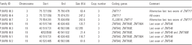
Co-ordinates given are from hg19.
Discussion
In family PJMR1, eight HBD regions were identified (>0.9 Mb), although the five regions on chromosome 3 are all likely part of a single large (97.4 Mb) HBD region. This region harbours the genes PPCB (propionacidaemia; MIM 606054) and SLC9A9 (autism susceptibility gene, AUTS16; MIM 613410). The remaining loci are not reported previously.
The PJMR2 family showed eight homozygous loci, four of which were more than 4 Mb in size, the largest one being 16 MB located on 7p12.1-q11.22 with stretch of 817 SNPs. Two AR ID genes leading to metabolic disruption, PSPH, and ASL, have been reported previously at this locus. The PSPH gene was sequenced by PCR and Sanger sequencing and revealed a homozygous variant, chr7:56087300C>T, NM_004577.3:c.268G>A, p.Gly90Ser, segregating with the phenotype in the family. This variant corresponds to a known SNP, rs75395437, with allele frequency of 0.45%. Amino-acid profiling for affected individuals in PJMR2 revealed plasma serine levels to be within the normal range, and it was concluded that this variant is unrelated to the phenotype. The 10q21.3-q22.1 locus harbours the known recessive phenylalaninaemia gene PCBD1. The14q23.2-q24.1 harbours GPHN – a gene recently linked to autism and schizophrenia – although the causative deletions were heterozygous, suggesting a dominant mode of inheritance (Reference Lionel, Vaags and Sato15). At the 16p13.3 locus, there are several good candidate genes, most notably the recently identified THOC6 gene, which cause a recessive ID syndrome with facies, as well as cardiac and urogenital anomalies [Beaulieu-Boycott-Innes syndrome (BBIS), MIM 613680]. Comparison with photographs of affected individuals from PJMR2 suggests that there are none of the syndromic facial features reported for BBIS.
The PJMR3 family showed two HBD regions of size of 4.192 and 2.301 Mb that are located almost adjacent to each other on chromosome 5p13.3-p13.2 and 5p13.2-p13.1, respectively, and thus may actually be part of a single larger HBD locus. The 5p13.3-p13.2 locus harbours c5orf42 (Joubert syndrome, JBTS17; MIM 614615) as well as the Cornelia de Lange syndrome (CDLS) gene NIPBL. The family show none of the dysmorphic features of CDLS; however, in order to exclude JBTS, magnetic resonance imaging of the patients’ brains would be needed, as milder symptoms, particularly given the young age of the affected family members, may easily be overlooked.
The analysis for family PJMR5 showed HBD on chromosome 11, 14, 15, and 17 (Table 2). The 11q locus harbours genes for a number of known recessive syndromes that include ID; however, the associated phenotypes make it unlikely that these are the aetiologically responsible genes for an apparently non-syndromic family, CHD8 (AUTS18).
Analysis for family PJMR6 gave two HBD regions, one on chromosome 4p15.1; however, this region contains no coding genes, and a second, larger region on chromosome 7q11.21. This locus overlaps with an HBD region for family PJMR2, including the ASL gene. As it is evident from the pedigree (Fig. 1), this family also is compatible with X-linked ID, and thus we also compared haplotypes across all six genotyped affected males, and identified two haplotype regions of interest – a 9.869 Mb locus at the pericentromeric region (Xp11.21-q21; rs6521410–rs471205) and a 1.092 Mb locus on Xq25 (rs149918–rs5976794). The former locus contains a number of genes including the gene ARHGEF9 – a known ID and epilepsy gene (MIM 300429); however, no seizures were reported for this family.
Continued.
Continued.
Pedigrees are shown alongside homozygosity-by-descent plots from HomozygosityMapper [HM; Seelow et al. (Reference Seelow, Schuelke, Hildebrandt and Nurnberg14)] for 11 consanguineous Pakistani intellectual disability families, PJMR1, 2, 3, 5, 6, 7, 8, 9, 10, 11, and 12. The HM output was reviewed at the genotype level for autozygosity (i.e., homozygosity shared between all affected individuals and unaffected individuals) and haploidentity; positive regions are marked with a red arrow, and are described in detail in Table 2.
Analysis for PJMR7 showed five HBD regions, three of which are adjacent to each other on chromosome 1. It is possible that intervening regions may also be HBD, but did not show up in the analysis, possibly because of poor genotype calls or coverage. If this is the case, the region would also include the known NS-ARID gene ACBD6 (Reference Najmabadi, Hu and Garshasbi16). This region also encompasses the gene GLUL, for which mutations can cause glutamine deficiency; however, it seems unlikely that this gene would be involved, as symptoms are severe and multi-systemic.
For PJMR8, we note eight HBD regions above 0.9 Mb in size. In one of these, on 1p12, the α-mannosidase gene MAN1A2 is located. The encoded enzyme plays a similar role to that of MAN1B1, for which a number of mutations have been identified in Pakistani ID families (MRT15) (Reference Rafiq, Kuss and Puettmann17). To date, mutations in MAN1A2 have not been associated with any phenotype, but it represents a very likely candidate gene for this family. Also of note, within the HBD region on 3p22.3-p14.2, a Bardet–Biedl syndrome (BBS) gene is located (L7TFL1, BBS17, MIM 606568). This also represents a good candidate, as milder BBS phenotypes may be difficult to distinguish from NS-ARID without magnetic resonance imaging. Another likely candidate is the phosphoserine aminotransferase 1 gene, PSAT1, on 9q21.2. However, the reported symptoms for the only published case were very severe (Reference Hart, Race and Achouri18); therefore, it would only be a likely candidate for NS-ARID if a less deleterious mutation was identified. PJMR8 is also compatible with X-linkage; therefore, haplotypes were compared across all five males affected using dChip, but no common haplotype was identified.
Analysis of HBD for PJMR9 showed a single large autozygous region spanning the centromere on chromosome 3. This region contains many genes including the ARL13B for Joubert syndrome and ARL6 gene for BBS. CNV analysis also shows a gain CNV spanning the zinc finger gene ZNF780B segregating among all the affected individuals run on the microarray. Validation of this among all family members would be required to include or exclude this as a plausible aetiological genetic variant. In addition, screening ethnically matched controls must be carried out to check whether this CNV is common in Pakistanis. PJMR10 also showed a single HBD region, in this case, on 11p. This region contains the MRT17 and MRT21 gene, PGAP2, which, although representing an excellent candidate gene for this family, has now been excluded by Sanger sequencing (data not shown).
HBD mapping for PJMR11 identified no genuine HBD regions. It may, thus, be likely that there may be an X-linked gene involved in this family. Analysis for PJMR12 – a family with just two affected siblings, and thus less power and likelihood of identifying a single HBD region – identified 11 regions, which include known genes such as BBS5 and SLC25A12 responsible for global cerebral hypomyelination syndrome, regions that overlap the mapped NS-ARID loci for MRT23 and MRT35 (MIM 624344 and 615162, respectively), and mapped quantitative trait loci for intelligence (Table 2).
As it is evident from Fig. 1, many of the top scoring regions from the HomozygosityMapper algorithm are actually excluded upon genotype analysis, whereas some regions score low, but appear upon inspection to be truly HBD. Thus, we may recommend that additional software be developed that can more accurately score HBD in microarray genotype data, and that would be useful for different microarray platforms and arrays.
These findings suggest that we may find a causative gene/mutation at the 7q11.21 locus (PJMR6). In addition, the regions 3p12.3-q13.11 and 11p15.5-p15.3, for PJMR9 and PJMR10, respectively, may be new loci for NS-ARID. Whole-exome sequencing (WES) is the logical next step to identify the aetiological gene and mutation for these families, and is currently underway. The HBD data generated here will help guide the WES analysis, allowing us to prioritise these regions in the search for the causative mutations.
Acknowledgements
The authors are thankful to the families who participated in this research. Muhammad Ayub and John Vincent made substantial contribution to conception and design of this study. Iltaf Ahmad, Attya Bhatti, and Muhammad Arshad Rafiq collected data and contributed in its analysis. Iltaf Ahmad, John Vincent, and Muhammad Ayub drafted the paper and all the other authors critically revised it for intellectual contribution. All the authors approved the draft for publication.
Financial support
This research was supported by a grant from the Canadian Institutes of Health Research (#MOP-102758).
Conflicts of interest
None.
Ethical standards
The authors assert that all procedures contributing to this work comply with the ethical standards of the relevant national and institutional committees on human experimentation and with the Helsinki Declaration of 1975, as revised in 2008, and the ethical standards of the relevant national and institutional guides on the care and use of laboratory animals.