INTRODUCTION
Male spider crabs (Majoidea sensu Ng et al., Reference Ng, Guinot and Davie2008) exhibit complex reproductive strategies related to the condition of the female (Jones & Hartnoll, Reference Jones and Hartnoll1997) and to their morphotype within the population (Sagi et al., Reference Sagi, Homola and Laufer1993; Laufer et al., Reference Laufer, Sagi and Ahl1994). They reach sexual maturity at the terminal moult, exhibiting a disproportionally large cheliped that plays an important role in mating displays (Donaldson & Adams, Reference Donaldson and Adams1989; Sainte-Marie et al., Reference Sainte-Marie, Gosselin, Sévigny and Urbani2008). Another male secondary sexual character, as in other Brachyura, is the modification as gonopods of the first and second pairs of pleopods (Beninger et al., Reference Beninger, Elner and Poussart1991). The gonopods are used in the transfer of spermatophoric masses to the female seminal receptacles during mating events (Bauer, Reference Bauer1986). These organs are rather stable and little affected by environmental variables (Chambers et al., Reference Chambers, Payne and Kennedy1980), a condition that underlies hypotheses on decapod phylogeny as they are significant taxonomic characters (Bauer, Reference Bauer1986; Martin & Abele, Reference Martin and Abele1986).
Early descriptions of Brachyura male gonopods were limited to the gross morphology under dissecting microscopes, although some studies have used scanning electron microscopy (SEM) for detailed observation of microstructures (Beninger et al., Reference Beninger, Elner and Poussart1991; Neumann, Reference Neumann1996; Beninger & Larocque, Reference Beninger and Larocque1998; Brandis et al., Reference Brandis, Storch and Türkay1999; Chen et al., Reference Chen, Cheng, Chen and Hsu2007; Rorandelli et al., Reference Rorandelli, Paoli, Cannicci, Mercati and Giusti2008). The first descriptions dealt only with the first pair of gonopods, although they are part of a complex spermatophore transfer mechanism together with the second pair (Beninger et al., Reference Beninger, Elner and Poussart1991; Brandis et al., Reference Brandis, Storch and Türkay1999). Knowledge concerning the internal anatomy and histology of brachyuran gonopods are mainly focused on the rosette glands located in the first pair (Beninger et al., Reference Beninger, Elner and Poussart1991), for which possible functions have been suggested: hardening of the cuticle (Johnson & Talbot, Reference Johnson and Talbot1987); or a role in reproduction as accessory sex glands (Diesel, Reference Diesel1989; Beninger et al., Reference Beninger, Elner and Poussart1991, Reference Beninger, Ferguson and Lanteigne1995; Beninger & Larocque, Reference Beninger and Larocque1998).
Morphological studies of majoid gonopods are scarce compared to the attention given to the reproductive biology of females (Hartnoll, Reference Hartnoll1965; Diesel, Reference Diesel1989; Beninger et al., Reference Beninger, Lanteigne and Elner1993; Sainte-Marie et al., Reference Sainte-Marie, Sévigny and Carpentier2002; Corgos & Freire, Reference Corgos and Freire2006; Barón et al., Reference Barón, Quiroga, Leal and González-Pisani2009). Brachyuran gonopores, or vulvae, may take one of three forms of closure: (1) a membrane opened by simple muscular action; (2) a soft membrane; or (3) a calcified operculum (Hartnoll, Reference Hartnoll1968, Reference Hartnoll2006; Thompson & McLay, Reference Thompson and McLay2005). Majoid females present vulvae of the concave type (Hartnoll, Reference Hartnoll1968). In mature individuals the opening of the vulva remains permanently soft (Diesel, Reference Diesel, Bauer and Martin1991).
Juvenile females, as well as males, are capable of locking their abdomen using a variety of structures, while in mature or ovigerous females those structures may remain functional, become non-functional scars, or disappear completely (Ng et al., Reference Ng, Guinot and Davie2008). Guinot & Bouchard (Reference Guinot and Bouchard1998) argue that the acquisition of a locking mechanism may be regarded as a synapomorphy for the Brachyura. They showed that in primitive podotremes the locking structures are associated to the coxae of thoracopods, while in eubrachyurans they have moved onto the sternal plate, consisting of a prominence on the fifth thoracic sternite and a socket on the sixth abdominal segment. The Majoidea shows the typical press-button system (Guinot & Bouchard, Reference Guinot and Bouchard1998).
Several majoid species occur in the Argentinean Biogeographic Province (south-western Atlantic; Boschi, Reference Boschi2000), among which Libinia spinosa (Milne Edwards, 1834) is the most abundant along the coast of Buenos Aires Province (Boschi et al., Reference Boschi, Fischbach and Iorio1992; Scelzo et al., Reference Scelzo, Martinez Arca and Lucero2002). Boschi (Reference Boschi1964) published line drawings of the first pair of gonopods of Libinia spinosa. The genitalia and gonadal stages of development were described in detail for both sexes through morphological and histological studies (Sal Moyano, Reference Sal Moyano2007; Sal Moyano et al., Reference Sal Moyano, Gavio and Cuartas2010a, b). Physiological maturity, as defined by the presence of spermatophores in the male vas deferens, occurs before morphological maturity. The latter is determined by the enlargement of the chelae at the terminal moult (Sal Moyano, Reference Sal Moyano2007; Sal Moyano et al., Reference Sal Moyano, Gavio and Maggi2010b). In contrast, the development of mature ovaries in females (physiological maturity) is synchronized with the enlargement of the abdomen at the terminal moult (morphological maturity) (Sal Moyano, Reference Sal Moyano2007; Sal Moyano et al., Reference Sal Moyano, Gavio and Maggi2010b). Reported here are results of investigations on the functional morphology of the copulatory system of L. spinosa based on: (1) the detailed micromorphology of the male gonopods using SEM and histological sections; (2) the female vulva and the abdomen holding system; and (3) differences between mature and immature individuals.
MATERIALS AND METHODS
Crabs were collected along the coast of Mar del Plata (38°S 57°33′W), Argentina, using trawl nets towed by commercial boats, between September 2006 and October 2007. Carapace width (CW) was measured with a digital caliper and used as the reference variable. Comparison of mature and immature individuals was conducted following Sal Moyano et al. (Reference Sal Moyano, Gavio and Maggi2010b); accordingly, males larger than 33.6 mm CW and females larger than 40.3 mm CW were considered physiologically mature.
The gonopods of 15 individuals ranging from 17.6 to 82 mm CW were removed and selected for SEM studies, while those of 28 males ranging from 34.3 to 71.4 mm CW were used for histological sections. The area surrounding the vulvae and holding systems of twenty females (8 to 52.3 mm CW) were dissected and used for SEM. Samples were fixed in 2.5% glutaraldehyde in 0.1 M sodium cacodylate buffer, at pH 7.2–7.4. Samples were dehydrated in a graded alcohol series, dried in hexamethyldisilazane replacing the critical-point, coated with gold-palladium and viewed with a JEOL 6460 microscope. For histological studies, samples were fixed in Bouin's solution for 48 hours. The fixed tissues were dehydrated in a series of graded alcohol solutions, cleared in xylene agent and embedded in paraffin. 5–7 µm serial sections were cut on a rotary microtome. Sections were mounted on glass slides and stained with standard haematoxylin–eosin and Masson's Trichrome for observation under light microscope.
The terminology used mostly follows that of Nishimura (Reference Nishimura1967), Hartnoll (Reference Hartnoll1975), Beninger et al. (Reference Beninger, Elner and Poussart1991), Neumann (Reference Neumann1996), Guinot & Bouchard (Reference Guinot and Bouchard1998) and Garm (Reference Garm2004).
RESULTS
External morphology of the gonopods
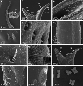
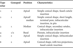
The first gonopod (G1) of mature males larger than 35.1 mm CW is composed of two articles: the basal protopodite and the long, slender distal endopodite that narrows towards its terminal end. The cuticle is ventro-laterally infolded forming a tube that functions as a seminal canal (Figure 1A). The apical tip exhibits two wing-like processes, sternal and abdominal, of different shape. The abdominal process is characterized by the presence of a sub-terminal seminal canal, expanded as a funnel and surrounded by a cuticle girdle of simple short setae, denominated as setae of type 1, characterized by a conical shape and fused-cuticle insertion (Figure 1B, C, D). The sternal process is closed, with a spatula-like ending; the folded cuticle bulges on its surface, having simple, grouped setae of type 1 (Figure 1B, D). Pits are arranged on the external surface of the gonopod, close to the tip of both processes and continuing to the median region. Short, conical setae with terminal pores and infracuticular insertion protrude from the pits; these are referred to as setae of type 2 (Figure 1C, E). The mid-region has two other different types of setae (types 3 and 4) distributed along the gonopod (Figure 1F). Type 3 setae are characterized by a conical shape, and have secondary setules with infracuticular insertions on the median part (Figure 1G). Setae of type 4 are pinnate, with pinnules growing randomly along its axis and with infracuticular insertion (Figure 1H). The latter extend to the basal region, forming dense groups (Figure 1H). Types and position of setae are summarized in Table 1.
Libinia spinosa, scanning electron micrographs of the first gonopod, G1. Mature males: (A) general structure of G1. Inset: transversal section of the median portion showing the cuticle infolded, forming the seminal canal (sc); (B) apical tip with wing-like processes, the sternal process (sp) showing cuticular bulges (cb) holding setae type 1, and the abdominal process (ap) with the seminal canal opening (sco); (C) detailed structure of the seminal canal opening (sco) of the abdominal process, showing setae of type 1 (s1) on its cuticle girdle and setae of type 2 (s2) on its external surfaces; (D) detailed structure of setae of type 1; (E) detailed structure of setae of type 2; (F) mid-region showing setae of type 3 (s3) together with setae of type 4 (s4); (G) detailed structure of setae of type 3, note the setules growing from its median region; (H) general view of grouped pinnate setae type 4. Inset: infracuticular insertion of setae of type 4. Immature males: (I) apical tip showing the shorter sternal (sp) and abdominal (ap) processes; the abdominal one is not funnel-shaped and its tip is closed; (J) surface of the gonopod showing cuticle secretions (cs) arising from the cuticle pores (arrow), and the setae of type 2 (s2); (K) detailed structure of cuticle secretions; (L) colonies of bacteria inside the seminal opening of mature male G1.

Position and characterization of the different types of setae encountered in the first (G1) and second (G2) gonopods of Libinia spinosa males.

The G1 of immature males smaller than 29.9 mm CW present incomplete development of the apical processes mainly the abdominal one, which corresponds to the seminal canal. The whole process is shorter, is not funnel-shaped and the tip is closed (Figure 1I). Secretions extruding through pores are observed over the entire surface of the gonopod (Figure 1J, K). The type and position of setae were similar in immature and mature individuals. Bacterial colonies were observed inside the seminal opening of the G1 abdominal process of some mature individuals (Figure 1L).
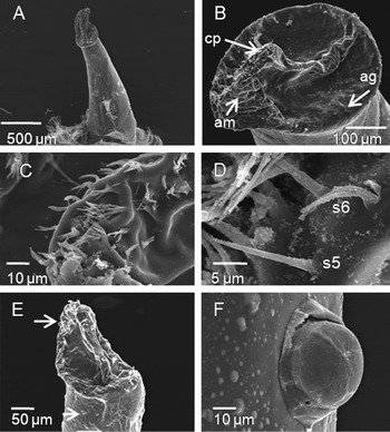
The second gonopod (G2) of mature males is approximately five times smaller than the G1, with the endopodite of a tubular shape (Figure 2A). The distal region consists of an apical girdle, an appendix masculina, and a central protuberance (Figure 2B). The apical girdle has isolated type 5 setae, characterized by conical form and infracuticular insertion. In the central protuberance, type 5 setae are found together with other types of setae, referred to as type 6 setae (Figure 2C, D). These are characterized by the presence of two projections at their base and fused-cuticle insertion. No setae were observed in the median region. The basal region is surrounded by setae of type 4. Types and position of setae are summarized in Table 1.
Libinia spinosa, scanning electron micrographs of the second gonopod, G2. Mature males: (A) general view of G2; (B) apical region showing the central protuberance (cp), the apical girdle (ag) and the appendix masculina (am); (C) detailed structure of the central protuberance holding setae of types 5 and 6; (D) detailed structure of setae of types 5 (s5) and 6 (s6); notice the two projections of setae of type 6. Immature males: (E) general view of G2 with cuticle secretions (arrows); (F) detailed structure of cuticle secretions.

No differences between mature and immature individuals were observed in the general morphology of the G2. Type and position of setae were similar in both mature and immature individuals. Cuticle secretions arising from cuticle pores were observed over the entire length of the G2 of immature males (Figure 2E, F), as in the G1.
Internal anatomy of the gonopods
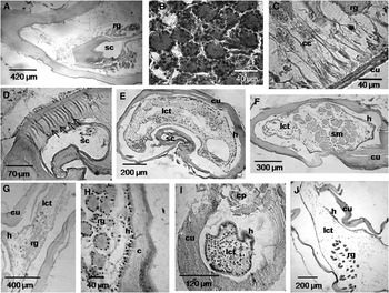
Cross-sections of the G1 showed the arrangement of the infolded cuticle which demarcates the seminal canal (Figure 3). Internally, in its apical region, rosette glands encircle the seminal canal (Figure 3A). Glands are composed of secretory cells with a basal nucleus, radially arranged around a central pore cell, and a canal cell that connects to the secretory surface (Figure 3B, C). Glandular ducts of the rosette glands are observed through the cuticle of the seminal canal (Figure 3D). The median region presents a thick cuticle and a subjacent hypodermis, characterized by a cubic epithelium and loose connective tissue (Figure 3E). The basal region shows the presence of striated muscle, surrounded by loose connective tissue (Figure 3F). Immature and mature individuals show differences in the arrangement and quantity of rosette glands. In immature males they are more abundant and arranged along the entire length of the G1 (Figure 3G, H), while they are restricted to the apical region, surrounding the seminal canal (Figure 3A) in mature individuals.
Libinia spinosa, histological sections of mature and immature male's gonopods. First gonopod, G1, of mature males: (A) transversal section of the apical tip showing the rosette glands (rg) surrounding the seminal canal (sc); (B) detailed structure of rosette glands; (C) detailed structure of the canal cells (cc) of rosette glands (rg) opening through the cuticle (cu); (D) pores (arrows) corresponding to the rosette glands traversing the cuticle of the seminal canal (sc); (E) transversal section of the median region showing the seminal canal (sc), cuticle (cu), hypodermis (h), and loose connective tissue (lct); (F) transversal section of the basal region showing the striated muscle (sm), cuticle (cu), hypodermis (h) and loose connective tissue (lct). G1 of immature males; (G) longitudinal section showing the presence of rosette glands (rg) distributed along the gonopod, cuticle (cu), hypodermis (h) and loose connective tissue (lct); (H) detailed structure of the rosette glands distributed along the gonopod, cuticle (cu) and hypodermis (h). Second gonopod, G2, of mature and immature males; (I) transversal section of the apical tip showing the central protuberance (cp), cuticle (cu), hypodermis (h) and loose connective tissue (lct); (J) longitudinal section showing the presence of rosette glands (rg) distributed along the gonopod, cuticle (cu), hypodermis (h) and loose connective tissue (lct).

The internal anatomy of both immature and mature G2 showed, in their apical regions, the central protuberance bordered by loose connective tissue and the hypodermis (Figure 3I). Only loose connective tissue was observed in the median portion, and striated musculature similar to G1 was recognized in the basal region. Longitudinal sections show the presence of rosette glands distributed along the entire length of the G2 (Figure 3J).
Morphology of the holding system and the vulvae
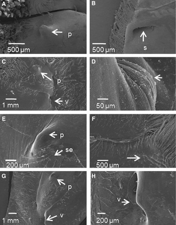
In immature females smaller than 36.1 mm CW the locking system is a typical eubrachyuran press-button composed by an acute cuticle protuberance of the fifth cephalothoracic sternite and a deep socket on the sixth abdominal segment (Figure 4A, B). The vulvae are located near the cuticle protuberance and are of biconcave shape (Figure 4C). The aperture is closed by integument on the ventral side, which bulges resembling an ‘operculum’ (Figure 4D). The holding system and vulvae are surrounded by long pinnate setae, similar to the setae of type 4 present in male gonopods.
Libinia spinosa, scanning electron micrographs of holding systems and vulvae of females. Immature females: (A) well developed sternal protuberance (p) of the holding system; (B) abdominal socket (s) of the holding system; (C) fifth cephalotoracic sternite showing the sternal protuberance (p) and the biconcave vulva (v); (D) detailed structure of the vulva with its aperture closed by integument that bulges (arrow). Mature females: (E) sternal protuberance (p) of the holding system fused with the cuticle and pinnate setae (se); (F) abdominal socket of the holding system fused with the cuticle (arrow); (G) fifth cephalotoracic sternite showing the sternal fused cuticle protuberance (p) and the biconcave vulva (v); (H) detailed of the open biconcave shape of the vulva (v).

In mature females, larger than 42.6 mm CW, the locking system is reduced; the protuberance and the abdominal socket are almost completely fused with the cuticle (Figure 4E, F). The vulvae are open, biconcave in shape, with a more inner flexible membrane hardly seen from the outer surface but easily displaced (Figure 4G, H). Long pinnate setae surround both structures.
DISCUSSION
Copulatory system: mechanism of insertion
Male first gonopods of higher brachyurans are characterized by the complete folding of the cuticle, forming a tube-shaped structure (Hartnoll, Reference Hartnoll1975; Beninger et al., Reference Beninger, Elner and Poussart1991; Neumann, Reference Neumann1996; Brandis et al., Reference Brandis, Storch and Türkay1999) that is inserted in the female vulvae during copulation (Guinot & Quenette, Reference Guinot and Quenette2005). The G1 transfers ejaculates into the female seminal receptacle. Coupled with the G2 it acts as a hydraulic pump to push the seminal fluid through the seminal canal (Beninger et al., Reference Beninger, Elner and Poussart1991). The apical tip of the G1 shows specific variation, and it is an important taxonomic character. While in the genera Chionoecetes (Beninger et al., Reference Beninger, Elner and Poussart1991), Maja (Neumann, Reference Neumann1996) and Inachus (Rorandelli et al., Reference Rorandelli, Paoli, Cannicci, Mercati and Giusti2008) the tip has a single ending, in Libinia spinosa the two wing-like processes, described in detail in this study, have a feature that has been also mentioned for Leurocyclus tuberculosus (Milne Edwards & Lucas, 1842) (Boschi, Reference Boschi1964). In L. spinosa each process has a particular shape and micro-structures, prompting the hypothesis that they have different roles during copulation. The form of the apical tip matches the biconcave shape of the female vulva, suggesting that both G1 processes are inserted during mating. Due to its funnel-like morphology, it is unmistakable that the abdominal process is responsible for the transfer of spermatophores through its seminal canal, clearly visible on the gonopod tip. The sternal process has a complex shape, consisting of a cuticular expansion on its closed terminal portion and a bulging site with simple setae of conical form, close to the proximal branching point of the gonopod's tip. The whole tip of the abdominal process could be inserted into the female vulva and anchored by the structures of the gonopod sternal process described earlier. This fit would keep the abdominal process, which holds the seminal canal, fixed in its position while the ejaculate is being transferred during the copula.
Contrasting hypotheses have been proposed to explain the specific role of morphological structures considered to be involved in the process of gonopod insertion. Jennings et al. (Reference Jennings, McLay and Brockerhoff2000) described the presence of a hook in the G1 process of the ocypodoid Macrophthalmus hirtipes (Heller, 1862), and suggested that it might help the male to be positioned during copulation. The presence and function of that hook resembles the sternal process described here for L. spinosa. Mature majoid females are characterized by the lack of opercula (Hartnoll, Reference Hartnoll1968), as shown also for L. spinosa in this study. It has been suggested that in Chionoecetes opilio (Fabricius, 1788) the insertion of the G1 process is involved in the removal of seminal receptacle contents corresponding to previous mating (Beninger et al., Reference Beninger, Elner and Poussart1991). In L. spinosa that function seems improbable because the histological study of female seminal receptacle revealed the presence of distinct sperm packets, indicative of multiple mating events without removal of previous ejaculate (Sal Moyano et al., Reference Sal Moyano, Gavio and Cuartas2010a).
External anatomy of the gonopods: function of the setae
Distribution of the setae on the gonopods was addressed in a number of studies dealing with brachyuran crabs. Beninger et al. (Reference Beninger, Elner and Poussart1991) attributed functions to the setae based on their position, structure and insertion in the cuticle in C. opilio. Setae of type 1 were found here in the apical tip of the G1 and were similar to the ‘pyramid-like’ setae described in the G2 of three species of the genus Maja (Neumann, Reference Neumann1996), the ‘cuspidate’ setae observed in the maxillae and maxillipeds of Cherax quadricarinatus (Martens, 1868) (Garm, Reference Garm2004) and the ‘tooth-like’ setae described for the G1's apical tip of Inachus phalangium (Fabricius, 1775) (Rorandelli et al., Reference Rorandelli, Paoli, Cannicci, Mercati and Giusti2008). Rorandelli et al. (Reference Rorandelli, Paoli, Cannicci, Mercati and Giusti2008) suggested that the function of these structures surrounding the seminal canal is to break down spermatophores during the transfer to the female seminal receptacle. Such a function seems implausible in L. spinosa due to the fact that setae of this type are located in the abdominal process, surrounding the seminal canal, but also in the sternal process. We suggest a mechanical function: they would allow keeping the gonopod positioned during copulation while inserted in the female vulva. The type 2 setae described here are similar to setae present in the G1 of species of Maja (Neumann, Reference Neumann1996) and in the antennae of the lobster Homarus americanus (Milne Edwards, 1837) (Derby, Reference Derby1982). Derby (Reference Derby1982) proposed that these setae, which emerge from a cuticle depression, have a sensory function. In L. spinosa the distribution of pits with setae of type 2, present only in the mid and apical portions of G1, would indicate that males are capable of sensing female condition during gonopod insertion, and that the setae would be protected from harmful events. Setae of type 3 have been also observed in C. opilio, for which Beninger et al. (Reference Beninger, Elner and Poussart1991) suggested a possible mechanoreceptor role related to their infracuticular insertion, helping to position the gonopod and facilitating the copula. The completely pinnate setae (type 4) observed at the basal region of the G1 of L. spinosa are morphologically similar to those described by Phillips & Macmillan (Reference Phillips and Macmillan1987), Beninger et al. (Reference Beninger, Elner and Poussart1991) and Garm (Reference Garm2004) for other crustaceans. These setae could rub against the abdomen during copulation, while those of the G2 could rub against the protopodite of the first gonopod (Beninger et al., Reference Beninger, Elner and Poussart1991).
The presence of bacterial colonies in the apical opening of the male gonopod seminal canal, reported here for L. spinosa, has been reported before in other species (Benhalima & Moriyasu, Reference Benhalima and Moriyasu2001), as well as inside the female seminal receptacle and on the vaginal wall (Beninger et al., Reference Beninger, Lanteigne and Elner1993; Elner & Beninger, Reference Elner and Beninger1995; Benhalima & Moriyasu, Reference Benhalima and Moriyasu2001). Their presence in C. opilio male gonopods indicates that they infect the female's seminal receptacles during mating (Benhalima & Moriyasu, Reference Benhalima and Moriyasu2001), a phenomenon that is likely to occur also in L. spinosa. In the case of female seminal receptacles the production of melanin by the epithelium has been involved in maintaining the bacterial population, being responsible for the exclusion of opportunistic microbes, or to provide substrates for the sperm to metabolize (Elner & Beninger, Reference Elner and Beninger1992; Beninger et al., Reference Beninger, Lanteigne and Elner1993; Jensen et al., Reference Jensen, Orensanz and Armstrong1996).
The G2 of L. spinosa is short and highly modified, as reported also for Chionocetes and Maja, consisting of an apical girdle, an appendix masculina, and a central protuberance which could represent a vestigial appendix interna. Hartnoll (Reference Hartnoll1975) and Beninger et al. (Reference Beninger, Elner and Poussart1991) suggested that the G2 would be involved in the direct transport of seminal fluid through the seminal canal due to its short size. The apical girdle could act as a seal in the seminal canal, allowing the seminal fluid to be hydraulically pushed down the canal (Beninger et al., Reference Beninger, Elner and Poussart1991). Neumann (Reference Neumann1996) compared the G2 morphology of three species of Maja with C. opilio, concluding that the functional interpretation proposed by Beninger et al. (Reference Beninger, Elner and Poussart1991) could apply to all Majoidea. A special case occurs in freshwater crabs of the genus Potamon, where the G2 is unusually long; ending in a long sclerotized tube, which receives the ejaculate from the lumen of the first gonopod, and transfers it directly to female genital ducts (Brandis et al., Reference Brandis, Storch and Türkay1999).
The type 5 setae, described here for the apical tip of G2, are similar to the long cuspidate setae with cup insertion described by Beninger et al. (Reference Beninger, Elner and Poussart1991) for the first gonopod of C. opilio and by Garm (Reference Garm2004) for the maxilla of C. quadricarinatus. These setae, of infracuticular insertion, could have a role in mechanoreception, probably detecting the movements of G1 during copula and allowing the insertion of G2 inside the seminal canal. Setae of type 6 resemble the complexly branched setae described by Neumann (Reference Neumann1996) as ‘star-shaped’. Because of the fused-cuticle insertion it is difficult to hypothesize on their function. Beninger et al. (Reference Beninger, Elner and Poussart1991) suggested that the non-articulating setae of the G2 probably assist in the pumping of seminal fluids during ejaculation.
Internal anatomy of the gonopods: rosette glands
The internal anatomy of both gonopods showed significant development of the musculature in the basal region, and in the presence of rosette glands. The striated musculature suggests that positioning of the gonopods is adjusted through basal movements. Rosette glands are present in the pleopods of both immature and mature females and in adult males of L. spinosa, as is also the case for the lobster H. americanus (Johnson & Talbot, Reference Johnson and Talbot1987) and the G1 of several species of Portunoidea, Cancroidea, Grapsoidea (Beninger & Larocque, Reference Beninger and Larocque1998) and Majoidea (Diesel, Reference Diesel1989; Beninger et al., Reference Beninger, Elner and Poussart1991). Secretions of these glands could play multiple functions. In females they would serve to maintain the eggs attached to the pleopods (Johnson & Talbot, Reference Johnson and Talbot1987), while in males they could participate in the formation of seminal fluid and sperm plugs (Beninger et al., Reference Beninger, Elner and Poussart1991, Reference Beninger, Lanteigne and Elner1993); they may participate in the moulting process, in both sexes (Gorvett, Reference Gorvett1946; Talbot & Zao, Reference Talbot and Zao1991). In this study, external glandular secretions were restricted to both gonopods of immature individuals, while internal rosette glands were found in mature and immature crabs. Diverse roles could be assigned to rosette glands in the G1. In immature males they are likely to play a role in the moulting process (Gorvett, Reference Gorvett1946; Stevenson, Reference Stevenson1961; Arsenault et al., Reference Arsenault, Clattenburg and Aiken1979) and in the hardening of the new cuticle after the moult (Johnson & Talbot, Reference Johnson and Talbot1987). Majoid crabs are characterized by the presence of a terminal moult, usually coinciding with the puberty moult, after which mature individuals cease moulting. For that reason the most likely function of the rosette glands in mature L. spinosa males may be their participation in the formation of seminal fluid, as they are arranged surrounding the seminal canal. The abundant glands, localized along the entire length of the gonopods of immature crabs, would be restricted after the puberty moult to the seminal canal, their role changing in the transition from the puberty to the adult stage.
In G2, as in G1, the presence of cuticle secretory products in immature individuals is an indication of their role in the moulting process, while in mature ones secretions would act as a lubricant fluid, facilitating the displacement of G2 inside G1 during ejaculation.
Female holding system and vulva
In the immature females of L. spinosa the abdominal locking system has a pointed button and a deep socket, but both structures change in the course of successive moults. The button remains smooth and the socket disappears in mature females, and so the abdomen cannot be fastened to the sternum, implying that females are morphologically ready to mate. According to metabolic advantage explanations, the presence of a locking system in immature females enhances their mobility (Ng et al., Reference Ng, Guinot and Davie2008). Most brachyuran mature females lack the locking system, and their abdomen enlarges to form a brood chamber carrying the developing eggs. An exception, for instance, is the Parthenopidae, in which females have a well developed locking system. This is intriguing because parthenopid females hold large egg masses (Ng et al., Reference Ng, Guinot and Davie2008).
Following the puberty moult, the vulvae reach the size required to permit insertion of the male gonopods during mating in the Majoidea (Hartnoll, Reference Hartnoll1965). In the case of L. spinosa not only the size of the gonopods but also their morphology would be essential for the intrusion of the two processes in the bifid gonopod tip. Immature females showed the presence of a bulged portion covering the posterior part of the vulva which, together with the locking device, would restrict the access of males and prevent mating.
Copulatory system of immature and mature individuals: relationship to morphological and physiological maturity
Males of Libinia spinosa mature physiologically at a small size, approximately at 33.6 mm CW (Sal Moyano, Reference Sal Moyano2007; Sal Moyano et al., Reference Sal Moyano, Gavio and Maggi2010b). Morphometric maturity is reached on average at 58.9 mm CW, following terminal moult (Sal Moyano, Reference Sal Moyano2007; Sal Moyano et al., Reference Sal Moyano, Gavio and Maggi2010b). It was observed that the morphological characters shown by the ‘mature gonopods’ appear coincidentally with the development of spermatophores inside the vas deferens. This character thus develops synchronously with the acquisition of physiological maturity, not with morphological maturity. In females, in contrast, physiological and morphometric maturity are reached simultaneously at an average size of 40.3 mm CW (Sal Moyano, Reference Sal Moyano2007; Sal Moyano et al., Reference Sal Moyano, Gavio and Maggi2010b), which coincides with the acquisition of the mature holding systems and vulvae described here. An experimental study of mating behaviour currently under way will shed new light on inferences made on the basis of comparative morphology.
ACKNOWLEDGEMENTS
We particularly thank Dr José María (Lobo) Orensanz for his thorough reading of the manuscript. His comments and suggestions helped us to improve this manuscript as well as our English writing. We gratefully acknowledge two anonymous referees whose critical comments improved this manuscript. This research was supported by the: ‘Biología de Crustáceos’ project of the Universidad Nacional de Mar del Plata, Argentina, to M.A.G. M.P.S.M. was supported by a fellowship from Consejo Nacional de Investigaciones Científicas y Técnicas (CONICET).