Introduction
Cryptic diversity can be defined as the presence of two or more species that are difficult to recognise based solely on morphology (Bickford et al. Reference Bickford, Lohman, Sodhi, Ng, Meier and Winker2006). Among aphids (Hemiptera: Aphididae), there are numerous instances where complexes of similar species have been recognised based on host associations and other biological differences. The evolutionary separation of host-associated lineages is facilitated by the nature of typical aphid life cycles, especially the alternation of several asexual generations that permit amplification of particular genotypes, with a single sexual generation, permitting a relatively narrow temporal window for genetic exchange. Well-known examples include the Aphis fabae Scopoli species group (Jörg and Lampel Reference Jörg and Lampel1996), and the group of species related to Aphis gossypii Glover and Aphis frangulae Kaltenbach (Vanlerberghe-Masutti and Chavigny Reference Vanlerberghe-Masutti and Chavigny1998; Margaritopoulos et al. Reference Margaritopoulos, Tzortzi, Zarpas, Tsitsipis and Blackman2006; Cocuzza and Cavalieri Reference Cocuzza and Cavalieri2014). The advent of DNA sequencing has provided a new tool for the analysis and interpretation of variation within and among species. In several cases, it has been noted that DNA barcodes (mitochondrial cytochrome oxidase sequence) for a species form multimodal clusters suggesting the possibility of undetected sibling species. For example, it was found that barcode sequence differences within Pentalonia Coquerel correspond to differences in morphology and host usage leading to the resurrection of Pentalonia caladii van der Goot as separate from Pentalonia nigronervosa Coquerel (Foottit et al. Reference Foottit, Maw, Pike and Miller2010b). Pemphigus Hartig samples identified as Pemphigus betae Doane have been shown to represent three species, one currently unnamed (Foottit et al. Reference Foottit, Floate and Maw2010a). Brachycaudus helichrysi (Kaltenbach) consists of two cosmopolitan groups with differences in life cycle (Piffaretti et al. Reference Piffaretti, Vanlerberghe-Masutti, Tayeh, Clamens, Coeur d’Acier and Jousselin2012).
Fitch (Reference Fitch1866) described Brysocrypta rhois Fitch from galls on sumac (Rhus Linnaeus; Anacardiaceae) collected at Salem, New York, United States of America. Walsh (Reference Walsh1867) proposed the genus Melaphis Walsh for this species. Theobald (Reference Theobald1918) described an apterous aphid, Truncaphis newsteadii Theobald, from moss in the United Kingdom, which Eastop (Eastop and Hille Ris Lambers Reference Eastop and Hille Ris Lambers1976; Eastop Reference Eastop1977) considered to represent the secondary host form of Melaphis rhois. Baker (Reference Baker1919) reared alate aphids from larvae collected on moss in West Virginia, United States of America, which he described as Melaphis minutus Baker, now also considered a synonym of M. rhois (Eastop and Hille Ris Lambers Reference Eastop and Hille Ris Lambers1976; Remaudière and Remaudière Reference Remaudière and Remaudière1997). The genus Melaphis thus currently contains the single species, M. rhois. It is the only North American member of subtribe Melaphidina (Eriosomatinae: Fordini), otherwise represented by six genera in Southeast Asia. All host-alternating Fordini use species in Anacardiaceae as primary hosts and all Melaphidina are believed to alternate between Rhus species and mosses (Moran Reference Moran1989; Blackman and Eastop Reference Blackman and Eastop2006, Reference Blackman and Eastop2017; Yang et al. Reference Yang, Chen, Feng and Chen2009). Moran (Reference Moran1989) and Ren et al. (Reference Ren, Zhong, Kurosu, Aodi, Ma, von Dohlen and Wen2013) have discussed the biogeographic aspects of this host association.
Yang et al. (Reference Yang, Chen, Havill, Feng and Chen2010) included Melaphis in a phylogenetic analysis of the Melaphidina and Li et al. (Reference Li, Jiang and Qiao2014) included it in an analysis of Eriosomatinae. The genus has been included in several studies treating the obligatory bacterial aphid endosymbiont Buchnera aphidicola Baumann (Enterobacteriaceae) (Moran et al. Reference Moran, Munson, Baumann and Ishikawa1993, Reference Moran, von Dohlen and Baumann1995; Clark et al. Reference Clark, Moran and Baumann1999; Nováková et al. Reference Nováková, Hypša, Klein, Foottit, von Dohlen and Moran2013).
Hebert et al. (Reference Hebert, Finston and Foottit1991) examined allozyme variation among Melaphis samples collected in Ontario, Canada, revealing two groups differing in allele frequency profiles. Preliminary DNA barcoding of Melaphis (sequences included in Foottit et al. Reference Foottit, Maw, von Dohlen and Hebert2008 and Pike et al. Reference Pike, Graf, Foottit, Maw, von Dohlen and Harpel2012) showed two haplotype clusters. This study takes an integrative taxonomic approach and combines molecular, morphological, and life history data in the examination of variation in this genus in order to assess the significance of these molecular observations.
Materials and methods
Galls were collected in September or early October of 1991–1994, and in 2000, 2008, and 2009, from 72 sites in southeastern Ontario and southwestern Québec, Canada. These sites include the majority of the locations sampled by Hebert et al. (Reference Hebert, Finston and Foottit1991). Collections were also made in Nova Scotia, Canada in 1993 and 2008 and at State College, Pennsylvania, United States of America in 2013. The aphids from each gall were removed and stored dry at −80 °C (1991–1994 samples) or in 95% ethanol (2000–2013 samples). Apterae on moss, sexuparae, sexual forms, and fundatrices were collected in southeastern Ontario and western Québec in spring of 1994 and 2014. This material was supplemented by additional collections from Québec (C. Pilon), DNA from collections in Georgia and Arizona, United States of America (N. Moran and C. von Dohlen), and by specimens collected from moss in Washington State, United States of America (K.S. Pike). Specimen collection data are available on the Barcode of Life Datasystems (Ratnasingham and Hebert 2007) as data set DS-MELAPHIS (www.boldsystems.org/index.php/Public_SearchTerms?query=DS-MELAPHIS).
Additional slide-mounted specimens from the Canadian National Collection of Insects, Arachnids, and Nematodes (CNC; Ottawa, Ontario, Canada) and the United States National Museum of Natural History aphid collection (USNM; Beltsville, Maryland, United States of America), including a syntype of M. minutus Baker, were also examined. Measurements for a second syntype of M. minutus Baker were taken from images available on the National Museum of Natural History, Department of Entomology (Washington, District of Columbia, United States of America) electronic collection database at http://collections.nmnh.si.edu/search/ento.
DNA sequencing
DNA was extracted from individual whole bodies using one of three methods: EDNA HiSpEx (Fisher Biotech, Perth, Australia) (following manufacturer’s directions, except using 1/8 of recommended volumes), leaving the body intact for subsequent mounting; modified CTAB/phenol/chloroform extraction; or procedures described in Hajibabaei et al. (Reference Hajibabaei, Singer, Hebert and Hickey2007) for those specimens processed by Biodiversity Institute of Ontario (BIO; University of Guelph, Guelph, Ontario, Canada).
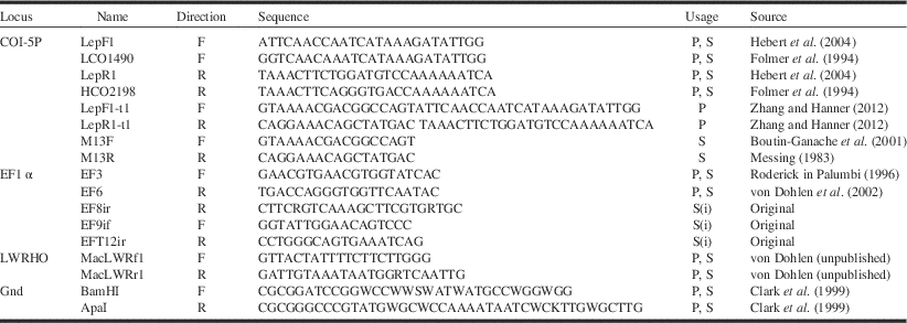
Mitochondrial cytochrome oxidase subunit I, 5' end (COI-5P; 658 base pairs after primer trimming; “DNA barcode region”) was amplified from one specimen from each gall (multiple specimens from some galls, especially those that had begun to open at the time of collection, in order to confirm within-gall uniformity). For subsets of specimens, fragments of the following loci were also amplified: nuclear elongation factor 1α (EF1α; 1046 base pairs), nuclear long-wavelength opsin (LWRHO; 1012 base pairs); and 6-phosphogluconate dehydrogenase (gnd; 892 base pairs) of the obligate bacterial endosymbiont B. aphidicola. Buchnera aphidicola is vertically transmitted transovarially (Douglas Reference Douglas1989; Rouhbakhsh et al. Reference Rouhbakhsh, Lai, von Dohlen, Clark, Baumann and Baumann1996) and thus at a whole-body level may be conceptually treated as a maternally inherited part of the genome of the host organism. Primers for amplification by polymerase chain reaction and sequencing are given in Table 1.
Primers used in this study.

Note: Direction: F=forward; R=reverse relative to sense strand. Usage: P=polymerase chain reaction; S=sequencing; S(i)=internal sequencing primer. M13F and M13R used to sequence amplicons produced using LepF1-t1 and LepR1-t1, respectively.
Direct sequencing of amplicons other than COI-5P was performed at the Ottawa Research and Development Centre, Agriculture and Agri-Food Canada, using BigDye Terminator v.3.1 Cycle Sequencing Kit (Applied Biosystems, now a part of Thermo Fisher Scientific, Watham, Massachusetts, United States of America) on an Applied Biosystems 3130xl Genetic Analyzer. COI-5P amplicons were sequenced at the Ottawa Research and Development Centre, National Research Council of Canada (Saskatoon, Saskatchewan, Canada), or Biodiversity Institute of Ontario.
Sequence assembly and alignments were done using Geneious 6.1.7 (Biomatters Limited, Auckland, New Zealand) or CodonCode Aligner Ver2.0.6 (CodonCode, Centerville, Massachusetts, United States of America) (sequences processed by the Biodiversity Institute of Ontario), with adjustments by hand as necessary.
Additional COI-5P sequences for Melaphis and as well as for East Asian members of Melaphidina were obtained from GenBank (accession numbers given on Fig. 2). Also retrieved from Genbank were EF1α sequences for Melaphis (accession numbers FJ215685, KF601614, KF601615, KF601630) and other Melaphidina (DQ499611–DQ499615, DQ499618, DQ499619), LWRHO sequences for Schlechtendalia Kirkaldy (JX559467, JX559468), and Buchnera gnd (accession AF129280 from Melaphis).
Resulting COI-5P sequences were digested in silico using Geneious 6.1.7 in order to find restriction enzymes providing fragment profiles reflecting observed sequence clustering patterns. Candidate restriction enzymes (Fermentas Life Sciences, now part of Thermo Fisher Scientific) were then tested on polymerase chain reaction products following manufacturer directions and results visualised using standard agarose gel electrophoresis.
Measurements
Representative specimens from galls were mounted on microscope slides in Canada Balsam using methods described at www.zoology.ubc.ca/~mawe/bcaphid.
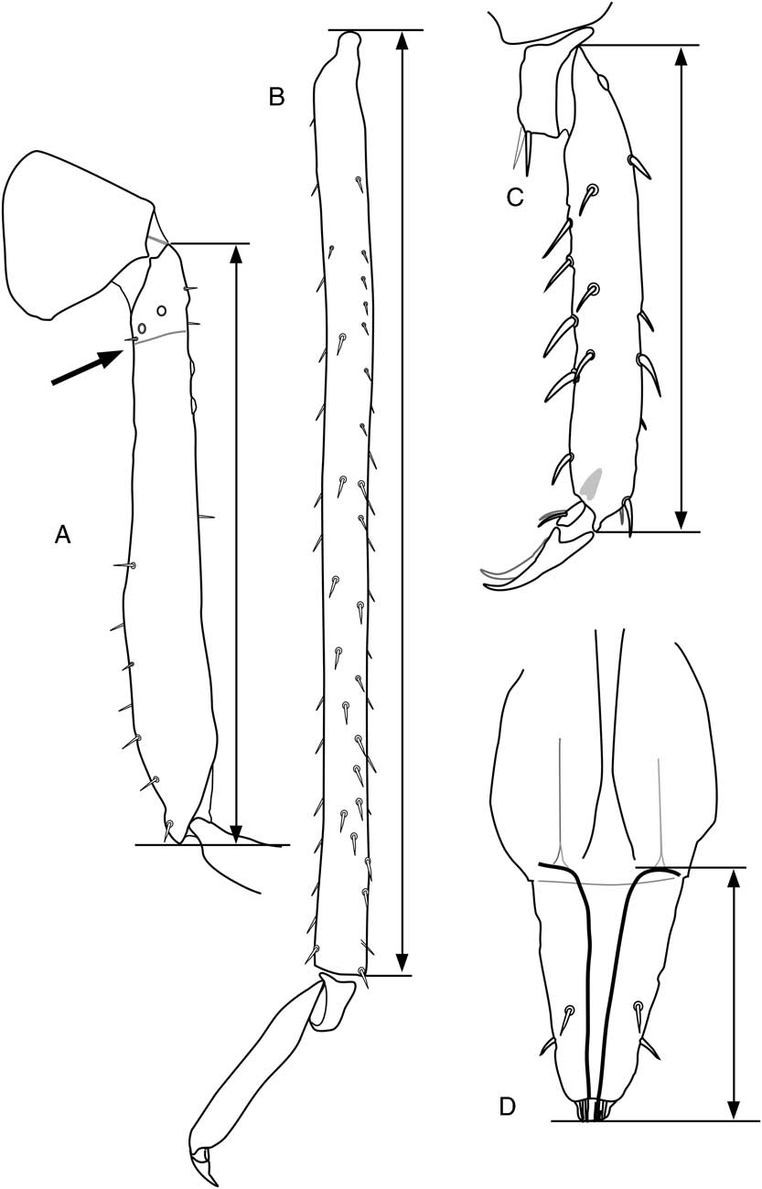
Slide-mounted specimens of winged individuals and wingless adults from mature galls were measured using the Measurement Module of Leica Application Suite (version 4.2; Leica Microsystems, Wetzlar, Germany) software from a live image captured from a Leica DFC450 camera attached to a Leica DM2500 microscope. The location of the femoro-trochantral junction was not apparent in all forms, and therefore “femur length” is taken as the linear distance from the ventro-proximal angle of the trochanter to the dorso-apical angle of the femur (Fig. 1A). Length of the tibia was taken as the greatest linear distance from the most proximal point of the basal condyle to the apico-ventral angle (Fig. 1B). Length of distitarsi excludes pretarsal structures (claws) but includes the apical condyle on which the claws articulate (Fig. 1C). For morphs in which the basitarsus and distitarsus are fused, the proximal point of the measurement was taken as the basi-dorsal angle of the fused tarsus. Length of the terminal antennomere includes the short terminal process. Ultimate rostral segment was measured on anterior face from apex to a point even with the insertions of the retractor muscles on the basal arches (Fig. 1D).
Endpoints for selected measurements. A. Metafemur+trochanter (femoro-trochanteral commissure at arrow). B. Metatibia. C. Distal metatarsus. D. Ultimate rostral segment.

All statistical analyses were performed in R v 3.0.2 (R Core Team Reference Ratnasingham and Hebert2014). Principal component analysis was done using function prcomp of the standard R stats package. Principal component analysis transforms a set of variables into a new set of uncorrelated variables (components) formed as linear combinations of the original variables, such that each successive variable accounts for the maximum amount of remaining total variation in the data set (Tabachnick and Fidell Reference Tabachnick and Fidell2001). A linear discriminant analysis (similar to principal component analysis, but operating on means of groups defined a priori), as implemented by the R functions lda and predict of package MASS (Venables and Ripley Reference Venables and Ripley2002) was used to establish appropriate discriminant functions for distinguishing groups.
Photographic illustrations were made using a DS-Fi1 camera mounted on an Eclipse E800 microscope (Nikon, Minato, Japan), and focal-plane stacks combined using CombineZM software (Alan Hadley, freeware, available at various software download sites) followed by manual editing.
Results
COI-5P
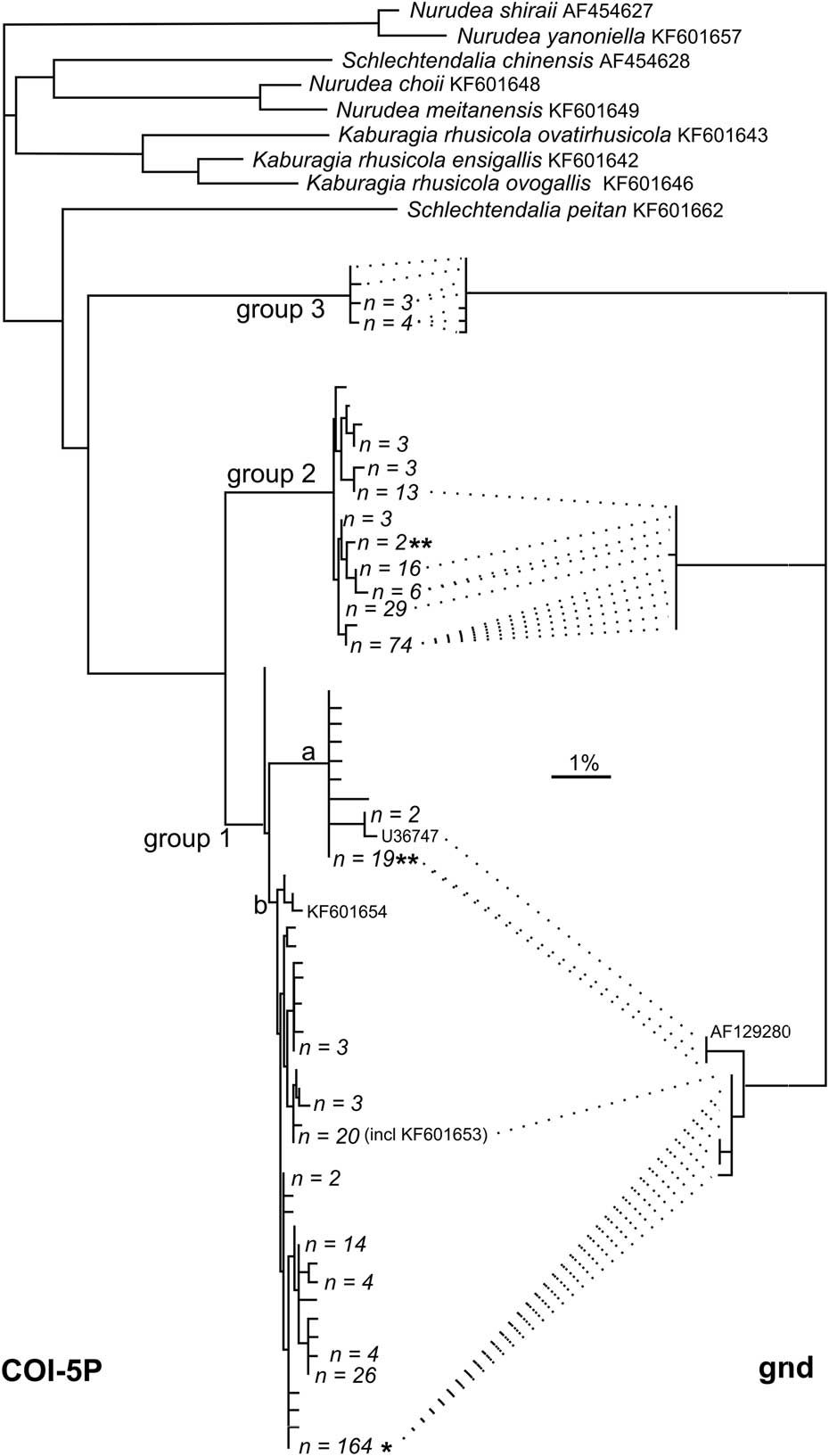
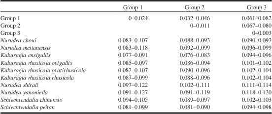
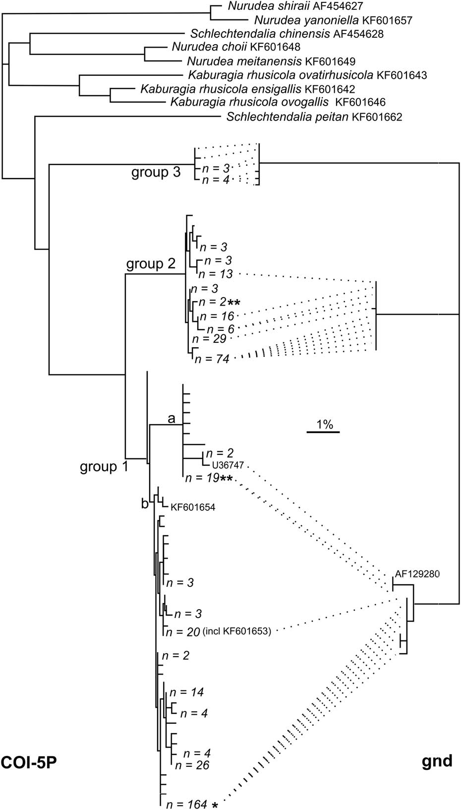
The 456 COI-5P sequences obtained for Melaphis samples show 83 variable sites that contribute to the recognition of three distinct clusters (hereafter referred to as COI-5P Groups 1, 2, and 3), shown in Figure 2. Groups 1 and 2 are geographically widespread, while specimens from Group 3 were collected only at State College, Pennsylvania. Minimum pairwise divergences (uncorrected distances) among groups, along with Asian Melaphidina, and maximum pairwise divergences within groups are given in Table 2. The three North American groups are more similar to each other than to any of the Chinese species. There are 28 fixed base differences differentiating Group 3 from Groups 1+2. Groups 1 and 2 are distinguished from each other by fixed differences at 13 positions, and have different bases dominant at six additional sites.
Neighbour-joining dendrogram summarising sequence divergence among sequences for Melaphis mitochondrial cytochrome oxidase subunit 1 (COI-5P, left side) and 6-phosphogluconate dehydrogenase (gnd, right side) for endosymbiont Buchnera aphidicola from Melaphis specimens. Placements of three Melaphis sequences and one Buchnera sequence from GenBank are indicated. Additional COI-5P sequences for other Melaphidina species are included for comparison. Terminal nodes for COI-5P represent unique haplotypes with the number of specimens associated with each haplotype indicated when greater than 1. All terminal nodes for gnd represent individual sequences. Dotted lines link gnd sequences with the COI-5P haplotype associated with the corresponding Melaphis specimen. Asterisks indicate placement of five specimens collected from mosses. Group 1 corresponds to sequences for specimens here assigned to Melaphis rhois and group 2 corresponds to Melaphis asafitchi. Letters “a” and “b” indicate subgroups of group 1.
Uncorrected pairwise divergence (range as per cent) within and among COI-5P groups of Melaphis, and comparison to Asian Melaphidina as represented by the GenBank accessions (from Ren et al. Reference Ren, Zhong, Kurosu, Aodi, Ma, von Dohlen and Wen2013) given in Figure 2.

Within Group 1, two subgroups, diverging by about 1%, may be recognised (labelled “a” and “b” in Fig. 2). Although Subgroup 1a is rare in collections in Canada (found by us in southern Ontario twice only; additional sequences in BOLD from Pukaskwa National Park, Ontario and Cape Breton, Nova Scotia are not shown), the majority of Group 1 specimens from Pennsylvania, and those from Arizona and Washington State belong to this subgroup.
Base changes predict three amino acid substitutions in Group 3 relative to Groups 1 and 2. Within Group 1, two specimens exhibit different single amino acid changes.
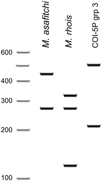
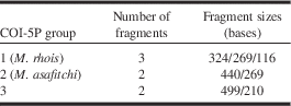
Digestion of the COI amplicon using restriction enzyme HinfI (palindromic 5-base recognition sequence GAWTC) yields a characteristic fragment profile for each COI-5P group (Table 3, Fig. 3) with only one exception. This digest thus provides a convenient means of identifying group membership. Other candidate enzymes yielded more fragments, making recognition of group membership less obvious, or provided several profiles for each sequence group.
Representation of fragment size HinfI restriction enzyme digest of COI-5P amplicon for each Melaphis haplotype group (in silico digest as implemented in Geneious 6.1.7, Biomatters).

Fragment sizes for Melaphis haplotype groups resulting from HInfI restriction enzyme digest of standard COI-5P (DNA barcode) polymerase chain reaction product (including primers).

COI sequences are available on Barcode of Life Datasystems in BOLD data set DS-MELAPHIS. Sequences are also available on GenBank (accession numbers EU701748–EU701750, GU667108–GU667239, GU667484, GU668188, GU668200, GU668828–GU668945, JN201208, and KY754285–KY754476).
gnd
Pairwise divergences among sequences for B. aphidicola gnd were strongly trimodal with well-separated clusters. These partitions (right side of Fig. 2) correspond to those defined by COI-5P. Of the 93 variant sites among the 892 base pairs of the trimmed amplicon, 48 base states are diagnostic for Group 3, 20 for Group 2, and 14 for Group 1. In addition, three sequences (including GenBank accession AF129280) differed somewhat (three base differences) from the remaining sequences in Group 1, corresponding to samples in COI-5P Subgroup 1a and thus reflecting the pattern exhibited by COI-5P. Sequences are available on GenBank with accession numbers KY694633 to KY694661.
EF1α
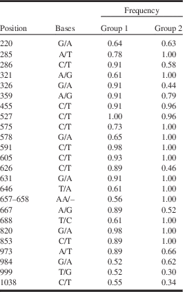
EF1α sequences showed eight nucleotide differences between the four sequenced specimens corresponding to COI-5P Group 3 and all other samples. There were no fixed differences between specimens assigned to COI-5P sequence Groups 1 and 2. However, there are differences in allele frequencies for the observed variant sites (Table 4). In particular, of the 24 variant sites among pooled Group 1 and Group 2 samples, there are 12 sites (including a two base deletion) at which at least 20% of Group 1 specimens differ from the consensus, while Group 2 specimens are uniform at these positions. This locus thus supports the recognition of Group 3 as a separate entity and is consistent with the partitioning of Group 1 from Group 2.
Elongation factor 1-alpha sequences of Melaphis species.

Note: Base frequencies at variant positions for specimens classified by COI-5P sequence group. Frequency is for first variant listed, with variant bases ordered with dominant allele among specimens of group 1 given first. “Position” is the base position in the consensus alignment of all trimmed sequences.
Translation of the protein-coding segments of the sequenced fragment revealed no changes in amino acid composition (all three groups).
EF1α sequences for samples corresponding to COI-5P Group 3 are more similar to the other North American samples (<2.5% uncorrected divergence) than to sequences in GenBank for the Chinese species (3.5–6.5% divergence). Sequences are available on GenBank with accession numbers KY694531 to KY694584.
LWRHO
The long-wavelength opsin amplicon contains four introns at the same position as found in other aphids (Ortiz-Rivas et al. Reference Ortiz-Rivas, Moya and Martínez-Torrres2004). The single opsin sequence corresponding to COI-5P Group 3 differed from the remaining sequences by about 1% (uncorrected distance 0.8–1.2%). Variation among Group 1+2 samples show divergences of 0–0.4%. There are no fixed differences separating Groups 1 and 2, with inter-group divergences of 0–0.4%. Three polymorphic sites are shared (including one synonymous substitution in exon 5). However, Group 2 is polymorphic at two positions at which Group 1 is uniform. Group 1 is polymorphic at one base position, and three individuals were heterozygous for a two base insertion/deletion in intron 2, while Group 2 samples are uniform at these sites. The sequence for Group 3 is much more similar to the other North American samples than to the available Schlechtendalia Lichtenstein species sequences (GenBank JX559467, JX559468, which differ from Group 3 sequence by 7.0% and 6.3%, respectively). The sequences are available on GenBank with accession numbers KY694585 to KY694632.
Morphometrics
Alate and apterous individuals with both five and six antennomeres were present. During larval development in aphids, early instars usually have fewer antennomeres than the adult form, with additional antennomeres being added by subdivision of the basal flagella. Thus, for antenna with five antennomeres, antennomere 3 was considered homologous with antennomeres 3+4 of antennae with six antennomeres, and thus the terminal two antennomeres of all individuals were always treated as antennomeres 5 and 6. In addition, six apterae had one antenna with four antennomeres (antennomeres 3–5 fused). When asymmetrical within an individual, the antenna with the larger number of antennomeres was measured. In addition to the characters shown in Table 5, protibia, metafemur (including trochanter), and, for alatae, various wing measurements were made. In alates, lengths of the protibia and metafemur were highly correlated (r 2>0.95) with the length of the metatibia and were excluded as redundant for purposes of multivariate analysis (but the length of the metafemur is included in descriptive measurements). Preliminary principal component analysis including wing measurements, as well as analysis of wings alone suggested that distribution of wing characters among individuals of the two COI-5P haplotype groups was similar. In addition, there was difficulty in consistently determining endpoints for wing measurements. Thus the wings were excluded from subsequent analyses. In apterae, correlation between the length of the metatibia and metafemur was lower and both measurements were retained.
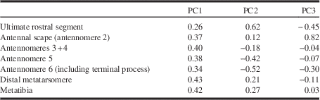
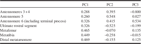
Loadings for first three components (PC) for principal component analysis of fundatrispuriae (alatae emerging from galls) of Melaphis.

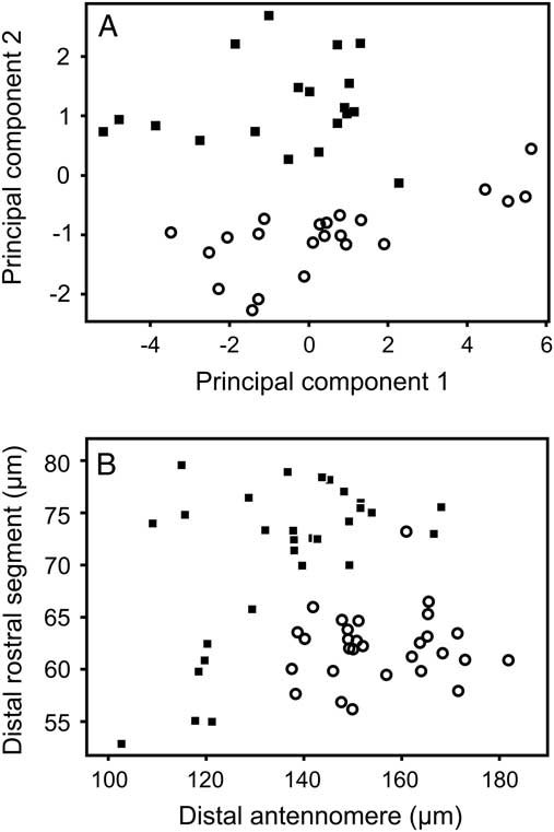
Component loadings for a principal component analysis for all alate specimens from galls (treating combined length of antennomeres 3 and 4 of antenna with six antennomeres as equivalent to length of fused antennomeres 3+4 of the antennae with five antennomeres) are given in Table 5. Component 1 is plotted against component 2 in Figure 4A, with points labelled a posteriori according to the COI-5P sequence group of the corresponding specimen. Component 1 represents a general size axis (all coefficients of similar magnitude and sign). Component 2 contrasts antennal length with rostral and leg measurements, and captures variation corresponding to differences between specimens of the two COI-5P sequence groups. As principal component analysis does not take into account any classificatory variables, the separation of individuals across the joint distribution according to sequence groups is a strong indication that the expression of variation within these groups is independent. No patterns were detected among the remaining components and these likely express combined individual variation and measurement error.
Ordination of alate individuals of Melaphis species from galls on Rhus. A. Ordination on first two components of principal component analysis. B. Bivariate plot of length of distal rostral segment against length of terminal antennomere. Points labelled a posteriori with symbols representing COI-5P sequence group: square, group 1; circle, group 2.

A second analysis including only specimens with at least one antenna with six antennomeres, with antennomeres 3 and 4 treated as separate variables, yielded similar results (not shown).
Figure 4B compares the length of antennomere 6 (A6) with length of the ultimate rostral segment for alatae from galls. For the most part, it is apparent that use of these two features in combination can be used as a simple classifier ([21 – [ultimate rostral segment]+0.34×[ultimate antennomere]] <0 for COI-5P Group 1 and >0 for Group 2) for the majority of specimens. The cluster of specimens at the lower left of the plot represent dwarfed individuals from a small dry gall growing at a dry location (Group 1 points at the right side of Fig. 4A also represent these individuals). No similarly dwarfed specimens of COI-5P Group 2 were available, so it is not yet clear if dwarfed specimens can be distinguished by this combination. If dwarfed individuals are excluded, then the rostral measurement alone (using a threshold of about 68 µm) is sufficient to separate the majority of individuals into the proper group. Based on application of a discriminant function analysis, all examined individuals of known haplotype may be separated by the value of the function [0.03×[total length of flagellum]−0.14×[ultimate rostral segment]−0.09×[distal metatarsomere]+3.0]; negative values correspond to COI-5P Group 1, positive values to Group 2.
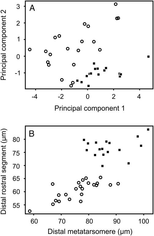
Component loadings for a principal component analysis of fundatrigeniae (combined length of separated antennomeres 3 and 4 treated as equivalent to length of fused antennomeres 3+4) are given in Table 6 and components 1 and 2 plotted in Figure 5A. As for alates, the haplotypes fall into separate regions of the joint distribution. Component 1 represents generalised size. Component 2 is dominated by contrast of the lengths of the ultimate rostral segment and leg parts with the lengths of the antennomeres. Component 3 is dominated by contrast between the basal flagellae (antennomeres 3+4) and the ultimate antennomere (antennomere 6). The two haplotypes also respond differently along this component but the separation is less distinct.
Ordination of apterous individuals of Melaphis species from galls on Rhus. A. Ordination on first two components of principal component analysis. B. Bivariate plot of length of distal rostral segment against length metatibia. Points labelled a posteriori with COI-5P sequence: square group, 1; circle, group 2.

Loadings for first three components (PC) for principal component analysis of fundatrigeniae (apterous offspring of fundatrix in galls) of Melaphis species.

The length of the ultimate rostral segment and the metatibia for adult apterous individuals in galls are shown in Figure 5B. There is no overlap in rostral length of individuals of the two haplotype groups and thus rostral length is diagnostic for this morph of these groups.
Phenology of gall maturation
Group 3 galls collected at State College, Pennsylvania, on 22 August 2013 contained only apterae and young larvae. In contrast, Group 1 and Group 2 galls from the same site and date contained mainly fourth instar alatoids or a mix of such instars and alates. Gall samples corresponding to Groups 1 and 2 do not show any difference in timing of gall maturation and production of alates; individuals within galls corresponding to each of these groups, collected on the same dates throughout September in 1994 and 2009, ranged from all alates, through mixes of alates and fourth instars, to galls containing all instars, often at the same site.
Taxonomic treatment
The concordance of various molecular loci and morphometric data suggests that the three main groups identified here represent distinct species. We treat Group 1 as Melaphis rhois, for reasons given later, and describe Group 2 as Melaphis asafitchi new species. The current generic classification of Melaphidina is based on the morphology of winged adults. As Group 3 has no available associated alate specimens, and its life history is unknown, we will defer taxonomic treatment of this group until additional material becomes available. The following generic description, other than the apterous second generation in the gall, is based only on specimens assignable to Groups 1 and 2.
Melaphis Walsh, Reference von Dohlen, Kurosu and Aoki1867
Type species. Brysocrypta rhois Fitch, Reference Fitch1866.
Synonym. Truncaphis Theobald, Reference Theobald1918 (type species Truncaphis newsteadii Theobald, Reference Theobald1918).
Life history. The Melaphis species described here have a complex life cycle involving alternation between Rhus species and mosses, with at least five parthenogenetic generations and one sexual generation, exhibiting nine functionally and morphologically distinct body forms: (1) the biparental fundatrix that initiates gall formation on the primary host with two functional forms – a vagile first instar and relatively sessile later instars; (2) the wingless offspring of the fundatrix (fundatrigenia, possibly more than one generation); (3) a winged generation that leaves the gall and migrates to the secondary host (fundatrispuria or emigrant); (4) one or more (at least facultatively) wingless generations on the secondary host (alienicola or exulis) with two different types of first instars: a vagile black form deposited by the fundatrispuria, and a yellow, less active form deposited by the adult alienicolae; (5) a winged generation that migrates from the secondary host to the primary host (sexupara or remigrant); (6) the sexual generation (male and sexual female) that lack wings and mouthparts, the female producing only one offspring. Unlike other host-alternating Eriosomatinae, migration to the primary host occurs in the spring, rather than the autumn; thus the sense of the terms “spring migrant” and “fall migrant” commonly applied to host-alternating aphid life cycles is reversed in this group, and will be avoided.
Migration from moss begins in mid-May in Ontario and west Québec, with sexuparae forming loose aggregations and depositing sexuals on previous year twigs of Rhus at the time that expansion of current year shoot buds begins. Small aggregations tend to occur within a few centimetres below the expanding leaf, but large aggregations may consist of >100 individuals scattered along 0.5 m of stem. There they deposit larvae of the sexual forms and die immediately thereafter. Although sexuals are initially found scattered along the stem, moulting individuals are more densely clustered close to the base of the expanding shoot (but usually still below the lowest of these shoots) suggesting a further active aggregation by first instar sexuals. The sexuals undergo four moults over a period of four or five days.
In most aphids, the sexual female lays an egg, which is the overwintering form. Moran (Reference Moran1989) postulated that eggs laid on Rhus in the spring remained dormant until the following spring in a biennial life cycle. However, the sexual female is in fact ovoviviparous. Complete development of the single embryo within the shell-less egg takes place within the body of the living sexual female, and the larval fundatrix emerges from the membranous egg immediately after deposition, as described by Takada (Reference Takada1991) for Schlechtendalia chinensis (Bell). Thus, the usual practice among aphid workers of using the term ovipara for the sexual female is inappropriate in this group.
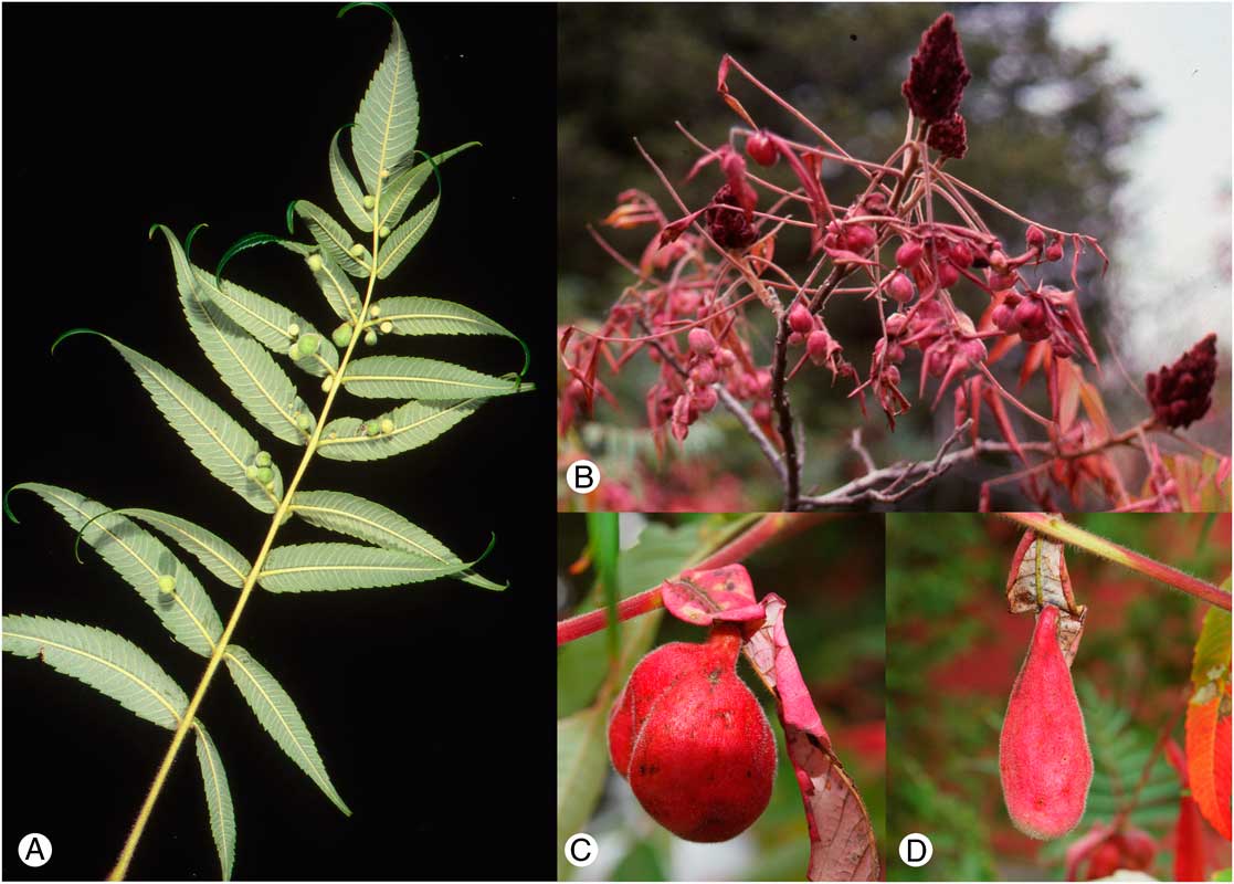
Deposition of larval fundatrices begins two to three weeks after copulation, i.e. in early to mid-June. The first instar fundatrix moves to the distal unexpanded leaves of the new shoot, where it may remain for several days, apparently initiating feeding only after the leaflets begin to unfold. Within one or two days of settling, feeding by the fundatrix on the upper (adaxial) surface of a leaflet results in the formation of an obvious pocket on the abaxial surface of the leaflet opening onto the adaxial surface. Subsequent instars of the fundatrix occur within the closed gall. As a consequence of aggregation by the sexuparae, galls tend to occur in clusters, so that a single compound leaf usually bears several galls (Fig. 6) and often many leaves of a single shoot are affected.
Galls of Melaphis species on Rhus typhina. A. Immature galls in early July, view of adaxial surface of leaf. B. Aggregation of mature galls, Odessa, Ontario, September 1992. C–D. Mature galls of M. asafitchi showing variations in shape, Marlborough Forest, Ottawa, Ontario, September 2014.

At least three generations develop within the gall, namely the fundatrix, at least one generation of apterae, and the alate emigrants. We have found living fundatrices containing embryos as late as mid-August. Adult alatae begin to appear within the gall in mid to late August (in North Carolina, alates collected as early as 7 August (slides in USNM)) and continue to develop throughout September; some alatoid nymphs may still be present in galls as late as early October. The gall opens by formation of longitudinal slits in the neck of the gall after the majority of individuals have become adult. In Ontario, migration to moss occurs over an extended period from the mid-September to early October, as the host leaves begin to senesce and fall; in Virginia, United States of America they “may be found flying at the last of October” according to Baker (Reference Baker1917). During this period, infested leaves are often apparent from a distance because they tend to senesce and redden earlier than other leaves of the same plant. Open galls are subject to heavy predation by Syrphidae (Diptera) larvae. The number of alates produced in a single gall is usually in the hundreds, but we have observed up to 3750 developing within large galls.
Apterae and developing sexuparae on moss develop within a loose cocoon-like wax cell. Summer generations on moss do occur; we have reared both sexuparae and larvae of a further moss-feeding generation from the same moss sample collected in early May, and Moran (Reference Moran1989) reports that she established “long-lived” colonies on moss. Populations in Europe currently assigned to Melaphis rhois appear to persist entirely on mosses (Heie Reference Heie1980) as do populations in Washington State (Pike et al. Reference Pike, Graf, Foottit, Maw, von Dohlen and Harpel2012). However, the required minimum number of generations on moss is unknown. Various stages in the life history of Melaphis species are illustrated in Supplementary Figure S1.
Host plants. Galls of Group 1 and Group 2 have both been found on both Rhus glabra Linnaeus and Rhus typhina Linnaeus; Group 3 galls have so far been collected only on R. glabra. Known moss hosts include Haplocladium microphyllum (Hedwig) Brotherus (Leskeaceae) (Moran Reference Moran1989), Dicranum scoparium Hedwig (Dicranaceae), Rhytidiadelphus loreus (Hedwig) Warnstorf (Hylocomiaceae) (Pike et al. Reference Pike, Graf, Foottit, Maw, von Dohlen and Harpel2012), and Polytrichum juniperinum Hedwig (Polytrichaceae) (our observations). We have observed feeding on the moss rhizoids in contact with the rock substrate, but the label on a slide from North Carolina, United States of America in the USNM states “feeding in leaf axils”.
Generic diagnosis. In North America, alate forms may be separated from other Fordini by the transverse, linear, secondary rhinaria; and from other Eriosomatinae by the combination of: lack of siphunculi, setae associated with the wax-gland plates outside the adjacent plate (Fig. 10G) (within the plate in Pemphigini), wax plates composed of an aggregation of similar facets (at least some wax plates with facets in a ring-like or rosette-like arrangement in many Eriosommatini), and the more-or-less concave inner margin of the pterostigma beyond radial vein (Fig. 12). Apterae on moss are distinguished by tarsal fusion and wax-gland pattern, as in the key by Pike et al. (Reference Pike, Graf, Foottit, Maw, von Dohlen and Harpel2012). Other apterous forms are easily distinguished by the obligatory association with Rhus.
Emigrants from the gall may be separated from Asian Melaphidina by the combination of short pterostigma (elongate and reaching wing apex in Schlechtendalia (Blackman and Eastop Reference Blackman and Eastop1994, Reference Blackman and Eastop2017)) and possession of numerous transverse, narrow rhinaria (versus fewer, broad and band-like or extensive plate-like rhinaria in most Asian species) with simple margins (spinulose margins in Nurudea meitanensis (Tsai and Tang)) (Yang et al. Reference Yang, Chen, Feng and Chen2009).
Generic description. General characteristics. Siphunculi absent. Ultimate rostral segment without accessory setae, but with anterior primary setae displaced basad. When facetted wax-gland plates are present, each with a single associated seta located outside of the plate.
All apterous morphs with compound eye absent (except for triommatidion) and without secondary antennal rhinaria.
All alate forms with apex of pterostigma acute, often with the posterior margin of the pterostigma beyond the origin of the radius slightly concave (Fig. 12); secondary rhinaria transverse-linear; in individuals with all four antennal flagellum fully developed, first flagellomere is the longest, second and third shortest and similar in length, fourth 0.6–0.9 times first.
Fundatrix, first instar (Fig. 7). In life dark with caudal wax filaments. Body sclerotic; dorsal setae relatively long, spine-like, longest as long as antennal pedicel; wax glands not aggregated into facets. Spiracles conspicuous, circular; thoracic spiracles about twice diameter of abdominal spiracles. Antenna with four antennomeres (i.e., with two flagellomeres). Scape broader than long, about 1.5 times diameter of pedicel. First flagellomere about as long as or a bit longer than pedicel; with a single subapical seta; primary rhinarium transversely oval. Ultimate flagellomere with terminal process about half length of basal part, with four terminal setae; basal part with two setae; primary rhinarium circular. Ultimate rostral segment with anterior primary setae displaced to middle of segment. Each tarsus (Fig. 10A) with basitarsus distinct, but broadly joined to distitarsus; apico-ventral basitarsal setae 2, long (reaching or slightly exceeding base of claw); apicodorsal setae of distitarsus about twice length of claw, curved, finely capitate; pretarsal setae (i.e., setiform parempodia on unguitractor plate) curved, reaching apex of claw; claw slender, attenuate, about one-third length of distitarsus.
Fundatrix of undetermined Melaphis species, first instar. A. Dorsal aspect of CNCHEM007770.1. B. Ventral aspect of CNCHEM007071.1. C. Lateral aspect of CNCHEM007770.1. Scale bar=100 µm.

Fundatrix, adult. In life, yellow, globular. All setae minute, inconspicuous. Wax glands not aggregated into facets, body with an even light wax coat. Antennae with four antennomeres, ultimate antennomere longest. Rhinarium of first flagellomere round, set basad of distal margin of flagellomere; terminal process of ultimate flagellomere about 0.25 length of basal part of flagellomere. Ultimate rostral segment short, 1.0–1.5 times longer than basal width. Trochanter fully fused to femur; tibia shorter than femur of same leg. Tarsi with basitarsus broadly fused to distitarsus, demarcation line distinct to obscure; two inconspicuous basitarsal setae about 0.2 time ventral length of basitarsus; apicodorsal setae of distitarsus about as long as distance from base of seta to base of claw; pretarsal setae obscure, less than thickness of unguitractor plate. Cauda with two setae.
Fundatrigenia (second generation aptera in gall). In life bright yellow, globular; smaller than fundatrix and with appendages relatively longer. Wax glands aggregated into facetted plates. Antenna with four to six antennomeres, usually five. Ultimate antennomere with terminal process <0.15 times base. Trochanter fused with femur without obvious demarcation. Basitarsus and distitarsus broadly joined but demarcation usually distinct; with two or three basitarsal setae. Claw about one-third length of distitarsus, with attenuate apex; pretarsal setae not apparent. Cauda usually with four setae (range 2–4).
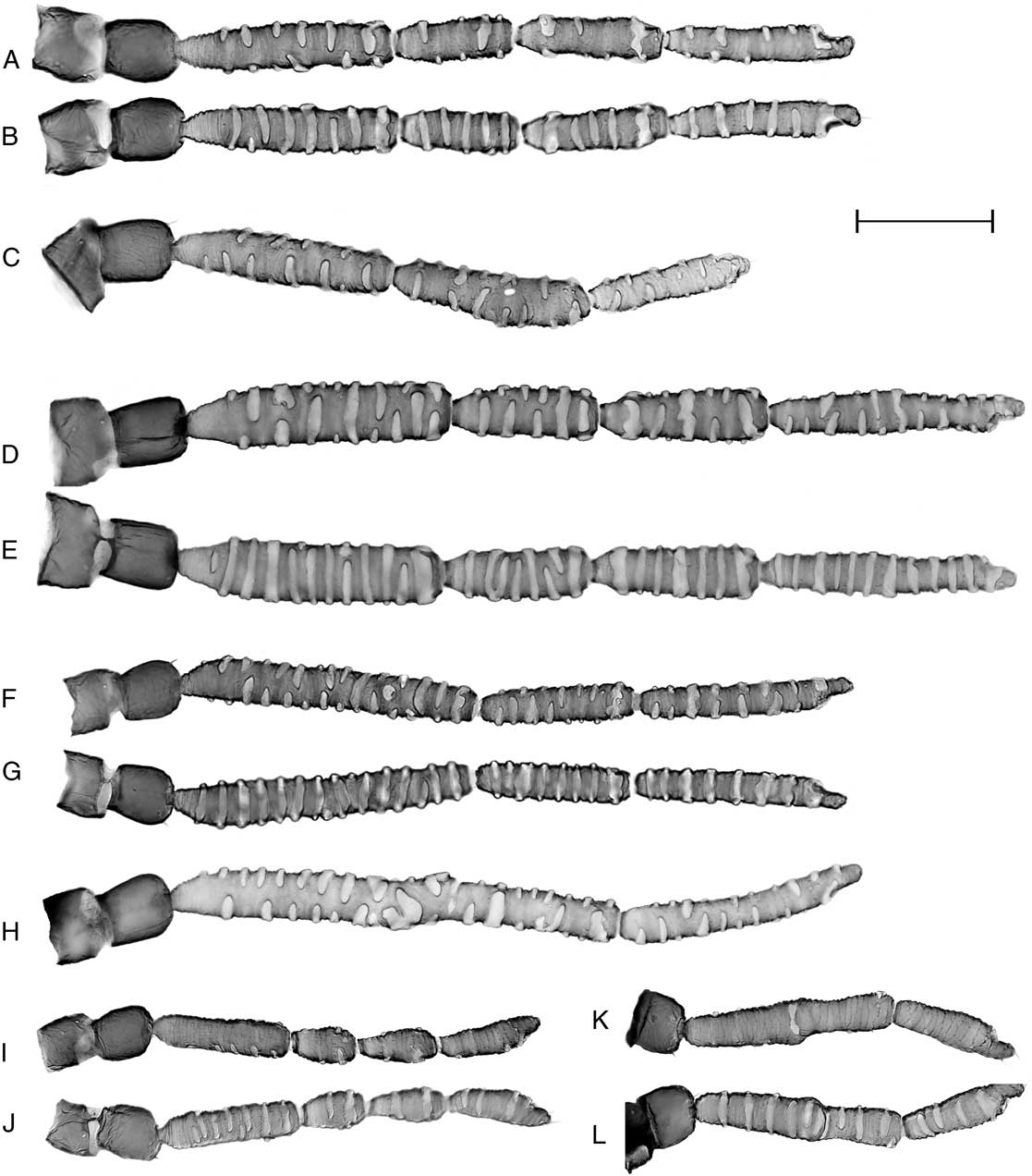
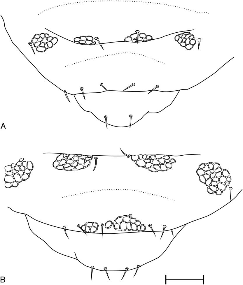
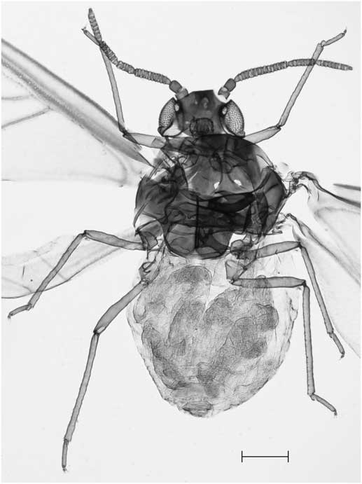
Fundatrispuria (alate emigrant from gall). In life, dark, abdomen dark green (overall appearance dark grey due to wax bloom), wings hyaline with green pterostigma. Body length 1.0–1.7 mm. Slide-mounted specimens with head and pterothorax darkly pigmented; legs (including coxa), antenna, cauda, genital, and anal plates pigmented but paler than head and pterothorax; pronotum lightly pigmented or hyaline; abdominal tergite 8 hyaline, or with lightly to distinctly pigmented scleroites at bases of setae; abdomen otherwise hyaline. Antenna (Fig. 8A–I) with six antennomeres (Fig. 8A–B, 8D–E), but often with first and second flagellomeres partially or completely fused to give an apparent antenna with five antennomeres (Fig. 8F–G) (often bilaterally asymmetrical in same individual; in almost all such antenna, at the point of fusion a hyaline area of cuticle is present on the dorsal surface and the rhinaria are distorted); occasionally with second and third (Fig. 8C) or first through third (Fig. 8H) flagellomeres fused; terminal process of distal flagellomere about 0.1 times length of base. Primary rhinarium of penultimate flagellomere fused with distal secondary rhinarium, often forming a broad band-like field; primary rhinarium of ultimate flagellomere sometimes fused with irregular patch-like or band-like distal secondary rhinarium and represented by sections of ciliate margin, or separate but largely enveloped by arms of an irregular patch-like or branched secondary rhinarium; accessory rhinaria of ultimate flagellomere usually remain separate. Secondary rhinaria present on all flagellomeres; transverse, usually narrow and separated by at least their own width, occasionally adjacent rhinaria anastomosing or fused into a broader band; extending up to three quarters of circumference of flagellomere, usually not extending onto mid-dorsal surface but occasionally forming a complete ring encircling the flagellomere. Basal half of basal flagellomere surface generally smooth, with obscure transverse rows of minute spinules distally; distal flagellomeres more distinctly spinulose (occasionally a row of spinules aligned with margin of a secondary rhinarium thus appearing as if the rhinarium has a spiculose margin). Trochantero-femoral suture incomplete, apparent only on anterioventral surface of mesothoracic and metathoracic legs and on posteroventral surface of prothoracic leg. Basitarsus most often with three, less often with two, apico-ventral setae; when three present the middle seta sometimes displaced basad, rarely with a single additional plantar seta. Plantar surface of distitarsus with five to nine pairs of setae. Full complement of facetted wax-gland plates distributed as follows: four pairs on head (but some or all are frequently missing or reduced to single facet, or the area represented by a roughened patch without distinct facets: one rarely expressed pair on anteroventral surface of head anterior to median ocellus, one infrequently expressed pair near anterodorsal margin of head, one pair between lateral ocelli, one pair near posterior margin of head), two pairs on pronotum, one pair near posteromedian margin of mesonotal lobes, one pair on metanotum (often absent), three pairs on each of abdominal terga 1–6, and two pairs on abdominal tergite 7, sometimes merging to form one or two continuous plates, zero to two pairs on abdominal tergite 8 (Fig. 9). Abdominal tergite 8 with four to nine setae (usually six to eight). Forewing with pterostigma not reaching wing apex; media unbranched, atrophied basally, sometimes also missing apically, or rarely not apparent for entire length; cubital veins approximate at base, usually complete to base. Hind wing with two transverse veins arising separately. Cauda with two to six setae, usually four.
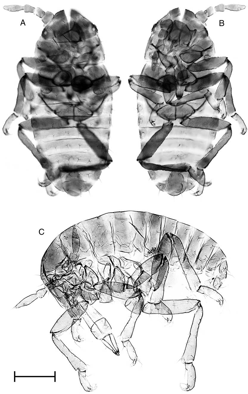
Antenna of alate forms of Melaphis species. A–H. Alatae from gall (fundatrispuriae). A–B. Melaphis rhois CNCHEM085826.6, dorsal and ventral aspect. C. Melaphis rhois CNCHEM085823.5, dorsal aspect showing fusion of second and third flagellomeres. D–E. Melaphis asafitchi CNCHEM085637.1 dorsal and ventral aspect. F–G. Melaphis asafitchi CNCHEM084142.9 dorsal and ventral aspect with fusion of first and second flagellomeres. H. Melaphis asafitchi CNCHEM085796.2, dorsal aspect with fusion of first to third flagellomeres. I–L. Melaphis asafitchi, sexupara: I–J. CNCHEM85936.26, dorsal and ventral aspect. K–L. CNCHEM85937.21, dorsal and ventral aspect, showing fusion of first to third flagellomeres. Scale bar=100 µm.

Apex of abdomen of fundatrispuriae of Melaphis species showing distribution of wax glands. A. Melaphis asafitchi CNCHEM085786.5, without wax glands on abdominal tergite 8. B. Melaphis rhois CNCHEM084807.4. Scale bar=100 µm.

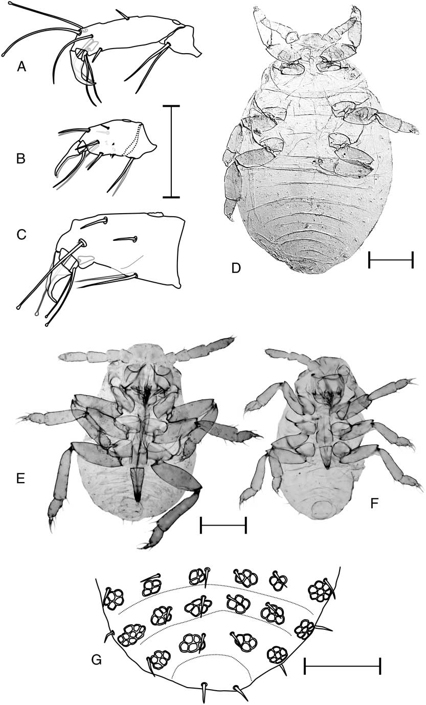
Alienicola (exulis), first instar (deposited by fundatrispuria) (Fig. 10E–F). Grey-black in life. Antenna with four antennomeres; rhinarium of basal flagellomere small, round, with distinct fringe of cilia; ultimate flagellomere with transverse rows of small spinules; rhinarium of ultimate flagellomere transversely oval, distinctly ciliate. Basitarsus broadly fused to distitarsus (Fig. 10C), but demarcation line distinct; ventral basitarsal setae 2, about as long as ventral length of basitarsus; distitarsus with a few transverse rows of spinules; apicodorsal setae of distitarsus reaching middle of claw; claw with distal part long and slender; pretarsal setae reaching end of claw. Body with full complement of wax plates (three pairs on head, two pairs on pronotum, two pairs on mesonotum and metanotum, three pairs on abdominal segments 1–7, zero to two pairs on abdominal segment 8) (Fig. 10G). Cauda with two setae.
Melaphis species. A–C. Metatarsus. A. Fundatrix first instar CNCHEM007071.1, species undetermined. B. Melaphis asafitchi, alienicola first instar from fundatrispuria CNCHEM085814.10. C. Sexual female first instar CNCHEM007375.1, species undetermined. D. Adult sexual female CNCHEM007771.1, postpartum, habitus, undetermined species. E–F. First instar alienicolae from fundatrispuriae. E. Melaphis rhois CNCHEM085811.4, habitus, ventral aspect. F–G. Melaphis asafitchi CNCHEM085814.10. F. Habitus, ventral aspect. G. Apex of abdomen, dorsal aspect, showing distribution of wax glands and setae. Scale bar for A–C, G=50 µm. Scale bar for D–F=100 µm.

Alienicola aptera (exulis; aptera on moss). In life, dull yellow to somewhat greyish yellow, surrounded by loose wax filaments; legs (including coxae) and antennae brown to dusky; front of head, cauda, anal, and genital plates dusky. Body with facetted wax plates (three pairs on head, two pairs on pronotum, two pairs on mesonotum and metanotum, three pairs on abdominal segments 1–7, zero to two pairs on abdominal segment 8). Antenna with fout to five antennomeres, ultimate antennomere longest. Metatibia shorter than, to about as long as, metafemur. Basitarsus broadly fused to distitarsus (Fig. 10B), separation sometimes at least partially demarcated by unpigmented line or irregular window along the line of fusion; ventral basitarsal setae 2, shorter than length of ventral surface of basitarsus; apicodorsal setae shorter than distance from base of these setae to basal articulation of claw. Cauda with two setae. Measurements given by Pike et al. (Reference Pike, Graf, Foottit, Maw, von Dohlen and Harpel2012) under M. rhois may be a composite of two species.
Alienicola, first instar (deposited by aptera alienicola on moss). Dull pale yellow in life (in contrast to dark colour of larvae deposited by fundatrispuria).
Sexupara (alate migrant from moss). Colour and wax-gland distribution similar to fundatrispuria. Antenna usually with five antennomeres (first apparent flagellomere resulting from fusion of first and second flagellomeres, usually with an unpigmented window on dorsal surface of composite flagellomere or other distortion at point of fusion); sometimes with a full complement of six antennomeres (Fig. 8I–J), sometimes with first three flagellomeres fused to give a four antennomere appearance (Fig. 8K–L). Antennomeres shorter than corresponding antennomeres of the fundatrispuria, with rhinaria fewer in number and generally extending no more half circumference of antennomere. Primary rhinarium of penultimate antennomere usually clearly differentiated from secondary rhinaria, round to transverse-oval with obscurely to distinctly ciliate margin, but sometimes similar to secondary rhinaria (transverse with smooth margin); primary rhinarium of ultimate antennomere small and round to large and oval, with distinctly ciliate margins. Basitarsus with two apico-ventral setae. Distitarsus shorter than in fundatrispuria; plantar surface with two or three pairs of setae. Cauda with two setae.
Sexual female (Fig. 10D). Colour in life dark olive to black. Body length of first instar about 620 µm, of adult about 550 µm. Apterous. Rostrum absent. Body setae very short, inconspicuous. Wax-gland plates obscure, often with only on to three facets. Legs short, femur about as long as tibia of same leg; femur and tibia both about 2.5 times longer than greatest width. Tarsomeres fully fused; claw short, blunt; apicodorsal seta of distitarsus long and curved (Fig. 10C), about twice as long as claw; ventral setae of basitarsus of adult shorter then ventral length of basitarus (longer in first instar); pretarsal setae of adult inconspicuous, about half as long as claw (but capitate and about 1.3 times longer than claw in first instar). Cauda with two setae.
Male. Similar to sexual female, body length of adult about 300 µm.
Gall. Initiated by feeding by the first instar fundatrix on upper surface of the lamina of a leaflet adjacent to major veins (not “am Mittelnerv”, i.e., “on midvein”, as stated by Mordvilko (Reference Mordvilko1935)). A spherical pocket extending above and below the blade forms around the fundatrix and subsequently expands below the blade, usually also with a conical projection above the blade, the opening becoming closed by trichomes at adaxial opening and by tissue expansion within neck. Immature gall globular (Fig. 6A), pale green, about 2.0–2.5 mm in diameter at the time the fundatrix undergoes its final moult to adult. Veins adjacent to gall become more or less swollen and adaxial surface reddened. Mature gall (Fig. 6B–D) globular to pyriform, unilocular, largely red in colour in exposed locations, but usually with pale yellow to yellow-green areas on the side protected from the sun, pale areas more extensive in shaded locations; up to 50 mm in length and up to 45 mm at greatest diameter. The gall surface is often irregular to weakly lobed. Galls distinctly bilobed to near the base occasionally occur, and very rarely galls have several deep lobes. It is not clear if these are the product of a single fundatrix (with lobes possibly resulting from a leaflet vein interfering with gall expansion) or result from the settling of more than one fundatrix at the same site; we have found recently initiated galls with more than one fundatrix, but only one surviving beyond first instar. Vestiture of the gall surface reflects the general vestiture of the host plant: those on Rhus glabra are largely glabrous while those on R. typina are finely setose.
Comments. Fitch (Reference Fitch1866) described the antenna of the alate form from the gall as having four antennomeres, with “the first joint slightly the shortest, and the second joint rather the longest”. Because Fitch states that the “second joint” is the longest, we presume he overlooked the scape due to retraction into the head in dried specimens, and that his “first joint” is the pedicel and the “second joint” is the apparent first flagellomere of a five-antennomere antenna. Walsh (Reference Walsh1867) established Melaphis as distinct on the basis of possession (chiefly) of four-antennomere antennae, and explicitly stated that “the typical two joints of the scape are soldered together” as are “the typical joints 1 and 2 of the flagellum”; Walsh did recognise, however, that almost a quarter of the 28 specimens he examined had one antenna with “five segments” (i.e., first two flagellomeres not fused). We have seen no specimens with fusion of the scape and pedicel, and believe that Walsh was also mistaken in this regard. We have seen several specimens having one antenna with four apparent antennomeres (the first, second, and third flagellomeres are more or less fused), but in this case either the “third joint” would be the longest if the scape is taken into account, or the antenna would be described as having three antennomeres if the scape is ignored.
Fitch (Reference Fitch1866) described material collected in early September well before galls had fully matured, as evidenced by his indication that each gall contained zero to four winged individuals and that the galls were closed. As noted by Walsh (Reference Walsh1867), the colours given by Fitch for the alate form in the gall are for teneral individuals.
The gender of Melaphis is feminine (Cortés Gabaudan et al. Reference Cortés Gabaudan, Nieto Nafría, Favret, Barbagallo, Sano and Stekolshchikov2011), by analogy with the conventionally established gender of Aphis. Thus, the correct spelling of “M. minutus” is M. minuta.
Melaphis rhois (Fitch, Reference Fitch1866)
Brysocrypta rhois Fitch, Reference Fitch1866 (original combination).
Neotype. Fundatrispuria (alate emigrant from gall), CNCHEM084187.2 (Fig. 11); United States of America: Pennsylvania: State College (Sunset Park); ex Rhus glabra; 22.viii.2013; R.G. Foottit. Deposited in Canadian National Collection of Insects, Arachnids, and Nematodes, Ottawa, Ontario, Canada (CNC). Neotype here designated.
Melaphis rhois, neotype, CNCHEM084187.2, habitus. Scale bar=100 µm.

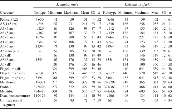
Diagnosis. Usually separable from M. asafitchi new species by longer ultimate rostral segment (usually exceeding 65 µm in all morphs sufficiently characterised; for fundatrispuriae, a value of [0.03×[total length of flagellum]−0.14×[ultimate rostral segment]−0.09×[distal metatarsomere]+3.0] is negative; abdominal segment 8 often with wax-gland plates. For isolated individuals with length of last rostral segment near 65 µm or fundatrispuriae with discriminant function value near 0, definitive diagnosis by mitochondrial COI-5P sequence is required.
Description. The following description includes only morphs for which individuals of known COI haplotype are available. See the generic description for general characteristics of the fundatrix, adult alienicola and sexual forms.
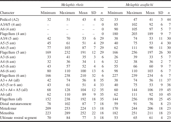
Fundatrigenia (second generation aptera in gall). As described for genus, with measurements in Table 7. Body length 0.9–1.2 mm. Antenna with five to six (usually five) antennomeres. Ultimate rostral segment >68 µm long.
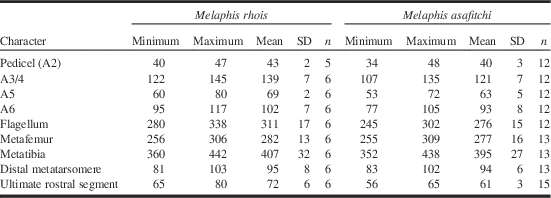
Summary of length measurements for Melaphis rhois and Melaphis asafitchi fundatrigeniae (apterae in gall on Rhus).

Note: Both antennae of each individual (if present) were measured; thus n for antennae is the total number of antennae measured rather than number of individuals. The prefix “A” signifies antennomere, where A1 is the scape and A3 is the first flagellomere. Apparent antennomere 3 of individuals with five antennomeres is considered homologous with fused antennomeres A3 and A4, thus subsequent antennomere is A5, and terminal antennomere of all antennae is A6. Antennomere lengths of different degrees of fusion (i.e., apparent 4, 5, and 6 antennomeres) are reported separately, as well as length for homologous extents for all antennae. A3/4 and A3/4/5 indicate fused antennomeres, while A3+A4, A3+A4+A5 and flagellum (=A3–A6) represent the total span of the indicated antennomeres whether fused or not. 5 ant and 6 ant indicate specimens with five or six antennomeres, respectively. A6 includes terminal process. Femur length includes trochanter. Measurements in micrometres.
Fundatrispuria (alate emigrant from gall). As described for genus, with measurements as given in Table 8 and rhinarial distribution in Table 9.
Summary of length measurements for of Melaphis rhois and Melaphis asafitchi fundatrispuriae migrating from Rhus to moss.

Note: See caption for Table 7 for labelling of antennomeres. Metafemur includes trochanter. Antennal and leg measurements for type specimens are for left/right appendages. All length measurements in micrometres.
Rhinarial (primary+secondary) counts for specimens fundatrispuriae of Melaphis rhois and Melaphis asafitchi.

Note: Labelling of antennomeres as in Table 7. Counts for types are for left/right antenna.
Expression of wax glands on abdominal tergite 8 (Fig. 9B) is variable; the basic pattern consists of two pairs of facetted plates as on tergite 7, with expression ranging from absent (in 62% of 190 specimens examined), through a few scattered single facets up to completely confluent fully developed plates covering much of the tergite (neotype with two pairs of distinct plates).
Of 300 specimens, 67% from 18 localities have both antennae with six antennomeres, 28% have one five-antennomere antenna, and five per cent of individuals have both antennae with five antennomeres (i.e., about 19% of all individual antennae have five antennomeres). Two individuals examined have one antenna with the second and third flagellomeres (rather than first and second) partly fused.
Alienicola, first instar (deposited by fundatrispuria) (Fig 10E). As described for genus. Body 330–450 µm long. First flagellomere about 1.6 (range 1.4–1.9) times length of pedicel. Rostrum reaching middle of abdomen, apex of third rostral segment reaches posterior margin of metacoxa; ultimate rostral segment 1.2 times length of ultimate rostral segment and 1.6 times length of metatarsus. Measurements of antennomeres, rostrum, and metathoracic leg parts given in Table 10.
Measurements for Melaphis rhois and Melaphis asafitchi first instar offspring of fundatrispuriae.

Note: Femur length includes trochanter. Measurements in micrometres.
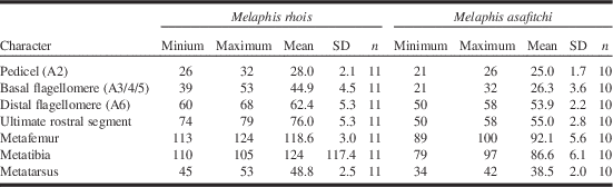
Sexupara (alate migrant from moss). As described for genus, with measurements in Table 11. Length of ultimate rostral segment >64 µm, average length of flagellum 311 µm.
Measurements for Melaphis rhois and Melaphis asafitchi sexuparae migrating from moss to Rhus.

Note: See Table 7 for terminology regarding antennomeres. Femur length includes trochanter. Measurements in micrometres.
Mitochondrial cytochrome oxidase subunit1, 5' region (DNA barcode). Sequence of specimen from same gall as neotype, Genbank accession KY754324. Additional sequence variants given in BOLD data set DS-MELAPHIS (www.boldsystems.org/index.php/Public_SearchTerms?query=DS-MELAPHIS).
Distribution. Widespread in United States of America and in southeastern Canada. Canada: southern Ontario, southern Québec, Nova Scotia. United States of America: Arkansas, Arizona, Georgia, New Jersey, Pennsylvania, Washington (all confirmed by COI-5P sequence); Kentucky, Maine, Massachusetts, New Hampshire, North Carolina (based on morphology only).
Hosts. Primary hosts: Rhus glabra, Rhus typhina. Confirmed secondary hosts: Haplocladium microphyllum (Moran Reference Moran1989), Dicranum scoparium (Pike et al. Reference Pike, Graf, Foottit, Maw, von Dohlen and Harpel2012).
Material examined. Fundatrispuriae (fp) and fundatrigeniae (fg) measured for morphometric analysis (see Supplementary Tables S1–S3 for additional details) – Canada: Ontario: Actinonolite, Price Conservation Area, 17.ix.2009; Beech Corners, Buckshot Lake, 25.ix.1993 and 25.xi.2008; Chaffeys Locks, 17.ix2009; Griffith, Matawatchan River, 24.ix.2009; Khartoum, 24.ix.2009; Maberley, Highway 7 at McGowan Lake, 16.ix.1993 and 17.ix.2009; Mallorytown, 17.ix.2009; Mount St. Patrick, English Road, 24.ix.2009; Ottawa, Marlborough Forest, 19.vi.1993; Prescott, Pruner Road, 13.vi.1993; Prince Edward County at Deseronto bridge, 17.ix.2009; Robertsville, 16.vi.1993 and 24.ix.2009; South Lake (Taylor/Berryton), 17.ix.2009; Westbrook, Highway 401 service centre, 13.vi.1993; Westport, Perth Road North near Lennon Road, 17.ix.2009. Québec: Parc de la Gatineau, Champlain Lookout, 19.ix.1993 and 17.ix.2009; Parc de la Gatineau, Hickory Trail, 10.ix.2001; United States of America: Pennsylvania: State College, 22.viii.2013.
Additional specimens – Canada: Ontario: Point Anne, Rhus typhina, 18.v.1993, E. Maw, 93EM-016 (5 sexuparae) [CNC]; Québec: Parc de la Gatineau, reared from Polytrichum, 31.v.1994, E. Maw, 94EM-015.1 (1 sexupara) [CNC]; Shawinigan, “sumack”, 23.viii.1939, L. Daviault (7 fg) [CNC]. United States of America: Kentucky: Greenup County, 24.ix.1971, D. Barnett, Ky-106, 72-1834 (1 fp) [USNM]; Maine: Orono, Rhus, 15.x.1935, E.M. Patch 18-35 (76 fp) [CNC]; Massachusetts: Quincey, sumac in galls, viii.1980 (4 fg) [USNM]; New Hampshire: Claremont, sumac, 15.ix.1917, Mosher, Q.13619 (5 fp) [USNM]; North Carolina: Fletcher, sumac, 23.ix.1960, C.F.S[mith], 60-1081 (3 fp) [USNM]; Pennsylvania: Philipsburg, sumac, 24.ix.1959, Pepper (4 fp) [USNM] .
Comments. According to Barnes (Reference Barnes1988), almost all aphid material from Fitch’s collection is currently housed in USNM and in New York State Museum (Albany, New York, United States of America). A few of his aphid specimens are also found in the Museum of Comparative Zoology (Harvard University, Cambridge, Massachusetts, United States of America). There is no existing material of Brysocrypta rhois examined by Fitch in any of these institutions. As it is unknown which species Fitch actually had at hand (possibly both) we have designated a neotype. COI-5P Group 1 is selected to carry the name M. rhois. Current evidence suggests that this is the more widespread species. The neotype was selected from among specimens collected at State College, Pennsylvania, as this is the nearest locality to Fitch’s original collections for which we have sequenced individuals. This specimen belongs to the subgroup (COI-5P Subgroup 1a) to which GenBank accession U36747 belongs. These decisions serve to maintain the usage of this name in previous phylogenetic and endosymbiont studies based on material collected by Moran in Arizona (Moran et al. Reference Moran, Munson, Baumann and Ishikawa1993, Reference Moran, von Dohlen and Baumann1995; Rouhbakhsh et al. Reference Rouhbakhsh, Lai, von Dohlen, Clark, Baumann and Baumann1996; Clark et al. Reference Clark, Moran and Baumann1999; Nováková et al. Reference Nováková, Hypša, Klein, Foottit, von Dohlen and Moran2013). The samples used by Yang et al. (Reference Yang, Chen, Havill, Feng and Chen2010), the sequence used by Li et al. (Reference Li, Jiang and Qiao2014), and the sequences generated by Ren et al. (Reference Ren, Zhong, Kurosu, Aodi, Ma, von Dohlen and Wen2013) belong to subgroup 1b. We interpret the subgroups as variants of a single species.
Melaphis asafitchi Foottit and Maw, new species
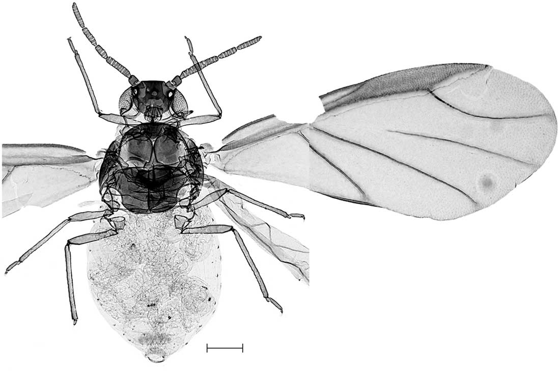
Type series. Holotype: fundatrispuria (alate emigrant from gall), CNCHEM085883.2 (Fig. 12); Canada: Ontario: Kemptville, G. Howard Ferguson Forest Station, 45.037°N 75.667°W; ex Rhus typhina; 17.ix.2009; E. Maw. Deposited in CNC. Paratypes: fundatripuriae (fp) and fundatrigeniae (fg) measured for morphometric analysis (see Supplementary Tables S1–S3 for additional details) – Canada: Ontario: Actinolite, Price Conservation Area, 17.ix2009 (12 fp); Beech Corners, Buckshot Lake, 25.ix.2008 (3 fp); Chaffeys Locks, 17.ix.2009 (3fp, 7 fg); Griffith, Matawatchan River, 24.ix.2009 (6 fp); Kemptville, Ferguson Forestry Station, 17.ix.2009 (8 fp); Khartoum, 24.ix.2009 (8 fp, 6 fg); Maberley, Highway 7 at McGowan Lake, 17.ix.2009 (9 fp, 2 fg); Mallorytown, 17.ix.2009 (12 fp); Mount St. Patrick, English Road, 24.ix.2009 (3 fp, 5 fg); Prince Edward County at Deseronto bridge, 17.ix.2009 (2 fg); South Lake (Taylor/Berryton), 17.ix.2009 (3 fp, 1 fg). Québec: Parc de la Gatineau, Champlain Lookout, 17.ix.2009 (8 fp); Parc de la Gatineau, Hickory Trail, 10.ix.2001 (3 fp); United States of America: Pennsylvania: State College, 22.viii.2013 (3 fp). Deposited in Canadian National Collection (CNC), National Museum of Natural History (USNM) (3 fp, 2 fg), and Natural History Museum (London, United Kingdom) (3 fp, 2 fg).
Melaphis asafitchi, holotype, CNCHEM085883.2, habitus. Scale bar=100 µm.

Diagnosis. Usually separable from M. rhois by the shorter ultimate rostral segment (usually less than 65 µm in all morphs sufficiently characterised; for fundatrispuriae, value of [0.03×[total length of flagellum]−0.14×[ultimate rostral segment]−0.09×[distal metatarsomere]+3.0] is positive; abdominal segment 8 without wax-gland plates. For isolated individuals with length of last rostral segment near 65 µm or fundatrispuriae with discriminant function value near 0, definitive diagnosis by mitochondrial COI-5P sequence.
Description. The following description includes only morphs for which individuals of known COI haplotype are available. See the generic description for general characteristics of the fundatrix, adult alienicola and sexual forms.
Fundatrigenia (second generation aptera in gall). As described for genus, with measurements as in Table 7. Body length 0.9–1.2 mm. Rostrum and legs relatively shorter and antennae relatively longer than in M. rhois. Ultimate rostral segment <68 µm long. Antenna with four to six (usually 5) antennomeres.
Fundatrispuria (alate emigrant from gall). As described for genus, with measurements as given in Table 8 and rhinarial distribution in Table 9. Wax-gland facets lacking on abdominal tergum 8 (all specimens examined). About 57% of 325 specimens from 20 galls have both antennae with six antennomeres, 30% have one five-antennomere antenna, and 14% of individuals have both antennae with five antennomere (i.e., about 28% of all individual antennae have five antennomeres). Two individuals had one antenna with the three basal flagellomeres fused, resulting in an apparent four-antennomere antenna.
Alienicola, first instar (deposited by fundatrispuria) (Fig. 10F–G). As described for genus. Body 320–425 µm long. First flagellomere about 1.1 (range 0.8–1.2) times length of pedicel. Rostrum reaching just beyond posterior margin of metacoxa, apex of third rostral segment not reaching posterior margin of metacoxa; ultimate rostral segment 1.0 times length of ultimate antennomere and 1.4 times length of metatarsus. Measurements of antennomeres, rostrum, and metathoracic leg parts given in Table 10.
Sexupara (alate migrant from moss). As described for genus, with measurements in Table 11. Ultimate rostal segment <66 µm. Mean length of antennal flagellum 276 µm.
Mitochondrial cytochrome oxidase subunit1, 5' region (DNA barcode). Holotype, GenBank KY754388. Additional sequence variants are available in BOLD data set DS-MELAPHIS (www.boldsystems.org/index.php/Public_SearchTerms?query=DS-MELAPHIS).
Distribution. Canada: southern Ontario, southern Québec, Nova Scotia (confirmed by COI-5P sequence); New Brunswick (based on morphology). United States of America: Pennsylvania, Washington (confirmed by COI-5P sequence); Illinois, Maine, Maryland, Minnesota, New York (based on morphology).
Hosts. Primary hosts: Rhus glabra, Rhus typhina. Confirmed secondary hosts: undetermined mosses (specimens included in Pike et al. Reference Pike, Graf, Foottit, Maw, von Dohlen and Harpel2012 as M. rhois).
Etymology. Named in honour of Dr. Asa Fitch (1809–1879), the first American professional entomologist, who first described a species of Melaphis.
Additional material examined. Canada: New Brunswick: Burton, 23.ix.1933, C.E. Atwood, 18788-31 (3 fp) [CNC]; Ontario: Chaffeys Lock, Clear Lake Road, Rhus typhina, 20.v.2014, E. Maw and B. Richardson, 2014EM-0015 (20 sexuparae); Québec: Cté Papineau, Notre Dame de la Salette, Rhus typhina, 12.ix.1981, L. Jobin (64 fp); Parc de la Gatineau, Rhus typhina, 21.v.1994, E. Maw, 94EM-018; 20.v.2014, 28.v.2014, E. Maw and B. Richardson, 2014EM-010 A,-0021 (35 sexuparae) [all in CNC]. United States of America: Illinois: Milan, galls on sumac, 16.viii.1885, accession number 7134 (1 fg, numerous L4 fp) [USNM]; Maine: Orono, Rhus glabra, 26.ix.1956, D. Folsom, 56-21 (6 fp, 1 fg) [CNC]; Maryland: Cabin John, Rhus glabra, 14.viii.1932, M.G. van Meter [USNM]; Glen Elg, sumac, 15.viii.1962, W.T. Johnson, Md 62-47, 62-22591 (4 fp) [USNM]; Geneld [?=Glen Elg], Rhus typhina, 16.viii.1962, W.T. Johnson (5 fp) [USNM]; Minnesota: Detroit Lakes, Rhus glabra, 3.ix.1984, V.H.J. Beregovey, 84-10669 (2 fp) [USNM]; Le Sueur, Rhus glabra, 21.ix.1907, G.G. Ainslie, number 199 (34 fp) [USNM]; New York: Ithaca, Rhus, 6.x.1924, M. Reidy, 514-24 (8 fp); West Point, Rhus glabra, 28.vii.1896, J.A. Williams (2 fg and numerous L4 fp) [USNM]; Pennsylvania: State College, Waddle, 25.viii.1962, Rhus glabra, J.O. Pepper, JOP62-51 (4 fp) [USNM]; Coraopolis, 12.ix.1916, sumac, Q-13214 (6 fp) [USNM].
Melaphis minuta Baker and Truncaphis newsteadii Theobald
The length of the ultimate segment of the rostrum of Baker’s material (83–84 µm) of Melaphis minuta is slightly beyond the upper end of the range of this measurement among other available sexuparae, and would suggest that it is the species with the longer rostrum, M. rhois. However, the length of the antennal flagellum in the single syntype seen and in the measurements given by Baker (Reference Baker1919) are less than the minimum we have observed for the species with the shorter antenna in the sexupara, M. asafitchi (Baker’s measurements seem to have a granularity of about 16 µm, but even inflating each of his measurements by 8 µm, the maximum value is about the same as the shortest length found for M. asafitchi, an unusual specimen with the first three flagellomeres fused). Thus, given this combination of characters, it is currently not possible to assign M. minuta to either of the two species discussed here. It is possible that it corresponds to our COI-5P Group 3, or is yet another species.
Melaphidine apterae on moss in the United Kingdom, originally described as Truncaphis newsteadii, and in Sweden are currently assigned to Melaphis rhois. However, given the sparse knowledge of the moss-inhabiting forms of both the North American and the Asian species, the extent of intraspecific variation is unknown, and the assignment of the European populations to one of the described species would be premature without supporting molecular evidence. Ren et al. (Reference Ren, Zhong, Kurosu, Aodi, Ma, von Dohlen and Wen2013) suggested the possibility that these populations are a relict of an indigenous European melaphidine species.
Discussion
DNA barcoding data for Melaphis specimens falls into four detectable clusters. Of these, one (Group 3) is supported by fixed differences in two nuclear genes, increasing the probability that the cluster represents a reproductively isolated population. Unfortunately, insufficient material of this group is available to characterise it morphologically. Groups 1 and 2 are both supported by the endosymbiont data, but support from the nuclear loci is weaker, with consistent differences in allele frequencies rather than any fixed differences. However, concordant morphometric partitioning supports the hypothesis that the two groups represent reproductively separate entities, and we have chosen to recognise them as separate species, M. rhois and M. asafitchi, respectively. The mitochondrial locus and the endosymbiont locus agree on the partitioning of subgroups 1a and 1b. Because both are matrilineally inherited, and the inter-group differences are not great, in the absence of additional sources of data, this observation may be interpreted either as an indication of reproductive isolation or as a reflection of past separation of populations and subsequent reintegration into a single interbreeding population. We have elected to accept the latter, but in future more extensive representation of subgroup 1a specimens and use of more sensitive nuclear markers may allow the recognition of these subgroups as distinct species. As noted earlier, it is possible that Group 3 corresponds to Melaphis minuta. In addition, a single collection of apterous specimens on moss in North Carolina (slide in USNM) differ in a number of respects from other Melaphis specimens on moss that we have seen, and it too may represent this, or another undescribed species. More extensive sampling and molecular analysis of Melaphis in the eastern United States of America is thus required to establish the true level of species diversity in this genus.
Definitions of genera among Melaphidina are based on morphology of fundatrispuriae and to some extent gall structure. The morphology of gallicolous apterae of Melaphidina is insufficiently characterised to differentiate genera based on this form. As this is the only form available for COI-5P Group 3 individuals, assignment of Group 3 to the genus Melaphis is speculative, and currently based on the strong similarity of the gall to that of M. rhois and M. asafitchi. Sequence similarity in all four loci examined here places this group more closely to the other North American species than to any known Asian species, suggesting that it is native. However, the collection site is adjacent to the Pennsylvania State University arboretum, so there is the possibly that it is an Asian introduction.
Both Melaphis species considered here accept both Rhus glabra and R. typhina as primary host plants. The range of these plant species overlap in the northeastern United States of America, but R. typhina tends to be more northeastern, and is the only species in most of southern Ontario, southern Québec, the Maritime Provinces of Canada, and northern New England in the United States of America. It is common south of the Great Lakes in a broad arc from Wisconsin southeast through West Virginia and northeast to New England, with southward extension in the Blue Ridge Mountains, and only scattered populations southwest of this arc as far as Mississippi River. Rhus glabra is widely distributed in the United States of America south of the Great Lakes east of 100° longitude, except most of the southeastern coastal plain, with only rare occurrences in southern Ontario, and with additional scattered populations in westward to southern British Columbia and Arizona, and into northern Mexico. Current knowledge suggest that there is a similar situation in Melaphis: M. asafitchi is common in the northeastern parts of the combined range, while M. rhois extends further south and west. It is tempting to speculate that M. asafitchi was originally associated with R. typhina and M. rhois with R. glabra and that the Melaphis species were derived by the same vicariance event that lead to speciation in this pair of Rhus species.
The southern extent of the distribution of M. asafitchi is not yet known. In addition to the distributions given under the species descriptions above, unassigned “Melaphis rhois” collections are recorded in the United States of America from Connecticut, Iowa, Kansas, Missouri, Nebraska, Ohio, South Carolina (Essig Reference Essig1943; Smith and Parron Reference Smith and Parron1978), and West Virginia (type locality for M. minuta). Both Melaphis species described here are currently abundant, at least in the northern parts of the joint range, but historical records are somewhat sparse despite the rather distinctive and obvious appearance of the galls: Fitch did not encounter sumac galls after 1857 (Fitch Reference Fitch1866); Hottes and Frison (Reference Hottes and Frison1931) knew of no collections in Illinois after 1885; the collection logbook of Edith Patch for the years 1904–1935 (housed in CNC) includes only seven collections from Maine and one from New York; and only three additional collections in CNC predate 1940. Rhus typhina is shade-intolerant and a coloniser of well-drained disturbed sites (Sullivan Reference Sullivan1994). It is now common along roadsides, fence lines and on abandoned farmland. It may have been present in the open oak savannas maintained by the indigenous peoples by fire in southern Ontario and other area of northeastern North America, but top growth of sumac is fire sensitive, reducing the chances of long-term establishment of large patches in such habitats. It is not included in historical records as a component of the flora of one such area (Rice Lake Plains; Catling et al. Reference Catling, Catling and McKay-Kuja1992) although it is currently common in that general area. The current available habitat for this plant is probably more abundant and connected than it was before European settlement. Our observations indicate that Melaphis species have relatively low vagility and dispersive ability. Fundatrispuriae emerging from galls and held in a container deposit larvae within hours and sexuparae on Rhus are empty of embryos within a day of arrival; thus it seems that they take flight only after embryonic development of their offspring is complete, leaving little time for long-distance dispersal. This supports the observations of high degrees of restricted gene flow by Hebert et al. (Reference Hebert, Finston and Foottit1991) based on allozyme variation. We speculate that the current, more connected distribution of the primary host has facilitated host finding during short distance flights, leading to an increase in frequency of occurrence.
Melaphis rhois and M. asafitchi are broadly sympatric and exhibit very similar habitat use. The mechanisms allowing segregation and sympatric maintenance of these species are not known. However, the strong spring aggregation of sexuparae on the same Rhus twig indicates that aggregation pheromones are involved, and these may be species specific. Although fall migrations of the two known species seem to coincide, there may be undetected temporal separation in the spring. Late heavy frosts (e.g., in early June in eastern Ontario in 2013) often kill the early flush of sumac foliage in exposed locations, favouring later migrants from moss in those years in which they occur.
The adaptive significance of the differential character most consistent across different segments of the life cycle, the difference in rostral length, is not known. Gall morphology for the two species is superficially identical, but it is possible that there is a difference in moss host taxa or feeding location.
In general, the sexual females of aphids deposit an overwintering egg with a rigid chorion, whereas parthenogenic forms are viviparous, with the membrane enveloping the embryo being removed before or during deposition. Hille Ris Lambers (Reference Hille Ris Lambers1950) described a form of ovoviviparity among alate parthenogenic forms depositing offspring on the secondary host (i.e., fundatrispuriae) of several Eriosomatini and of Pemphigus protospirae Lichenstein, and this may be the norm among Eriosomatinae. In these cases the offspring is deposited while still enveloped in the membranous egg, with eclosion occurring shortly thereafter. An unusual feature of the life history of Melaphis species and of Schlechtendalia chinensis (and likely other Melaphidina) is the occurrence of a similar form of ovoviviparity by the sexual female.
A number of questions remain regarding the life cycle of Melaphis (and other Melaphidina). The number of apterous generations within the gall is not known. According to Sasaki (Reference Sasaki1910) the fundatrix of Schlechtendalia chinensis produces a limited number of apterous offspring, which then produce another generation of apterae which show some morphological differences from the first generation fundatrigeniae. The mature gall of S. chinensis consists of a number of lobes branching from a common chamber and Sasaki states that the first generation of fundatrigeniae are found dispersed among these lobes. If this first generation plays a role in inducing this secondary gall structure, then other melaphidine species with lobed or multilocular galls may also show this generational succession. However, in Melaphis species, which generally produce a simple gall form, the fundatrix can continue to produce larvae through the summer and we have noticed no differentiation among its apterous descendents.
The details of the portions of the life cycle occurring on moss are also unclear. Tsai and Tang (Reference Tsai and Tang1946) report that that the larvae of Schlechtendalia peitan (Tsai and Tang) deposited on moss by emigrants from the gall enter an overwinter diapause and develop into sexuparae in the spring. Takada (Reference Takada1991) implies the same for S. chinensis, and Yang et al. (Reference Yang, Chen, Feng and Chen2009) attribute this life cycle characteristic to Melaphidina in general. Although compression of life cycles is known in Eriosomatinae (such as the elimination of the secondary host forms, so that the alate form emerging from the gall is the sexupara), this succession of two functionally distinct winged generations, if confirmed, would be unique for an eriosomatine aphid. In Melaphis, adult apterous alienicolae do occur on moss, although the nature and number of generations on moss has not been determined. Several trajectories are conceivable: (1) given the protracted period over which fundatrispuriae leave the gall, early-born alienicolae mature in the autumn and produce larvae that diapause and develop into the sexuparae in the following spring, while development of later-born individuals is delayed, overwintering as early instars and contributing to summer moss generations; (2) the first generation of alienicolae regularly or facultatively produce larvae of both sexuparae and of additional alienicolae; (3) all larvae deposited by the fundatrispuriae produce subsequent generations of alienicolae, with first sexuparae not produced until the second spring (biennial life cycle, possibly facultatively persisting in part as alienicola for additional years); (4) offspring of the fundatrispuriae obligatorily produce a second generation (in late autumn or in early spring), which then facultatively produces sexuparae or futher alienicolae; or (5) some larvae deposited by fundatrispuriae develop directly into sexuparae, as reported for the Asian species, in parallel with one of the other pathways.
Acknowledgements
The authors thank Larissa Ho, Jean-Paul Nadeau, Tanya Selander, Tian Wu (Agriculture and Agri-Food Canada, Ottawa), and the staff of the Biodiversity Institute of Ontario (University of Guelph) for DNA extraction and sequencing; Jean-Paul Nadeau and Bradley Richardson for slide mounting and measuring specimens; Gary Miller and Debbie Creel (Systematic Entomology Laboratory, United States Department of Agriculture, Beltsville, Maryland) for the loan of specimens. Funding provided in part by the Genomics Research and Development Initiative of the Government of Canada.
Supplementary material
To view supplementary material for this article, please visit https://doi.org/10.4039/tce.2017.57