INTRODUCTION
Fascioliasis, or infection with the liver fluke species Fasciola hepatica and Fasciola gigantica, is recognized by the World Health Organisation as a neglected tropical disease. It is estimated that 7 million humans are infected and many more millions are at risk of infection (Robinson and Dalton, Reference Robinson and Dalton2009). The majority of these people live in the developing world. The parasites also infect farm animals causing billions of pounds of agricultural losses worldwide and providing a reservoir for the human disease (Boray, Reference Boray1994). The treatment of choice for both animals and humans is the benzimidazole derivative triclabendazole. This drug is effective against both adult and juvenile flukes. However, in the past decade, resistance to triclabendazole has been reported in liver flukes infecting farm animals (Brennan et al. Reference Brennan, Fairweather, Trudgett, Hoey, McCoy, McConville, Meaney, Robinson, McFerran, Ryan, Lanusse, Mottier, Alvarez, Solana, Virkel and Brophy2007; Brockwell et al. Reference Brockwell, Elliott, Anderson, Stanton, Spithill and Sangster2014). It is highly probable that resistance will soon emerge in liver flukes which infect humans. Consequently, there is considerable interest in identifying and characterizing liver fluke proteins as potential drug targets. There has been recent revival of interest in targeting metabolic enzymes in the chemotherapeutic control of infectious diseases (Srinivasan and Morowitz, Reference Srinivasan and Morowitz2006). This has resulted in a number of selective inhibitors of metabolic enzymes from unicellular parasites (for example Aronov et al. Reference Aronov, Suresh, Buckner, Van Voorhis, Verlinde, Opperdoes, Hol and Gelb1999; Rodriguez-Romero et al. Reference Rodriguez-Romero, Hernandez-Santoyo, del Pozo Yauner, Kornhauser and Fernandez-Velasco2002; Enriquez-Flores et al. Reference Enriquez-Flores, Rodriguez-Romero, Hernandez-Alcantara, De la Mora-De la Mora, Gutierrez-Castrellon, Carvajal, Lopez-Velazquez and Reyes-Vivas2008; Durrant et al. Reference Durrant, Urbaniak, Ferguson and McCammon2010; Friedman et al. Reference Friedman, Durrant, Pierce, McCorvie, Timson and McCammon2012; Soares et al. Reference Soares, Sesti-Costa, da Silva, de Souza, Ferreira, Santos Fda, Monteiro, Leitao and Montanari2013).
Uridine diphosphate (UDP)-galactose 4′-epimerase (UDP-glucose 4′-epimerase, galactowaldenase, GALE, EC 5.1.3.2) catalyses the conversion of UDP-galactose to UDP-glucose (McCorvie and Timson, Reference McCorvie, Timson, Taniguchi, Honke, Fukuda, Narimatsu, Yamaguchi and Angata2014). This reaction, which reverses the stereochemistry at position 4 of the sugar moiety, is required for the Leloir pathway of galactose catabolism (Leloir, Reference Leloir1951). In addition to this catabolic role, the enzyme also has biosynthetic roles. GALE helps maintain the cellular pools of UDP-galactose and UDP-glucose along with the structurally related sugars UDP-N-acetylgalactosamine and UDP-N-acetylglucosamine (Daenzer et al. Reference Daenzer, Sanders, Hang and Fridovich-Keil2012). These compounds are precursors for the synthesis of the oligosaccharide units of glycoproteins and glycolipids.
GALE enzymes are typically homodimeric with 2 active sites and 2 tightly bound NAD+ cofactors. The reaction mechanism requires the NAD+-dependent oxidation of the C4-OH of the sugar to a ketone, rotation of the sugar moiety and re-reduction of the ketone to a hydroxyl in the opposite configuration (Maitra and Ankel, Reference Maitra and Ankel1971). The oxidation/reduction cycle is assisted by a tyrosine residue (Tyr-157 in human GALE, HsGALE) in the active site (Thoden et al. Reference Thoden, Wohlers, Fridovich-Keil and Holden2000).
In humans, lack of GALE activity resulting from mutations in the gene coding for the enzyme causes type III galactosaemia (Holton et al. Reference Holton, Gillett, MacFaul and Young1981; Timson, Reference Timson2006). Type III galactosaemia varies considerably in severity, depending on the mutations present in the individual. The most severe forms result in progressive damage to the kidneys, liver and brain with consequent physical and cognitive disabilities (Holton et al. Reference Holton, Gillett, MacFaul and Young1981; Fridovich-Keil et al. Reference Fridovich-Keil, Bean, He, Schroer, Pagon, Bird, Dolan and Stephens1993; Walter et al. Reference Walter, Roberts, Besley, Wraith, Cleary, Holton and MacFaul1999; Fridovich-Keil and Walter, Reference Fridovich-Keil, Walter, Valle, Beaudet, Vogelstein, Kinzler, Antonarakis and Ballabio2008). These severe consequences for the organism resulting from reduced GALE activity suggest that the enzyme may be a potential target for novel therapeutics in pathogens such as F. hepatica. GALE has been shown to be essential for normal development in Drosophila melanogaster (Sanders et al. Reference Sanders, Sefton, Moberg and Fridovich-Keil2010; Daenzer et al. Reference Daenzer, Sanders, Hang and Fridovich-Keil2012). Although the molecular pathology of type III galactosaemia is not completely elucidated, the fundamental molecular cause has been shown to be protein misfolding which results in reduced enzymatic activity, reduced cofactor binding or an increased propensity to aggregate (Wohlers and Fridovich-Keil, Reference Wohlers and Fridovich-Keil2000; Timson, Reference Timson2005; Chhay et al. Reference Chhay, Vargas, McCorvie, Fridovich-Keil and Timson2008; Bang et al. Reference Bang, Nguyen, Trinh, Kim, Song and Song2009; McCorvie et al. Reference McCorvie, Wasilenko, Liu, Fridovich-Keil and Timson2011, Reference McCorvie, Liu, Frazer, Gleason, Fridovich-Keil and Timson2012). A reduction in GALE activity results in a build-up of galactose 1-phosphate, which is believed to be toxic in high concentrations (Mayes et al. Reference Mayes, Miller and Myers1970; Tsakiris et al. Reference Tsakiris, Michelakakis and Schulpis2005). Cellular galactose concentrations also increase, most likely leading to increased free radical production; the biochemical mechanism of this is uncertain (Lai et al. Reference Lai, Elsas and Wierenga2009; Jumbo-Lucioni et al. Reference Jumbo-Lucioni, Hopson, Hang, Liang, Jones and Fridovich-Keil2013). In addition, altered protein and lipid glycosylation may occur in type III galactosaemia (Fridovich-Keil et al. Reference Fridovich-Keil, Bean, He, Schroer, Pagon, Bird, Dolan and Stephens1993).
Therefore, it is a reasonable assumption that selective inhibition of GALE from the liver fluke would be detrimental to the organism. In Trypanosoma spp., which cannot take up galactose and have an absolute requirement for GALE for the production of UDP-galactose, loss of GALE activity leads to defects in cell growth and division accompanied by reduced amounts of galactose moieties in cell surface mucins (Roper et al. Reference Roper, Guther, Milne and Ferguson2002; MacRae et al. Reference MacRae, Obado, Turnock, Roper, Kierans, Kelly and Ferguson2006; Urbaniak et al. Reference Urbaniak, Turnock and Ferguson2006b ). Therefore, considerable efforts have been made to identify specific inhibitors of this organism's GALE (Shaw et al. Reference Shaw, Bond, Roper, Gourley, Ferguson and Hunter2003; Alphey et al. Reference Alphey, Burton, Urbaniak, Boons, Ferguson and Hunter2006; Urbaniak et al. Reference Urbaniak, Tabudravu, Msaki, Matera, Brenk, Jaspars and Ferguson2006a ; Durrant et al. Reference Durrant, Urbaniak, Ferguson and McCammon2010; Friedman et al. Reference Friedman, Durrant, Pierce, McCorvie, Timson and McCammon2012). Here we describe, for the first time, the biochemical characterization of F. hepatica GALE (FhGALE), the in silico identification of potential inhibitors and the in vitro testing of these compounds against FhGALE and HsGALE.
MATERIALS AND METHODS
Cloning, expression and purification of FhGALE
The FhGALE coding sequence was amplified by PCR using primers based on sequences in the F. hepatica expressed sequenced tag (EST) and transcriptome libraries (Ryan et al. Reference Ryan, Hoey, Trudgett, Fairweather, Fuchs, Robinson, Chambers, Timson, Ryan, Feltwell, Ivens, Bentley and Johnston2008; Young et al. Reference Young, Hall, Jex, Cantacessi and Gasser2010). The amplicon was inserted into the Escherichia coli expression vector, pET46 Ek-LIC (Merck, Nottingham, UK) by ligation-independent cloning according to the manufacturer's instructions. This vector inserts nucleotides encoding a hexahistidine tag at the 5′-end of the coding sequence. The complete coding sequence was obtained by DNA sequencing (GATC Biotech, London, UK). FhGALE protein was expressed in, and purified from, E. coli Rosetta (DE3) (Merck) using the same method as previously reported for F. hepatica triose phosphate isomerase and glyceraldehyde 3-phosphate dehydrogenase (Zinsser et al. Reference Zinsser, Hoey, Trudgett and Timson2013b ).
Bioinformatics
Selected GALE protein sequences were aligned using ClustalW and a neighbour-joining tree constructed using MEGA5.0 (Larkin et al. Reference Larkin, Blackshields, Brown, Chenna, McGettigan, McWilliam, Valentin, Wallace, Wilm, Lopez, Thompson, Gibson and Higgins2007; Kumar et al. Reference Kumar, Nei, Dudley and Tamura2008; Tamura et al. Reference Tamura, Peterson, Peterson, Stecher, Nei and Kumar2011). The molecular mass and isoelectric point of the protein were estimated using the ProtParam application in the ExPASy suite of programs (Gasteiger et al. Reference Gasteiger, Hoogland, Gattiker, Duvaud, Wilkins, Appel, Bairoch and Walker2005).
Molecular modelling and computational identification of potential inhibitors
The predicted protein sequence of FhGALE was submitted to Phyre2 in the intensive mode to generate an initial molecular monomeric model of the protein (Kelley and Sternberg, Reference Kelley and Sternberg2009). Two copies of this model were aligned using PyMol (www.pymol.org) to the subunits of Burkholderia pseudomallei GALE structure (PDB ID: 3ENK) and NAD+ molecules from this structure inserted in order to generate an initial dimeric model. This was energy minimized using YASARA to generate the final substrate free dimeric model (Krieger et al. Reference Krieger, Joo, Lee, Lee, Raman, Thompson, Tyka, Baker and Karplus2009). The minimized model was realigned to 3ENK and UDP-glucose molecules from this structure inserted. This initial ligand bound structure was then minimized using YASARA. The final, minimized models with and without UDP-glucose bound are provided as Supplementary material to this paper.
For docking studies the final, minimized model containing UDP-glucose and NAD+ molecules was prepared. Docking was performed with 2 different tools – AutoDock Vina (Trott and Olson, Reference Trott and Olson2010) and Schroedinger's Glide (Friesner et al. Reference Friesner, Banks, Murphy, Halgren, Klicic, Mainz, Repasky, Knoll, Shelley, Perry, Shaw, Francis and Shenkin2004, Reference Friesner, Murphy, Repasky, Frye, Greenwood, Halgren, Sanschagrin and Mainz2006; Halgren et al. Reference Halgren, Murphy, Friesner, Beard, Frye, Pollard and Banks2004). Ligands and cofactors were removed from the AutoDock Vina receptor files. The AutoDock script (Morris et al. Reference Morris, Huey, Lindstrom, Sanner, Belew, Goodsell and Olson2009) prepare_receptor4.py was used to prepare the final receptor pdbqt files after the UDP-glucose coordinates had been removed. The docking box centre was chosen based on the PA phosphate coordinates of the UDP-glucose molecule. For the Glide screens, the receptors were prepared using the tools provided in the Maestro Protein Preparation Wizard and the Glide Receptor Grid Generation. Only the UDP-glucose ligand was removed from the Glide receptor grid files, while the cofactor was left in the binding site.
The virtual screen was performed using the National Cancer Institute (NCI) diversity set III, a subset of the full NCI compound database. Ligands were prepared using LigPrep, adding missing hydrogen atoms, generating all possible ionization states, as well as tautomers. The final set used for virtual screening contained 1013 compounds. Docking simulations were performed with both AutoDock Vina (Trott and Olson, Reference Trott and Olson2010) as well as Glide (Friesner et al. Reference Friesner, Banks, Murphy, Halgren, Klicic, Mainz, Repasky, Knoll, Shelley, Perry, Shaw, Francis and Shenkin2004, Reference Friesner, Murphy, Repasky, Frye, Greenwood, Halgren, Sanschagrin and Mainz2006; Halgren et al. Reference Halgren, Murphy, Friesner, Beard, Frye, Pollard and Banks2004). The AutoDock script (Morris et al. Reference Morris, Huey, Lindstrom, Sanner, Belew, Goodsell and Olson2009) prepare_ligand4.py was used to prepare the ligand pdbqt files for the AutoDock Vina screens. A docking grid of size 28·0×28·0×28·0 Å, centred on the position of the ligand in the active site, was used for docking. For Glide docking, all compounds were scored with the Glide XP scoring function. The individual AutoDock Vina and Glide rankings were combined to form a consensus list of compounds that scored well with both methods, a method that has been reported previously to be successful in virtual screening (Lindert et al. Reference Lindert, Zhu, Liu, Pang, Oldfield and McCammon2013).
Developmental Therapeutics Program (DTP) compounds
DTP compounds were obtained from the NCI/DTP Open Chemical Repository, USA (http://dtp.cancer.gov) in 3 mg aliquots. Initial stocks were created by dissolving these solids to 10 mm in 100% dimethyl sulfoxide (DMSO). Compounds that do not dissolve immediately were shaken overnight at 37 °C. Initial stocks were diluted 10-fold in 50 mm N-2-hydroxyethylpiperazine-N-2-ethane sulphonic acid (HEPES)-OH, pH 7·5 to give working stocks. Initial and working stocks were stored at −20 °C.
Analytical methods
Protein–protein cross-linking of FhGALE was carried out with bis(sulphosuccinimidyl)suberate (BS3) (Partis et al. Reference Partis, Griffiths, Roberts and Beechey1983). Protein (50 μ m) was incubated at 37 °C for 5 min before the addition of BS3 (1·6 mm); where required NAD+ or UDP-galactose was included at a final concentration of 1 mm. Reactions were allowed to proceed for 30 min at 37 °C and were stopped by the addition of an equal volume of SDS-PAGE loading buffer (120 mm Tris–HCl pH 6·8, 4% (w/v) SDS, 20% (v/v) glycerol, 5% (w/v) bromophenol blue and 1% (w/v) dithiothreitol (DTT)). Reactions were analysed by 10% SDS-PAGE.
Analytical gel filtration and thermal scanning fluorimetry were carried out as previously described (Banford et al. Reference Banford, Drysdale, Hoey, Trudgett and Timson2013; McCorvie et al. Reference McCorvie, Gleason, Fridovich-Keil and Timson2013; Zinsser et al. Reference Zinsser, Farnell, Dunne and Timson2013a , Reference Zinsser, Hoey, Trudgett and Timson b , Reference Zinsser, Hoey, Trudgett and Timson2014). Protein concentrations were estimated by the method of Bradford using bovine serum albumin as a standard (Bradford, Reference Bradford1976). Intrinsic fluorescence measurements were made using a Spectra Max Gemini XS fluorescence plate reader controlled by SOFTmax PRO software. FhGALE (15 μ m) samples (200 μL) were made up in triplicate in FluoroNunc black 96 well plates and excited at 280 nm. Fluorescence emission spectra between 310 and 500 nm were collected. Samples containing buffer only were also measured as used a background measurements and were subtracted from the protein spectra to generate the final spectra.
Enzyme kinetic analysis
The activity of FhGALE was measured by coupling the reaction to the NAD+-dependent oxidation of UDP-glucose catalysed by UDP-glucose dehydrogenase (Ng et al. Reference Ng, Donnell, Hodgman and Bergren1967). The coupling enzyme was prepared as previously described (McCorvie et al. Reference McCorvie, Wasilenko, Liu, Fridovich-Keil and Timson2011). Reactions (150 μL) were set up in triplicate in 96 well plates and contained 10 mm HEPES-OH, pH 8·8, 10 mm NAD+, 2·4 μ m UDP-glucose dehydrogenase and a variable concentration of UDP-galactose (0–1 mm). Reactions were incubated at 37 °C for 10 min and then initiated by the addition of FhGALE (16 nm). The absorbance was monitored at 340 nm in a Multiskan Spectrum platereader (Thermo Scientific) at 37 °C for 30 min. The linear part of the progress curve was used to estimate the initial rate and these initial rates plotted against the concentration of UDP-galactose. These data were fitted to the Michaelis–Menten equation by non-linear curve fitting as implemented in GraphPad Prism 6.0 (GraphPad software, CA, USA) (Michaelis and Menten, Reference Michaelis and Menten1913; Marquardt, Reference Marquardt1963). All points were weighted equally.
DTP compounds were initially screened, in triplicate, at a concentration of 100 μ m in the presence of 16 nm FhGALE and 1 mm UDP-galactose using the coupled enzyme assay described above. Those compounds which caused a significant reduction (P<0·05 in Student's t-test (Student, 1908)) in the rate were then tested for their ability to inhibit the coupling enzyme UDP-glucose dehydrogenase (reactions contained 0·17 mm UDP-glucose, 1·3 mm NAD+, 100 μ m DTP compound in 1% (v/v) DMSO/50 mm HEPES-OH, 1·2 μ m human UDP-galactose dehydrogenase in a total volume of 150 μL and absorbance measured at 37 °C for 15 min at 340 nm). Any compounds which caused significant inhibition (P<0·05 in Student's t-test) of this enzyme were eliminated from the screen. The remaining compounds were tested for their ability to cause dose-dependent inhibition of FhGALE and human GALE (HsGALE; prepared as described previously (Timson, Reference Timson2005)). Reactions (150 μL) contained 1% DMSO/50 mm HEPES-OH, pH 7·5 buffer, 2·4 μ m UGDH, 10 mm NAD+, 0·2 mm UDP-galactose and 1 nm–100 μ m DTP compound. Reaction mixes were pre-incubated at 37 °C for 10 min and were then initiated by addition of FhGALE (16 nm) or HsGALE (24 nm). The absorbance at 340 nm was monitored in triplicate in 96 well plates at 37 °C for 30 min with measurements taken every 8 s. Initial rates were calculated from absorbance vs time plots and the relative rate (compared to the uninhibited rate) derived. This relative rate was plotted against the common logarithm of the inhibitor concentration and the IC50 for each compound determined by non-linear curve fitting (GraphPad Prism 6.0).
RESULTS AND DISCUSSION
FhGALE is a dimeric protein
The coding sequence for FhGALE (GenBank: KF700240) encodes a polypeptide of 343 amino acid residues, an estimated, monomeric molecular mass of 38 kDa and a predicted isoelectric point of 6·75. The most similar protein sequence is that of Clonorchis sinensis GALE (72% identity) and protein sequence analysis shows that FhGALE clusters with other trematode GALEs (Supplementary Figure S1).
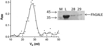
The protein could be expressed in, and purified from, E. coli (Supplementary Figure S2a). Cross-linking with BS3 showed that the protein is likely to be a homodimer in solution (Supplementary Figure S2b). In common with human and Saccharomyces cerevisiae GALE proteins, higher order oligomers were also observed in this cross-linking experiment (Supplementary Figure S2b (Timson, Reference Timson2005; Scott and Timson, Reference Scott and Timson2007; McCorvie et al. Reference McCorvie, Liu, Frazer, Gleason, Fridovich-Keil and Timson2012). This was further demonstrated by analytical gel filtration analysis which estimated a solution molecular mass for the protein of 71 kDa, consistent with a globular homodimer (Fig. 1).
Dimerization of FhGALE. FhGALE (90 μ m) was analysed by gel filtration using a Sephacryl S-300 (Pharmacia) column of total volume (V t) 46·0 mL and void volume (V 0, estimated using blue dextran) 14·6 mL. The protein eluted in 1 major peak (left) with an elution volume (V e) of 28·5 mL corresponding to a K av of 0·428 and a molecular mass of 71 kDa. That this peak contained FhGALE was confirmed by SDS-PAGE (right). L, FhGALE as loaded onto the column; 28 and 29, material from the fractions corresponding to 28 and 29 mL. The sizes of molecular mass markers (M) are shown to the left of the gel in kDa.

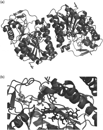
A molecular model of the dimeric protein was generated (Fig. 2). FhGALE is structurally similar to HsGALE (PDB: 1EK6 (Thoden et al. Reference Thoden, Wohlers, Fridovich-Keil and Holden2000)) with a root mean squared deviation (rmsd) of 1·378 Å over 4421 equivalent atoms. It is slightly less similar to Trypanosoma brucei GALE (PDB: 1GY8 (Shaw et al. Reference Shaw, Bond, Roper, Gourley, Ferguson and Hunter2003)) with an rmsd of 2·067 Å over 3671 equivalent atoms. By comparison with HsGALE, it was predicted that Tyr-155 is the catalytically important tyrosine in FhGALE.
Predicted 3-dimensional structure of FhGALE. (a) The overall predicted fold of the FhGALE dimer with the NAD+ cofactor shown in red. (b) A close up of the active site of FhGALE showing the NAD+ cofactor in red, the product, UDP-glucose, in pink and the catalytically important Tyr-155 residue in orange.

FhGALE binds NAD+, is stabilized by substrate binding and has Michaelis–Menten kinetics
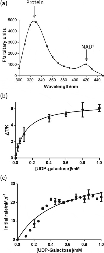
It has been demonstrated previously that the presence of NAD+ in human GALE causes a small, but detectable Förster resonance energy transfer (FRET). Irradiation of the protein at 280 nm results in a large tryptophan emission peak at ~330 nm. However, some of the emission transfers to the NAD+ which emits at ~420 nm (McCorvie et al. Reference McCorvie, Liu, Frazer, Gleason, Fridovich-Keil and Timson2012). A similar effect was observed with FhGALE (Fig. 3a).
FhGALE ligand binding and kinetics. (a) The intrinsic fluorescence spectrum of FhGALE (excitation 280 nm) shows 2 peaks, a major one at ~330 nm corresponding to the aromatic residues in the protein and a smaller one at ~420 nm corresponding to FRET from the NAD+ cofactor. (b) The change in melting temperature (ΔT m) of FhGALE (3·0 μ m) depends on the UDP-galactose concentration. Each point represents the mean of 3 independent determinations and the error bar represents the s.e.s of these means. (c) The substrate dependence of the initial rate was determined and plotted as a function of substrate concentration (see Materials and methods). Each point represents the mean of 3 independent determinations and the error bar represents the s.e.s. of these means.

In the absence of added ligands, the melting temperature (T m) of recombinant FhGALE (3·0 μ m) was 49·5±0·8 °C. UDP-galactose increased the T m in a concentration-dependent manner and the apparent dissociation constant (K D,app) was estimated to be 0·13±0·01 mm (Fig. 3b). NAD+ had no measurable effect on the T m (data not shown).
The activity of FhGALE shows a saturatable, hyperbolic dependence on UDP-galactose concentration (Fig. 3c). The K m for UDP-galactose is 470±90 μ m which is higher than the human enzyme (69 μ m (Timson, Reference Timson2005)). The turnover number was estimated to be 2·3±0·2 s−1, which is substantially lower than that for the human enzyme (36 s−1 (Timson, Reference Timson2005)).
FhGALE is inhibited by a range of different types of compound
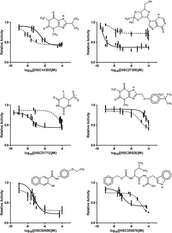
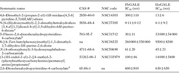
The molecular model of FhGALE was used to identify possible binding molecules from the NCI DTP repository. A virtual screen of the NCI diversity set III against the model was conducted using both AutoDock Vina (Trott and Olson, Reference Trott and Olson2010) and Schroedinger's Glide (Friesner et al. Reference Friesner, Banks, Murphy, Halgren, Klicic, Mainz, Repasky, Knoll, Shelley, Perry, Shaw, Francis and Shenkin2004, Reference Friesner, Murphy, Repasky, Frye, Greenwood, Halgren, Sanschagrin and Mainz2006; Halgren et al. Reference Halgren, Murphy, Friesner, Beard, Frye, Pollard and Banks2004). The individual docking results were combined into a consensus list of compounds that scored well with both methods. The top 40 hits from that list were obtained and used in in vitro testing (Supplementary Table 1). Of these compounds, 31 were soluble in 1% (v/v) DMSO/50 mm HEPES-OH. Twelve compounds reduced the rate of NADH production in an initial screen and 1 increased it; of these 1 (NSC91378) has been shown to affect the rate of the coupling enzyme UDP-glucose dehydrogenase and was not studied further. Six compounds (NSC13575, 16722, 50690, 91355, 116702 and 139105) gave unreliable or inconsistent results in concentration-dependent assays and so were eliminated. The ability of the remaining 6 compounds to inhibit FhGALE and HsGALE was then tested. All 6 compounds inhibited both enzymes (Fig. 4, Table 1). One, NSC27305, gave a sub-nanomolar IC50 under the conditions tested. Four more (NSC14303, NSC31712, NSC50690 and NSC335979) had submicromolar IC50 values and only 1 (NSC36525) had an IC50 in the millimolar range. This identification of high-affinity binders in the first round of screening gives us confidence that additional inhibitors in the nanomolar range can be discovered in future.
Six novel inhibitors of FhGALE. In each case, the solid line represents the fit to the data for FhGALE and the dashed line represents the fit for HsGALE. Each point represents the mean of 3 independent determinations and the error bar represents the s.e.s. of these means.

IC50 values for FhGALE inhibitors

a 2′,3′-O-isopropylidene-6-mercaptopurine riboside.
b 5-Fluoroorotate.
c (N-[(benzyloxy)carbonyl]leucyltryptophan).
d Orotate.
NSC27305 and NSC50690 inhibited both enzymes to approximately the same extent (as judged by their respective IC50 values). NSC14303 and NSC36525 inhibited the human enzyme to a greater extent than the liver fluke one. However, 2 compounds, NSC31712 and NSC335979, inhibited FhGALE several orders of magnitude more strongly than HsGALE (Fig. 4, Table 1). Both these compounds also affected the thermal stability of FhGALE, reducing the T m in a concentration-dependent manner (Supplementary Figure S3). Destabilization can result from the compound preferentially binding to partially unfolded forms of the protein (Cooper et al. Reference Cooper, Nutley, Wadood, Harding and Chowdhury2001). The K D,app values for NSC31712 and NSC335979 were 1·9±0·9 and 0·21±0·02 mm, respectively. It was not possible to get meaningful fits to the T m data with HsGALE and these compounds.
NSC31712 is 5-fluoroorotate (5-fluoro-2,6-dioxohexahydropyrimidine-4-carboxylate, 5FOA; CAS# 703-95-7). This compound is widely used in yeast molecular biology to select for transformants (Boeke et al. Reference Boeke, LaCroute and Fink1984). It has also been tested as a potential anti-cancer and anti-malarial agent (Heidelberger et al. Reference Heidelberger, Griesbach, Montag, Mooren, Cruz, Schnitzer and Grunberg1958; Heath et al. Reference Heath, Lopez, Stern and Papahadjopoulos1985; Rathod et al. Reference Rathod, Khatri, Hubbert and Milhous1989; Muregi et al. Reference Muregi, Kano, Kino and Ishih2009; Riviere et al. Reference Riviere, Kieler-Ferguson, Jerger and Szoka2011). In these applications, the target is not GALE; 5FOA can be converted to 5-fluorouracil which then inhibits thymidylate synthase – an enzyme required for the synthesis of thymidine monophosphate (dTMP) (Chaudhuri et al. Reference Chaudhuri, Montag and Heidelberger1958; Heidelberger et al. Reference Heidelberger, Griesbach, Montag, Mooren, Cruz, Schnitzer and Grunberg1958; Boeke et al. Reference Boeke, LaCroute and Fink1984). In malarial parasites, 5FOA also directly targets and inhibits thymidylate synthase (Rathod et al. Reference Rathod, Leffers and Young1992).
NSC335979 is the peptide derivative, N-[(benzyloxy)carbonyl]leucyl tryptophan (3-(1H-indol-3-yl)-2-[[4-methyl-2-(phenylmethoxy carbonylamino)pentanoyl]amino]propionate; CAS# 5326-00-3). Only limited pharmacological characterization is available for this compound: of the 114 bioassays currently recorded in PubChem (http://pubchem.ncbi.nlm.nih.gov/assay/assay.cgi?cid=333588), 113 showed no activity and 1 was considered inconclusive.
The fluorine atom in 5FOA is critical for selectivity over HsGALE
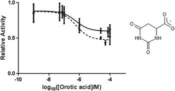
Docking of 5FOA into the FhGALE model structure, followed by alignment of the human structure revealed no structural differences in the predicted binding site which would be likely to account for the selectivity (data not shown). Interestingly, when orotic acid (which lacks the fluorine atom of 5FOA but is otherwise structurally identical) was tested as an inhibitor of both FhGALE and HsGALE, no selectively was observed (Fig. 5, Table 1).
Unlike 5FOA, orotic acid does not distinguish between FhGALE and HsGALE. The solid line represents the fit to the data for FhGALE and the dashed line represents the fit for HsGALE. Each point represents the mean of 3 independent determinations and the error bar represents the s.e.s. of these means.

Concluding remarks
Overall, FhGALE has similar properties to human GALE and the majority of other GALE enzymes characterized to date. It is a homodimer in solution and is stabilized by substrate binding. Despite these similarities, selective sub-micromolar inhibition could be obtained with 2 structurally different compounds. Further in silico and biochemical work will be required to elucidate the molecular basis of this specificity. This will facilitate the further development of selective FhGALE inhibitors. FhGALE may also have potential as a vaccine target: recent work has shown that Schistosoma mansoni GALE is located on the worm's tegument and that vaccination with the recombinant protein provides some protection against infection (Liu et al. Reference Liu, Shi, Yang, Cao, Shi, Li, Liu, Lin and Jin2012). Furthermore, this tegumental location may also indicate that the protein is involved in the production of the complex oligosaccharides found in the glycocalyx. If so, inhibition of FhGALE would have at least 2 negative consequences for the parasite: disruption of intermediary metabolism similar to that observed in type III galactosaemia and reduced ability to produce tegumental glycoproteins and glycolipids. These glycosylated molecules have been shown to be important in infection and host immune system evasion (Hanna, Reference Hanna1980; Duffus and Franks, Reference Duffus and Franks1981; Wilson et al. Reference Wilson, Wright, de Castro-Borges, Parker-Manuel, Dowle, Ashton, Young, Gasser and Spithill2011; Vukman et al. Reference Vukman, Adams, Metz, Maurer and O'Neill2013). In this context, it is interesting to note that galactose moieties have been observed in tegumental glycoproteins (Hanna, Reference Hanna1976). These observations and our identification of selective inhibitors of FhGALE suggest that attempts to disrupt galactose metabolism in F. hepatica may be a useful line of enquiry in the search for novel drugs to control fascioliasis.
SUPPLEMENTARY MATERIAL
To view supplementary material for this article, please visit http://dx.doi.org/10.1017/S003118201400136X.
ACKNOWLEDGEMENTS
We thank Prof. Aaron Maule (Queen's University, Belfast) for access to a qPCR machine used for protein T m determinations. S. L. was supported by the American Heart Association (12POST11570005) and the Center for Theoretical Biological Physics. We would like to thank Prof. Dr J. Andrew McCammon for helpful discussions. This work was supported in part by grants to the McCammon lab from the National Institutes of Health, the National Science Foundation, the Howard Hughes Medical Institute, the National Biomedical Computation Resource and the NSF Supercomputer Centers. Computational resources were supported, in part, by the National Science Foundation grant PHY-0822283, the Center for Theoretical Biological Physics.