INTRODUCTION
Neospora caninum is an apicomplexan parasite, which causes neoporosis in cattle and dogs. The disease is characterized by abortion, stillbirth or calves born with congenital infection, and neuromuscular dysfunction in dogs (Dubey and Lindsay, Reference Dubey and Lindsay1996; Hemphill, Reference Hemphill1999; Dubey et al. Reference Dubey, Barr, Barta, Bjerkas, Björkman, Blagburn, Bowman, Buxton, Ellis, Gottstein, Hemphill, Hill, Howe, Jenkins, Kobayashi, Koudela, Marsh, Mattsson, McAllister, Modry, Omata, Sibley, Speer, Trees, Uggla, Upton, Williams and Lindsay2002). Although phylogenetically closely related to Toxoplasma gondii, N. caninum differs from T. gondii with regard to its ultrastructure (Hemphill et al. Reference Hemphill, Vonlaufen, Naguleswaran, Guetg, Srinivasan and Alaeddine2004), antigenic composition (Howe and Sibley, Reference Howe and Sibley1999; Hemphill et al. Reference Hemphill, Vonalufen and Naguleswaran2006), and host cell recognition (Naguleswaran et al. Reference Naguleswaran, Cannas, Keller, Vonlaufen, Björkman and Hemphill2002, Reference Naguleswaran, Müller and Hemphill2003), all most likely contributing to its host-range specificity and distinct pathology (Dubey et al. Reference Dubey, Barr, Barta, Bjerkas, Björkman, Blagburn, Bowman, Buxton, Ellis, Gottstein, Hemphill, Hill, Howe, Jenkins, Kobayashi, Koudela, Marsh, Mattsson, McAllister, Modry, Omata, Sibley, Speer, Trees, Uggla, Upton, Williams and Lindsay2002). However, the infective stages of both species are capable of invading, and proliferating within, a wide range of tissues and cell types.
The physical interaction between host cells and members of the apicomplexa involves the sequential secretion from specialized organelles named micronemes, rhoptries and dense granules. Corresponding secreted proteins mediate adhesion to the host cell surface, host cell entry, formation of an intracellular parasitophorous vacuole in which the parasites reside, and modification and maturation of this vacuole (Mauel, Reference Mauel1996; Black and Boothroyd, Reference Black and Boothroyd2000; Carruthers et al. Reference Carruthers, Hakansson, Giddings and sibley2000a, Reference Carruthers, Sherman and Sibleyb; Tomley and Soldati, Reference Tomley and Soldati2001; Binder and Kim, Reference Binder and Kim2004; Mercier et al. Reference Mercier, Adjogble, Daubener and Delauw2005). An increasing body of literature demonstrates the important role of proteases in host cell adhesion and invasion by apicomplexan parasites. Toxoplasma and Plasmodium express serine and cysteine proteases required for attachment and penetration. These proteases are responsible for trimming of terminal peptides or segmentation of adhesive proteins, originating from micronemes, into multiple fragments. This eventually leads to activation of these adhesins and mediates improved binding activities to host cell surface receptors. The apically discharged adhesins then form a bridge connecting host receptors with the internal motility machinery comprised of a parasite-derived actin-myosin system (Dobrowolsky et al. Reference Dobrowolsky, Carruthers and Sibley1996; Buscaglia et al. Reference Buscaglia, Coppens, Hol and Nussenzweig2003; Jewett and Sibley, Reference Jewett and Sibley2003; Carruthers and Blackman, Reference Carruthers and Blackman2005).
Although the mechanisms leading to host cell adhesion and invasion in apicomplexans are generally regarded as conserved across the phylum, we have previously reported on distinct differences between Toxoplasma and Neospora with regard to the utilization of host cell receptors and susceptibilities to protease inhibitors. For instance, pre-treatment of Toxoplasma tachyzoites with protease inhibitors such as phenanthroline, E64 and pepstatin severely affected host cell interactions, while treatment with PMSF, a serine protease inhibitor, lead to a significant reduction in host cell invasion (Conseil et al. Reference Conseil, Soete and Dubremetz1999; Naguleswaran et al. Reference Naguleswaran, Müller and Hemphill2003). Conversely, neither cysteine-, metallo- nor serine protease inhibitors affected the host cell interaction of N. caninum tachyzoites, but invasion was drastically reduced by the aspartic protease inhibitor pepstatin-A (Naguleswaran et al. Reference Naguleswaran, Müller and Hemphill2003). Nevertheless, in N. caninum micronemes a subtilisin-like serine protease (NcSUB1; Louie and Conrad, Reference Louie and Conrad1999; Louie et al. Reference Louie, Nordhausen, Robinson, Barr and Conrad2002), homologous to the corresponding enzyme TgSUB1 in T. gondii (Miller et al. Reference Miller, Binder, Blackman, Carruthers and Kim2001), has been identified, and a putative role of this protease in host cell invasion has been discussed.
Investigations on host cell surface glycosaminoglycans as potential receptors demonstrated that N. caninum tachyzoites preferentially bound to host cell surface chondroitin sulfates, while T. gondii tachyzoites preferentially adhered to heparan sulfate glycosaminglycans on the host cell surface (Naguleswaran et al. Reference Naguleswaran, Cannas, Keller, Vonlaufen, Björkman and Hemphill2002, Reference Naguleswaran, Müller and Hemphill2003). Although cell surface-associated terminal sialic acid residues are known to be important elements in host cell recognition, binding and invasion by viruses, bacteria and protozoan parasites (Vimr and Lichtensteiger, Reference Vimr and Lichtensteiger2002), few reports exist on the role of sialic acid moieties during Neospora infection. Treatment of Vero cell monolayers with sialidase did not result in any notable alteration of the adhesive or invasive capacities of N. caninum tachyzoites (Naguleswaran et al. Reference Naguleswaran, Müller and Hemphill2003). This was confirmed by Vonlaufen et al. (Reference Vonlaufen, Guetg, Naguleswaran, Muller, Bjorkman, Schares, von Blumroeder, Ellis and Hemphill2004) who demonstrated that enzymatic removal of terminal sialic acid residues from either the host cell or the parasite surface increased the invasive capacities of bradyzoites, but not tachyzoites.
In this study, we found that pre-treatment of N. caninum tachyzoites with the sialidase inhibitor 2-deoxy-2,3-dehydro-N-acetylneuraminic acid reduced host cell adhesion of bradyzoites and to a lesser degree, also of tachyzoites, and incubation of tachyzoites with fetuin significantly increased the adhesive capacity of N. caninum tachyzoites to Vero cell monolayers. This pointed towards an as yet undefined role that sialic acid might play in the process of host cell recognition. Thus, N. caninum tachyzoite extracts were subjected to fetuin-agarose affinity chromatography, in order to isolate proteins potentially interacting with fetuin and/or sialic acid residues, and which could play a role in the host-parasite relationship. We here report on the characterization of this fetuin-binding fraction.
MATERIALS AND METHODS
Buffers and reagents
Unless otherwise stated, all reagents, chemicals and tissue culture media were purchased from Sigma (St Louis, MO).
Tissue culture and parasite purification
Cultures of Vero cells were maintained in RPMI 1640 medium (Gibco-BRL, Basel, Switzerland) supplemented with 10% FCS, 2 mm glutamine, 50 U of penicillin/ml and 50 μg of streptomycin/ml at 37°C with 5% CO2 in tissue culture flasks. Cultures were trypsinized at least once a week. Neospora caninum tachyzoites of the Nc-Liverpool isolate (Barber et al. Reference Barber, Holmdahl, Owen, Guy, Uggla and Trees1995) were maintained in Vero cell monolayer cultures during the time of which FCS was replaced with IgG-free horse serum. N. caninum tachyzoites were harvested as described previously (Hemphill, Reference Hemphill1996).
Induction of N. caninum tachyzoite-to-bradzoite stage conversion
In vitro stage conversion was carried out in sodium nitroprusside (SNP)-treated Vero cells according to the procedure described by Vonlaufen et al. (Reference Vonlaufen, Guetg, Naguleswaran, Muller, Bjorkman, Schares, von Blumroeder, Ellis and Hemphill2004). Briefly, Vero cell monolayers grown on glass cover-slips in 24-well tissue culture plates were infected with 105/ml tachyzoites of the Nc-Liverpool isolate. The infected monolayers were cultured at 37°C with 5% CO2 for 8 days with daily addition of 17 μm SNP.
Protein extraction and fetuin-agarose affinity purification
Proteins were extracted from 1×10⁸/ml freshly isolated tachyzoites in an extraction buffer (100 mm phosphate, pH 6·8 containing 0·1% Triton X-100 and 1 mm PMSF) on ice by vortexing for 5 min. The protein suspension was centrifuged at 10 000 g for 30 min to remove the insoluble debris and the supernatant was used for fetuin agarose affinity chromatography. Fetuin-agarose was equilibrated with 100 mm phosphate buffer (pH 6·8) and the protein extract was loaded onto the affinity column. The column was washed extensively with 100 mm phosphate buffer (pH 6·8) containing 500 mm NaCl to remove non-specifically bound proteins, and the fetuin-binding protein fraction was eluted with 100 mm methylmannoside in 100 mm phosphate buffer (pH 6·8), aliquoted and stored at −80°C.
SDS-PAGE, immunobloting and affinity purification of antibodies
The fractions eluted from fetuin-agarose affinity chromatography and N. caninum total extract were precipitated in methanol-chloroform (Wessel and Fluegge, Reference Wessel and Fluegge1984) and separated by SDS-PAGE under reducing conditions. Protein bands were visualized by silver staining. Immunoblots were prepared by electrophoretically transferring SDS-PAGE-separated proteins onto nitrocellulose, and non-specific binding sites were blocked in 3% bovine serum albumin (BSA) in TBS-Tween (20 mm Tris-HCl pH 7·6, 150 mm NaCl, 0·3% Tween 20) overnight at 4°C. Blots were labelled with anti-N. caninum hyperimmune serum (Hemphill et al. Reference Hemphill, Gottstein and Kaufmann1996) diluted in TBS-Tween/0·3% BSA for 2 h at room temperature. After extensive washing in TBS-Tween, bound antibodies were visualized using goat anti-rabbit-alkaline phosphatase conjugate (Promega) according to the instructions provided by the manufacturer. Affinity purification of monospecific antibodies directed against the 65 kDa protein (NcFBP) out of a polyclonal anti-N. caninum antiserum was carried out as previously described (Sonda et al. Reference Sonda, Fuchs, Gottstein and Hemphill2000). Antibodies were supplemented with 0·3% BSA, aliquoted and stored at −20°C. For immunoblotting, affinity-purified antibodies were used at a dilution of 1:200.
Immunofluorescence localization of NcFBP
Immunofluorescence staining was performed as described by Vonlaufen et al. (Reference Vonlaufen, Guetg, Naguleswaran, Muller, Bjorkman, Schares, von Blumroeder, Ellis and Hemphill2004) with tachyzoites and bradyzoites grown in Vero cell monolayers on glass cover-slips in 24-well tissue culture plates, either in the absence or presence of SNP. The following primary antibodies were diluted in PBS/0·3% BSA: (i) NcmAb-4, a monoclonal antibody directed against the immunodominant tachyzoite surface antigen NcSAG1 (Björkman and Hemphill, Reference Björkman and Hemphill1998) was used at 1 μg/ml; (ii) affinity-purified polyclonal rabbit anti-NcFBP antibody was diluted at 1:1 dilution; (iii) mAbCC2, a rat mAb reacting with a T. gondii cyst wall protein (Gross et al. Reference Gross, Bormuth, Gaissmaier, Dittrich, Krenn, Bohne and Ferguson1995), was diluted at 1:100. Incubation with primary antibodies was performed for 1 h, followed by 3 washes in PBS, 5 min each. Bound antibodies were detected with the appropriate tetramethylrhodamine isothiocyanate (TRITC)- or fluorescein isothiocyanate (FITC)-conjugated secondary antibodies. Finally, the preparations were washed in PBS 3 times for 5 min each and incubated in the fluorescent dye Hoechst 33258 (1 μg/ml in PBS) for 2 min, rinsed again in PBS and mounted in Fluoroprep (Biomerieux S.A.). All specimens were viewed on a Nikon Eclipse E 800 digital confocal fluorescence microscope. Processing of images was performed using the Openlab 2.0.7 software (Improvision).
Transmission electron microscopy (TEM) and immunogold labelling
For TEM, N. caninum-infected Vero cell cultures were fixed in 100 mm sodium cacodylate, pH 7·2, containing 2% paraformaldehyde for 30 min at 20°C. LR-White-embedding and on-section labelling was performed essentially as previously described (Vonlaufen et al. Reference Vonlaufen, Guetg, Naguleswaran, Muller, Bjorkman, Schares, von Blumroeder, Ellis and Hemphill2004), Sections were incubated in affinity-purified anti-FBP diluted 1:1 in PBS/0·3% BSA for 1 h. After washing in 5 changes of PBS, 2 min each, the goat anti-rabbit antibody conjugated to 10 nm gold particles (purchased from Amersham, Zürich, Switzerland) was applied (Vonlaufen et al. Reference Vonlaufen, Guetg, Naguleswaran, Muller, Bjorkman, Schares, von Blumroeder, Ellis and Hemphill2004). Finally, grids were stained with lead citrate and uranyl acetate and were subsequently viewed on a Phillips 400 transmission electronmicroscope operating at 80 kV.
Gelatin zymography
Protein fractions were precipitated with 80% pre-cooled acetone for 30 min on ice. The protein was resuspended in native sample buffer and separated on 10% SDS-PAGE containing 0·1% gelatin. After electrophoresis, gels were washed twice with 2·5% Triton X-100 for 30 min to remove the SDS and to re-nature the proteins. After rinsing with water, gels were incubated overnight at 37°C in an incubation buffer (50 mm Tris, pH 7·6, containing 50 mm NaCl and 10 mm CaCl2) with gentle shaking. In some experiments, 1 mm 1,10-phenanthroline or 1 mm ZnCl2 were added to the incubation buffer. Following overnight incubation, gelatinolytic activity was visualized with Coomassie brilliant blue G250 stain as clear bands on a blue background.
In vitro secretion assay
Freshly purified tachyzoites (108/ml) were resuspended in Earl's balanced salt solution (EBSS) and were incubated either on ice or at 37°C for 10 min. In some experiments, a cocktail of protease inhibitors (1 mm PMSF, 0·5 mm 1,10-phenanthroline, 0·02 mm E64 and 0·1 mm pepstatin-A) was added. After incubation, parasites were centrifuged at 2000 g and the supernatant was re-centrifuged at 10 000 g for 10 min at 4°C. Proteins arising from equal number of parasites from supernatants and pellets were separated on SDS-PAGE and immunoblots were performed using anti-NcFBP antibodies. Antibodies directed against NcMIC4, a secretory N. caninum microneme protein (Keller et al. Reference Keller, Riesen, Naguleswaran, Vonlaufen, Stettler, Leepin, Wastling and Hemphill2004) were included as a control.
Assessment of the effects of fetuin, anti-NcFBP antibodies and the sialidase inhibitor 2-deoxy-2,3-dehydro-N-acetylneuraminic acid on N. caninum tachyzoite and bradyzoite host cell interaction
Freshly purified tachyzoites (105) were resuspended in 200 μl of RPMI 1640 medium. The suspension was supplemented either with fetuin (1 mg/ml) and incubated at 37°C for 30 min, or with 50 μl affinity-purified anti-NcFBP antibodies (diluted 1:1), or anti-beta-galactosidase (control) antibodies and incubated at 4°C (Hemphill, Reference Hemphill1996; Naguleswaran et al. Reference Naguleswaran, Alaeddine, Guionaud, Vonlaufen, Sonda, Jenoe, Mevissen and Hemphill2005). In some experiments, parasites were pre-incubated with fetuin (1 mg/ml) for 30 min at 37°C and subsequently re-incubated with affinity purified antibodies at 4°C for 30 min. In other assays, the sialidase inhibitor 2-deoxy-2,3-dehydro-N-acetylneuraminic acid (10 μm) was added for 30 min at 37°C to tachyzoites and bradyzoites, followed by a centrifugation step to remove the sialidase inhibitor. The treated parasites were then allowed to interact with the Vero cell monolayer for 30 min at 37°C. The unbound parasites were washed with medium and tachyzoites associated with host cells were quantified by LightCycler PCR according to the method of Müller et al. (Reference Müller, Vonlaufen, Gianinazzi, Leib and Hemphill2002). In some experiments, the PDTC-PCR-based adhesion/invasion assay according to Naguleswaran et al. (Reference Naguleswaran, Müller and Hemphill2003) was used to specifically quantify intracellular and extracellular tachyzoites and bradyzoites.
NcFBP – Vero cell co-sedimentation assays
These assays were done as previously described (Naguleswaran et al. Reference Naguleswaran, Cannas, Keller, Vonlaufen, Björkman and Hemphill2002; Keller et al. Reference Keller, Riesen, Naguleswaran, Vonlaufen, Stettler, Leepin, Wastling and Hemphill2004). Freshly trypsinized, non-adherent Vero cells were fixed in 2·5% glutaraldehyde in EBSS for 30 min, followed by post-fixation in 0·5% OsO4 in 100 mm sodium phosphate buffer, pH 7·2. Subsequently, free aldehyde groups were blocked by incubation in 100 mm ethanolamine (pH 8) at 4°C overnight. Triton X-100 extracts of N. caninum tachyzoites were prepared by incubating 5×10⁸ freshly isolated N. caninum tachyzoites in 2 ml of PBS containing 1% Triton X-100 for 5 min at 4°C, followed by centrifugation at 10 000 g for 30 min at 4°C. Pre-fixed Vero cells (106) were then incubated in 250 μl of the Triton X-100 extracts for 2 h at 4°C. Subsequently the preparations were centrifuged at 10 000 g for 5 min, and the supernatants were collected and processed for SDS-PAGE and immunoblotting. The pellets were washed in PBS 3 times and were finally resuspended in SDS-PAGE sample buffer. Equal amounts of non-bound (supernatants) and bound (pellets) proteins were loaded onto gels. Immunoblotting was performed as above.
Binding of NcFBP to defined glycosaminoglycans
Binding assays were carried out as described (Naguleswaran et al. Reference Naguleswaran, Müller and Hemphill2003; Keller et al. Reference Keller, Riesen, Naguleswaran, Vonlaufen, Stettler, Leepin, Wastling and Hemphill2004). Ninety-six-well ELISA plates were coated with heparin, dextran, dextran sulfate, or chondroitin sulfate A, B, or C (CSA, CSB, CSC) (5 mg/ml) for 12 h at 4°C. Plates were washed and incubated with 100 μl of the Triton X-100 extracts of N. caninum tachyzoites for 2 h at 4°C. Specimens were then washed in PBS and nonspecific binding sites were blocked for 2 h with 1·5% BSA and 50 mm glycine in PBS. Bound NcFBP was detected using affinity-purified antibodies against NcFBP (diluted 1:50 in antibody dilution buffer), followed by the respective secondary antibodies conjugated to alkaline phosphatase. Antibody binding was visualized by using p-nitrophenylphosphate as a substrate and absorbance was measured at 405 nm on a Dynatech MR7000 enzyme-linked immunosorbent assay (ELISA) reader.
Proteolytic assays
For proteolysis of protein substrates, 200 μl of the eluted N. caninum fetuin-binding protein fraction and 20 μg of each fetuin, bovine immunoglobulin G and bovine serum albumin in 200 μl of incubation buffer were incubated at 37°C for 12 h. As a control, each protein substrate was incubated in incubation buffer and elution buffer. Protein degradation was assessed by SDS-PAGE followed by silver staining.
RESULTS
Involvement of sialic acid in the interaction between N. caninum and host cells
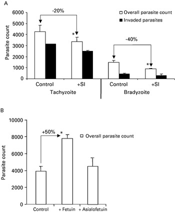
Pre-treatment of N. caninum with the sialidase inhibitor 2-deoxy-2,3-dehydro-N-acetylneuraminic acid resulted in a reduction in the number of parasites adhering/invading Vero cell monolayers, namely by 20% for tachyzoites and 40% for bradyzoites (Fig. 1A). The percentage of actually invaded parasites remained at similar levels independent of the treatments, i.e. 73% for the untreated tachyzoite control, 72% for the sialidase inhibitor-treated tachyzoites, and 28% and 30% for untreated bradyzoites and sialidase inhibitor-treated bradyzoites, respectively. This suggested that sialic acid could be involved in parasite attachment to host cells, but not necessarily in invasion. Fetuin, a liver-produced negative acute-phase protein rich in sialic acid content, was added to tachyzoites prior to letting them interact with Vero cell monolayers, and this resulted in a 2-fold increased number of parasites binding and/or invading the host cells (Fig. 1B). Addition of desialylated asialofetuin did not have any effect (data not shown), suggesting that the increase in host cell associated tachyzoites could be attributed to the presence of sialic acid residues.
Sialic acid residues are involved in Neospora caninum host cell interaction. (A) Incubation of N. caninum tachyzoites and bradyzoites with the sialidiase inhibitor 2-deoxy-2,3-dehydro-N-acetylneuraminic acid leads to a 20% and 40% reduction of parasites adhering to their host cells, respectively. However, the percentage of parasites invading the host cells remains the same. (B) The presence of fetuin leads to a massive increase (50%) of tachyzoites binding to host cells. The significance of the differences among the control and experimental assays was determined by Student's t-test using the Microsoft Excel program. P values <0·05 were considered statistically significant and are marked with *.

The N. caninum fetuin-binding fraction is constituted by a single protein of 65 kDa, which is expressed at the apical portion of N. caninum tachyzoites
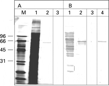
Analysis of the fetuin-binding protein fraction of N. caninum tachyzoites by SDS-PAGE under reducing conditions and silver staining revealed the presence of a single fetuin binding protein band of 65 kDa, which was named NcFBP. (Fig. 2A, lane 2). When proteins were separated under non-reducing conditions, silver staining failed to detect any protein (Fig. 2A, lane 3). Immunoblotting showed that the 65 kDa band was the only protein recognized by a hyperimmune serum directed against N. caninum tachyzoites (Fig. 2B, lane 2), but no immunolabelling could be observed when SDS-PAGE was carried out under non-reducing conditions (Fig. 2B, lane 3). Subsequently, affinity purification on the 65 kDa band yielded monospecific anti-NcFBP antibodies, and these specifically recognized the 65 kDa band on immunoblots of Neospora extracts (Fig. 2B, lane 4).
Analysis of fetuin-binding fraction by SDS-PAGE and immunoblotting. (A) Fetuin-affinity chromatography of Neospora caninum extracts was analysed by reducing (lanes 1, 2) and non-reducing (lane 3) SDS-PAGE and stained by silver stain. Lane 1, N. caninum Triton X-100 soluble extract prior to affinity chromatography, lane 2, eluted fraction containing the fetuin-binding protein (NcFBP), lane 3, eluted fraction separated under non-reducing conditions. (B) Immunoblot of the same fractions as in panel A. Lanes 1–3 were labelled with anti-N. caninum antiserum and detected by goat anti-rabbit antibody conjugated to alkaline phosphatase. Lane 4 shows an immunoblot of N. caninum Triton X-100 soluble extract labelled with affinity-purified anti-NcFBP antibodies and detected by goat anti-rabbit antibody conjugated to alkaline phosphatase.

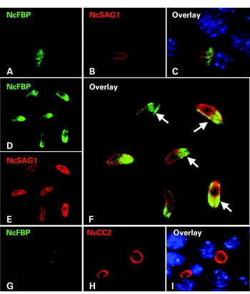
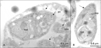
The affinity-purified antibodies localized the protein to the apical portion of both intracellular (Fig. 3A–C) and extracellular (Fig. 3D–F) tachyzoites, and closer inspection showed that at least a major portion of this protein expressed in these parasites was present on the tachyzoite surface (Fig. 3F). No cross-reaction of anti-NcFBP antibodies with host cells could be detected. Immunofluorescence labelling of parasites undergoing in vitro tachyzoite-to-bradyzoite stage conversion indicated that the expression of NcFBP during stage conversion was greatly reduced, or that the protein was modified in a way that rendered epitopes less accessible (Fig. 3G–I). Attempts to determine the exact subcellular localization of NcFBP by immunogold-TEM were hampered by the fact that anti-NcFBP antibodies recognized fixation-sensitive epitope(s): immunogold labelling of paraformaldehyde/glutaraldehyde–fixed samples did not yield any staining (data not shown). However, TEM of sections of infected cells fixed in 2% paraformaldehyde confirmed the predominant apical localization of NcFBP in both intracellular and extracellular tachyzoites. Gold particles appeared to bind preferentially to vesiculated structures at the anterior end resembling micronemes (see Fig. 4A), while rhoptries were not labelled (Fig. 4B).
Localization of the fetuin-binding protein. Immunofluorescence labelling of Neospora caninum tachyzoites grown in Vero cells (A–C) and purified parasites (D–F). Parasites were stained with anti-NcFBP antibodies and fluorescein isothiocyanate-conjugated anti-rabbit IgG (green) and with NcSAG1 antibody and tetramethyl rhodamine isothiocyanate-conjugated anti-mouse IgG (red). Nuclei were labelled with Hoechst 22358. (E–G) In vitro-generated bradyzoites exhibiting cyst wall staining with mAb CC2 (red) and down-regulated expression of NcFBP (green). Arrows in (C) and (D) highlight tachyzoites with NcFBP surface labelling.

Immunogold labelling of Neospora caninum-infected Vero cells employing anti-NcFBP antibodies. Cells were fixed in 2% paraformaldehyde and embedded in LR-White resin. Note the arrows pointing towards gold particles mostly associated with the apical part of the tachyzoites. Nuc, nucleus; pv, parasitophorous vacuole; con, conoid; rop, rhoptries.

NcFBP is continuously released by N. caninum tachyzoites irrespective of the temperature
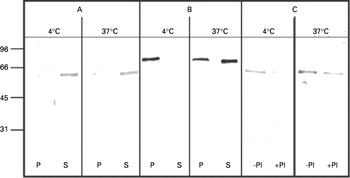
Experiments were undertaken to investigate whether NcFBP was part of the secretory fraction of N. caninum tachyzoites. Thus, purified tachyzoites were incubated either at 4°C or 37°C in EBSS, and parasite pellets and medium supernatants were collected and analysed by immunoblotting (Fig. 5A). The results showed that NcFBP was released or shed into the medium supernatant irrespective of the incubation temperature. As a control, the same samples were probed with anti-NcMIC4 antibodies, which detect a soluble microneme protein, NcMIC4 (Keller et al. Reference Keller, Riesen, Naguleswaran, Vonlaufen, Stettler, Leepin, Wastling and Hemphill2004). NcMIC4 could only be detected in the supernatant sample previously incubated at 37°C (Fig. 5B), indicating that active secretion has taken place. We incubated tachyzoites at 4°C and 37°C in the presence and absence of a protease inhibitor cocktail consisting of PMSF, pepstatin, E64 and phenanthroline, which would cover all 4 classes of proteases. Immunoblotting demonstrated that the release of NcFBP into the medium supernatant at both high and low temperature was strongly impaired (Fig. 5C), indicating that protease activity was required for release of NcFBP. However, when protease inhibitors were used individually, no effect was observed, indicating that more than one type of protease is involved in the release of NcFBP into the medium (data not shown).
Immunoblots of fractions secreted by tachyzoites incubated at 4°C or 37°C. Blots A and C were stained with anti-NcFBP antibodies and blot B was stained with anti-NcMIC4 antibodies. (S) Corresponds to the released fraction into the supernatant and the (P) corresponds to the pellet fraction. Blot C shows the release of NcFBP in the absence (−) or presence (+) of a cocktail of protease inhibitors (PI). Note the reduced release of NcFBP in the presence of a cocktail of protease inhibitors.

NcFBP binds to Vero cells and influences tachyzoite-host cell interaction
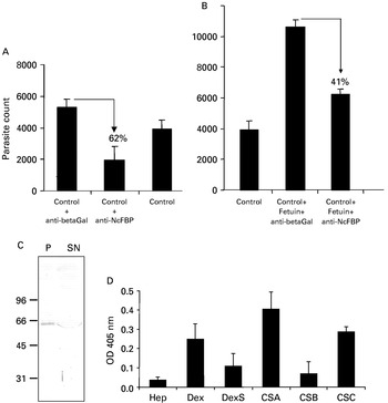
Anti-NcFBP antibodies had a profound negative impact on host cell adhesion by reducing the adhesion/invasion of tachyzoites to Vero cell monolayers by 62% compared to the control anti-beta galactosidase antibodies (Fig. 6A). In a further experiment, pre-incubation of N. caninum tachyzoites with fetuin enhanced the adhesion to Vero cells by 50% compared to the control, and this enhanced adhesive effect was reduced by 40%, when parasites pre-incubated with fetuin were re-incubated with anti-NcFBP antibodies prior to host cell interaction (Fig. 6B). Since anti-NcFBP antibodies reduced the adhesion of tachyzoites to Vero cell monolayers, we investigated whether NcFBP binds to the Vero cell surface. Triton X-100 extracts were incubated with pre-fixed Vero cells, and following centrifugation, co-sedimented and unbound proteins were subjected to SDS-PAGE and immunoblot analysis. Staining with anti-NcFBP antibodies showed that a substantial amount of NcFBP co-precipitated with the fraction bound to Vero cells (Fig. 6C). Moreover, solid-phase binding assays were performed to investigate whether glycosaminoglycans could potentially be involved in binding of NcFBP. ELISA wells were coated with dextran, dextran sulfate, heparin and CSA, CSB, and CSC. As shown in Fig. 6D, NcFBP exhibited an increased binding to CSA, CSC and dextran, while CSB, heparin and dextran sulfate did not bind NcFBP.
NcFBP interacts with host cells, and antibodies inhibit tachyzoite adhesion. The parasites were quantified by real-time PCR, and parasite count per PCR-reaction is given in the y-axis. Black columns indicate the overall number of parasites interacting with their host cell. Percentages given above the black columns indicate the reduction in the overall parasite number with respect to the control. Error bars indicate standard deviation. (A) Pre-treatment of tachyzoites with anti-NcFBP antibodies resulted in an adhesion inhibition of 62% compared to a control comprised of an irrevelant anti-beta-galactosidase antibody. (B) Pre-treatment of tachyzoites with fetuin increased the adhesion by 50% compared to the control. Pre-treatment of tachyzoites with fetuin followed by incubation with anti-NcFBP antibodies resulted in an adhesion inhibition of 41%. (C) Co-precipitation assay demonstrating binding of NcFBP to pre-fixed Vero cells. The immunoblot was stained with anti-NcFBP antibodies. Lane P (pellet) represents the protein fraction bound to Vero cells; lane SN (supernatant) represents non-bound proteins. (D) Binding of NcFBP to immobilized heparin, dextran, dextran sulfate, CSA, CSB, and CSC. Binding of NcFBP was assessed by labelling with anti-NcFBP antibodies and alkaline phosphatase-conjugated secondary antibodies. Note that NcFBP binds preferentially to dextran, CSA and CSC but not to other glycosaminoglycans. OD 405=optical density at 405 nm.

The fetuin-binding fraction of N. caninum tachyzoites contains metalloprotease activity directed against glycosylated proteins
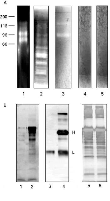
Since it was demonstrated earlier that human matrix metalloproteases bind to fetuin (Ochieng et al. Reference Ochieng, Warfield and Green1995), we investigated whether metalloprotease activity was present in the fetuin-binding fraction of N. caninum tachyzoites. Thus, total Neospora extract, secreted fraction of N. caninum tachyzoites, and fetuin-agarose eluted fraction, were examined by gelatin substrate zymography. The fetuin-agarose binding fraction exhibited 2 distinct protease bands at 96 kDa and 140 kDa, whereas the secreted fraction and the total Neospora extract revealed multiple proteolytic bands of different intensities, some of which co-migrated with the 2 bands of 96 kDa and 140 kDa in the fetuin-binding fraction (Fig. 7A). Fetuin-affinity chromatography of Vero cell extracts performed under identical conditions followed by zymography did not reveal any bands with protease activity (data not shown). In the presence of 1 mm of the zinc chelator 1,10-phenanthroline, the enzymatic activities in the fetuin-binding fraction were completely abrogated. Further, addition of an excess of Zn2+ to the incubation buffer completely inhibited the proteolytic activities. Proteolytic activity was not affected by the presence of PMSF, pepstatin and E64, respectively (data not shown). This showed that the presence of the 2 bands was attributable to metalloprotease activity.
Proteolytic activity of fetuin-binding fractions. (A) Gelatin substrate zymography. Lane 1, N. caninum Triton X-100 soluble extract, lane 2, secreted fraction of N. caninum tachyzoites at 4°C, lane 3, eluted fraction containing NcFBP, lanes 4 and 5, eluted fraction containing NcFBP incubated with 1,10-phenanthroline and 1 mm ZnCl2, respectively were electrophoresed on a 0·1% gelatin containing 10% polyacrylamide gel and further processed as described in the Materials and Methods. (B) Proteolytic degradation of glycosylated and non-glycosylated substrates of NcFBP. All reaction mixtures were incubated at 37°C for 12 h and degradation products were analysed by SDS-PAGE and visualized by silver stain. Lane 1, fetuin +NcFBP, lane 2, fetuin, lane 3, bovine immunoglobulin G +NcFBP, lane 4, bovine immunoglobulin G, lane 5, bovine serum albumin (BSA)+NcFBP, lane 6, BSA. Note the degradation of fetuin and bovine immunoglobulin G heavy chain (H). BSA and bovine immunoglobulin G light chain (L) remain undegraded.

To address the question whether fetuin, besides binding metalloprotease, could also act as a substrate, fetuin was incubated at 37°C, either in the presence or absence of the N. caninum fetuin-binding fraction, and the samples were analysed by SDS-PAGE and silver staining (Fig. 7B). When fetuin was incubated without the fetuin-binding fraction, the major protein band plus several other bands of lower Mr were visible. After incubation of fetuin in the presence of N. caninum fetuin-binding proteins, the fetuin was largely digested, reflecting strong proteolytic activity. Identical experiments using bovine immunoglobulin G as a substrate revealed that the glycosylated heavy chain was completely digested, whereas the non-glycosylated light chain remained largely unaffected (Fig. 7B). In contrast, bovine serum albumin, which represents a non-glycosylated substrate, was not affected by incubation with the fetuin-binding protein fraction (Fig. 7B). These experiments indicated that the metalloprotease activity was largely targeted towards glycoproteins, while non-glycosylated polypeptides remained largely unaffected.
DISCUSSION
Sialic acids are a structurally complex family of 9-carbon monosaccharides, that are involved in diverse activities related to glycoproteins and glycoconjugates, including host-pathogen interactions (Kelm and Schauer, Reference Kelm and Schauer1997; Vimir and Lichtensteiger, Reference Vimr and Lichtensteiger2002). The fact that functional inhibition of sialidase activity in Neospora caninum negatively affects tachyzoite host cell adhesion, but not invasion (see Fig. 1A), indicates, that sialic acid could also play a role in the physical relationship between N. caninum and its host cell. In the presence of fetuin, a highly sialylated glycoprotein with a triantennary glycan part that contains galactose, mannose, glucosamine and galactosamine as sugar molecules (Johnson and Heath, Reference Johnson and Heath1986), N. caninum tachyzoites bound much more efficiently to host cells compared to controls (Fig. 1B). It is likely that fetuin plays a similar role in adhesion as previously reported for laminin and Toxoplasma gondii (Furtado et al. Reference Furtado, Cao and Joiner1992a) where laminin was proposed to act as a bridging molecule, interacting with both host and parasite cell surface constituents such as the integrin receptor alpha6 beta 1 in human fibroblasts (Furtado et al. Reference Furtado, Slowik, Kleinman and Joiner1992b). Fetuin is regularly contained in FCS, which is used as an additive in culture medium, and in this context could contribute for efficient host cell interaction in vitro. However, fetuin is mainly a fetal protein in the sense that the highest concentrations are found in serum and body fluids of embryos and fetuses (Terkelsen, Reference Terkelsen, Jahnen-Dechent, Nielsen, Moos, Fink, Nawratil, Muller-Esterl and Mollgard1998), and this could be of relevance in vivo as neosporosis is mostly of concern in relation to fetal infection and disease in neonates.
In this study, we report on the characterization of a fetuin-binding fraction of N. caninum tachyzoites. In this fraction, silver staining revealed the presence of a single protein band of 65 kDa, and zymography demonstrated profound metalloprotease activity in 2 bands of 96 and 140 kDa, respectively. This is the first description of metalloproteases identified in N. caninum.
A 65 kDa fetuin-binding protein (NcFBP) is involved in host cell attachment of N. caninum tachyzoites
The purification of a single 65 kDa protein (NcFBP) was evident upon separation of the fetuin-binding fraction by SDS-PAGE under reducing conditions, while under non-reducing conditions this protein could not be resolved by silver staining nor by immunoblotting. Several attempts to identify the 65 kDa NcFBP by tandem mass spectrometry have not been successful (data not shown), most likely due to the fact that the sequence of the Neospora genome is not fully available to date, and the expression level of this protein could be low and thus the corresponding sequence not well represented in the current EST-database. Further studies are required to elucidate the exact molecular identity of NcFBP. NcFBP was the only protein in the fetuin-binding fraction that was recognized by polyclonal anti-N. caninum antiserum, allowing the preparation of affinity-purified anti-NcFBP antibodies that exhibited high specificity on immunoblots. Immunofluorescence showed that the protein was concentrated at the apical part of intracellular, and the apical part as well as the surface of extracellular, tachyzoites. This was largely confirmed by immunogold TEM, which indicated that NcFBP was a component of microneme-like vesicles. This suggested that NcFBP could be transported onto the parasite surface and then released into the exterior medium. Indeed, secretion assays employing extracellular tachyzoites demonstrated that NcFBP was released into the medium supernatant independently of the metabolic state of the parasites, e.g. at high (37°C) as well as at low (4°C) temperature. Release of NcFBP did not simply reflect leakage by dead parasites, as another secretory and soluble protein, NcMIC4 (Keller et al. Reference Keller, Riesen, Naguleswaran, Vonlaufen, Stettler, Leepin, Wastling and Hemphill2004), was not present in the 4°C supernatants. Also other micronemal proteins such as NcMIC1, NcMIC2 and NcMIC3 had been reported earlier to be secreted in substantial amounts at 37°C, but not at 4°C (Lovett et al. Reference Lovett, Howe and Sibley2000; Naguleswaran et al. Reference Naguleswaran, Cannas, Keller, Vonlaufen, Schares, Conraths, Bjorkman and Hemphill2001; Keller et al. Reference Keller, Riesen, Naguleswaran, Vonlaufen, Stettler, Leepin, Wastling and Hemphill2004). This suggests that surface-associated NcFBP reaches the medium supernatant by a process distinct of the one described for other secretory components of apicomplexans, whereas the secretory events involving microneme and rhoptry proteins represent active processes requiring metabolic energy and protease activity on part of the parasite (Carruthers, Reference Carruthers1999; Joiner and Roos, Reference Joiner and Roos2002; Binder and Kim, Reference Binder and Kim2004). Nevertheless, release of NcFBP into the medium was also dependent on functional protease machinery, as the addition of a cocktail of protease inhibitors inhibited the release of the protein. In fact, release of NcFBP into the medium supernatant appears to be more efficient at 37°C compared to 4°C, but the mechanisms involved in NcFBP shedding remain to be elucidated.
NcFBP is likely to be one of the factors mediating tachyzoite host cell attachment, since (i) antibodies directed against NcFBP inhibited parasite adhesion, and (ii) also the enhanced binding of tachyzoites to host cells in the presence of fetuin was largely neutralized by anti-NcFBP antibodies (iii) NcFBP can bind to the surface of Vero cells, and (iv) also binds to sulfated glycosaminoglycans, especially chondroitinsulfate A and C, which contain terminal sialic acid residues. Chondroitin sulfates, have earlier been shown to act as receptors for N. caninum tachyzoite adhesion to host cells (Naguleswaran et al. Reference Naguleswaran, Cannas, Keller, Vonlaufen, Björkman and Hemphill2002, Reference Naguleswaran, Müller and Hemphill2003). In this context it is interesting to note that earlier studies on Toxoplasma by Gross et al. (Reference Gross, Hambach, Windeck and Heesemann1993) had demonstrated the uptake of fetuin by T. gondii. Using fetuin-agarose chromatography of T. gondii tachyzoite extracts, they had identified a fetuin-binding protein of 15 kDa that specifically recognized fetuin glycan structures. In addition, Monteira et al. (Reference Monteiro, Soares and de Souza1998) demonstrated the involvement of host cell surface sialic acid residues in the process of host cell invasion by Toxoplasma gondii.
The N. caninum fetuin binding fraction contains metalloproteinase activity
The fetuin-binding fraction contains metalloprotease activity, which is evidenced by gelatin substrate zymography with the 2 bands of 96 and 140 kDa. The findings on metalloprotease activity are not overall surprising, as it has been shown previously that besides interacting with sialidases (Nok and Rivera, Reference Nok and Rivera2003), fetuin also binds metalloproteases. For instance, Ochieng et al. (Reference Ochieng, Warfield and Green1995) demonstrated that gelatinase A (MMP2) and B (MMP9) were both specifically bound to fetuin.
A profound role for metalloproteases in the interaction of protozoan parasites with host cells was reported in different studies. The gp63 surface metalloprotease in Leishmania promastigotes has been shown to be involved in the attachment of promastigotes to the fibronectin receptor of macrophages (Brittingham et al. Reference Brittingham, Chen, McGwire, Chang and Mosser1999). Further, pre-incubation of Trypanosoma cruzi trypomastigotes with affinity-purified antibodies against Tcgp63 reduced the invasion rate of Vero cells by 50% (Cuevas et al. Reference Cuevas, Cazzulo and Sanchez2003). In a recent study a reduced invasion of Plasmodium falciparum merozoites into red blood cells was seen in the presence of 1,10-phenanthroline (Kitjaroentham et al. Reference Kitjaroentham, Suthiphongchai and Wilairat2005). Although phenanthroline was previously not found to be effective in influencing N. caninum tachyzoite host cell adhesion or invasion (Naguleswaran et al. Reference Naguleswaran, Müller and Hemphill2003), it is still possible that N. caninum metalloproteinases play an important role in these processes, and additional investigations using other, less toxic and more specific, inhibitors need to be carried out.
Degradation of bovine immunoglobulin G by the metalloprotease activity found in fetuin-binding fractions could reflect a possible mechanism of N. caninum tachyzoites to evade the host immune response. In addition, digestion of glycosylated proteins such as fetuin and immunoglobulin G heavy chain suggests that the metalloproteases preferentially act on glycosylated proteins.
In conclusion, fetuin-binding fractions of N. caninum tachyzoites contain (i) a 65 kDa protein that could play a role in mediating the contact between the parasite and its host cell, and (ii) metalloproteinases that are targeted towards glycoproteins. It is presently not clear, whether the 2 metalloproteinase bands and NcFBP are identical or related proteins, or whether they represent completely different entities. Inspection of the N. caninum EST database for the occurrence of metalloproteases revealed only 4 entries coding for 3 proteins with similarities to metalloproteases, one of them coding for a putative sialoglycoprotease (Genbank No. CF274269; CF371306; CF597981). The corresponding genes are being cloned and respective proteins are currently investigated for their possible role in host-parasite interactions.
We acknowledge Alexander J. Trees and Diana Williams for providing the Neospora caninum Liverpool isolate for this work. We also acknowledge Jonathan Wastling and Sanya Sanderson for MS/MS analysis. This study was financially supported by the Foundation Research 3R, the Swiss National Science Foundation (Grant No. 3100A0-112532/1), and a grant obtained through the European Union from the Swiss Secretariat of Education and Research (SBF C03.0007). Many thanks are addressed to Norbert Müller for critically reading the manuscript and many helpful discussions.